

 Мышцы • Мышечная ткань – одна из 4 типов тканей в организме и вместе с нервами, кровеносными сосудами и различными видами соединительной ткани образует мышцу – орган, который известен большинству из нас.
 3 типа мышечной ткани Сердечная мышца Скелетная мышца Гладкие мышцы
 Функции скелетных мышц 1. Организация движений – – 2. Движение тела и его частей в окружающей среде Поддержание позы – – – 3. Сокрашение мышц позволяет нам поддерживать вертикальную позу. Мышцы шеи поддерживают вертикальное положение головы. Когда мы стоим, мышцы ног удерживают нас на двух ногах. Термогенез – Образование тепла. Выделяется при мышечной дрожи – непроизвольном сокращении мышц.
 Функции гладких мышц – Движение лимфы по лимфатичеким сосудам – Движение пищи (и пищевых остатков) через желудочно-кишечный тракт – Движение желчи из желчного пузыря в пищеварительный тракт – Движение мочи по мочевым путям – Движение сперматозоидов у мужчин и яйцеклеток у женщин – Движение новорожденных по родовым путям – И т. д.
 Функции сердечной мышцы • Обеспечивает сокращение сердца и перекачивание крови
 Свойства мышечной ткани 1. Возбудимость 2. Сократимость 3. Проводимость
 Скелетная мышца как орган • В скелетной мышце преобладает мышечная ткань, но также находятся нервы, сосуды и соединительная ткань разных типов. • Целая мышца окружена слоем плотной соединительной ткани
 Кровеносные сосуды и нервы • Каждая скелетная мышца содержит один нерв, артерию и одну или несколько вен
 Ультраструктура мышечного волокна • Каждая скелетная мышечная клетка называется мышечное волокно – Волокно имеет диаметр до 100 мкм и в длину могут превышать 30 см – Большая длина мышечного волокна - результат слияния сотен эмбриональных клеток миобластов – Каждое мышечное волокно имеет множество ядер (почему? )
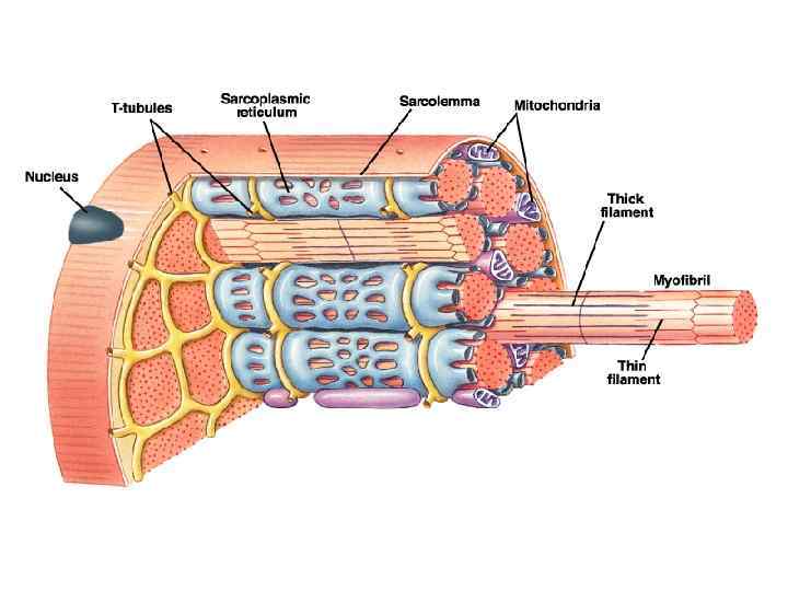
 Мембрана мышечного волокна называется сарколемма Цитоплазма мышечного волокна называется саркоплазма Впячивания сарколеммы внутрь клетки образуют систему трубочек В саркоплазме находится много митохондрий (зачем? ), много гранул гликогена, миофибриллы и саркоплазматический ретикулум
 Саркоплазматический ретикулум • СР – это эндоплазматическая сеть мышечной клетки • Выполняет функции депо для кальция • СР окружает все миофибриллы мышечного волокна.
 Миофиблиллы • Каждое мышечное волокно содержит удлиненные структуры, называемые миофибриллы, которые простираются на всю длину клетки. Каждая миофибрилла состоит из 2500 протофибрилл или миофиламентов. Это нити сократительных белков актина и миозина. • Миозиновые миофиламенты толстые, актиновые тонкие.
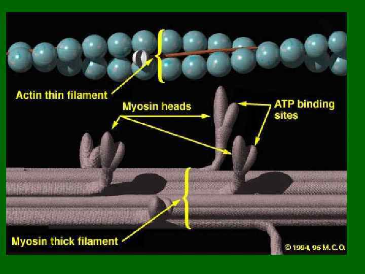
. • Толстые миофиламенты состоят из белка") Миолфиламенты • 2 типа миофиламентов (толстые и тонкие). • Толстые миофиламенты состоят из белка миозина Молекула миозина имеет отростки с головками Около 300 молекул миозина соединяясь вместе образуют один толстый миофиламент
Миолфиламенты • 2 типа миофиламентов (толстые и тонкие). • Толстые миофиламенты состоят из белка миозина Молекула миозина имеет отростки с головками Около 300 молекул миозина соединяясь вместе образуют один толстый миофиламент
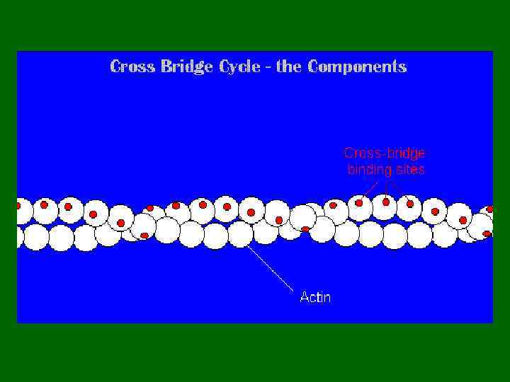
 • Тонкий миофиламент состоит из 3 разных типов белков : актина, тропомиозина и тропонина.
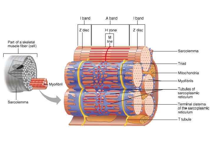
 Миофибриллы • Каждая миофибрилла состоит из тысяч повторяющихся участков, которые называются саркомер (на рис. внизу) • Каждай саркомер содержит тонкие и толстые филаменты – Участки только тонких филаментов (розовые нити) – Участки только толстых филаментов (сиреневые нити) – Участки, содержащие и тонкие и толстые филаменты
 Саркомер • Саркомер ограничен двумя белковыми структурами, которые называются Z пластинки. • Участки саркомера содержащие толстые филаменты называется А-диски (анизотропные). В центре А-диска видна светлая H зона, содержащая только тонкие филаменты и тонкая М-линия
") Участок саркомера, содержащий только тонкие филаменты называется I диск. Под микроскопом выглядит светлым (изотропный) I диск относится к В центре Н зоны видна M линия, которая 2 саркомерам. образована цепями опорных белков, к которым крепятся миозиновые миофибриллы
Участок саркомера, содержащий только тонкие филаменты называется I диск. Под микроскопом выглядит светлым (изотропный) I диск относится к В центре Н зоны видна M линия, которая 2 саркомерам. образована цепями опорных белков, к которым крепятся миозиновые миофибриллы
 Поперечные разрезы миофибриллы
 Поперечно-полосатые мышечные волокна
 • В каждом мышечном волокне есть система Tтрубочек, цистерн и продольных трубочек Т-трубочки и СР Каждая Т-трубочка окружена терминальными цистернами, образуя т. н. триаду, состоящую из 2 цистерн и одной Т-трубочки
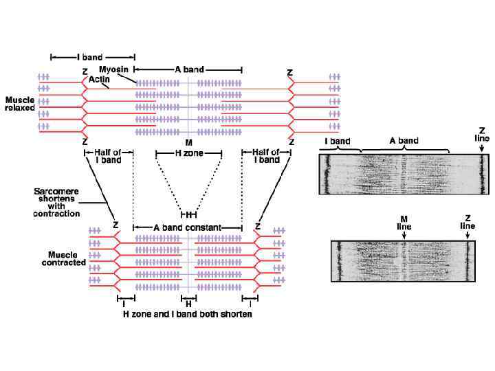
 Мышечное сокращение: Теория скользящих нитей – Тонкие филаменты скользят вдоль толстых филаментов. Мышечное волокно сокращается
 укорачивается.") Филаменты скользят друг относительно друга и саркомер (и мышечная клетка) укорачивается.
Филаменты скользят друг относительно друга и саркомер (и мышечная клетка) укорачивается.
 Электронно-микроскопическая фотография саркомера до и после сокращения.
 Скользящие филаменты • Все саркомеры мышечного волокна сокращаются одновременно. Это приводит к сокращению волокна. От количества сокращающихся волокон зависит сила сокращения целой мышцы.
 Нервно-мышечный синапс
 Медиатор в нервномышечном синапсе - ацетилхолин. Постсинаптическая мембрана содержит холинорецепторы") Нервно-мышечный синапс (концевая пластинка) Медиатор в нервномышечном синапсе - ацетилхолин. Постсинаптическая мембрана содержит холинорецепторы
Нервно-мышечный синапс (концевая пластинка) Медиатор в нервномышечном синапсе - ацетилхолин. Постсинаптическая мембрана содержит холинорецепторы
 Нервно-мышечный синапс
 • Распространение ПД по мышечному волокну
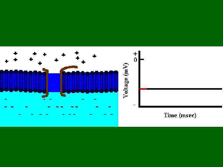
 Возбуждение мышечного волокна ПД распространяется в обе стороны по мышечному волокну. Так как Т-трубочки это впячивания сарколеммы, ПД распространяется по ним.
 Механизм мышечного сокращения • The T-tubular sarcolemma contains voltage sensitive proteins (red arrow in the picture below) that change their conformation in response to a significant Vm. – These are physically linked to calcium channels in the SR membrane – Upon Vm, the voltage sensors change their conformation. This mechanically opens the Ca 2+ channels in the SR membrane.
 Механизм мышечного сокращения
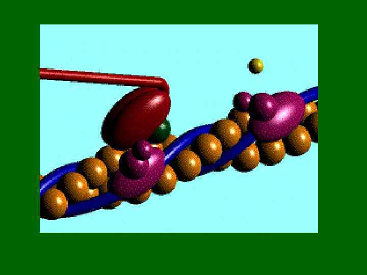
 В состоянии покоя, тропомиозин блокирует присоединение мостиков миозина к нитям актина Когда ноны кальция начинают выходить. из СР, молекула тропонина изменяет свою форму таким образом, что освобождает активные центры актина от тропомиозина Contraction
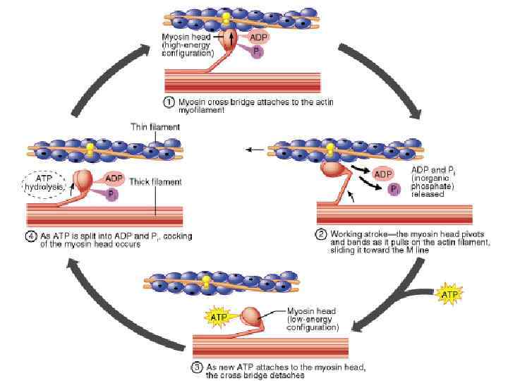
 Сокращение • К этим центрам присоединяются головки миозина. • На головках миозина есть каталитические центры. расщепляющие АТФ до АДФ и неорганического фосфата.
 Сила сокращения • Зависит от: 1. Количества мостиков, которые переходят в активное состояние, 2. Количества миофибрилл в мышечном волокне 3. Количества сокращенных мышечных волокон
 Расслабление мышц • Кальциевый насос мембраны СР работает постоянно, закачивая кальций из саркоплазмы обратно в СР • Кроме того периода, пока Ach соединен с рецепторами мышцы. • ACh выделяется окончаниями моторного нейрона во время его возбуждения • ACh не остается долго соединенным с постсинаптическими рецепторами. • Он быстро отсоединяется, затем соединяется снова, или, что более вероятно, гидролизуется ферментом ацетилхолинэстеразой, который встроен в сарколемму или находится в свободном состоянии в синаптический щели
 Раслабление • Когда стимуляция мышцы прекращается, кальциевые насосы закачивают Са+ в СР. • Са + становится недоступным для тропонина и тропомиозин меняет конфигурацию, закрывая активные центры актина. • Мышца приобретает первоначальную длину за счет эластичности элементов соединительной ткани, сокращения мышц антагонистов и силы гравитации. Но есть и другой способ вызвать расслабление мышцы. Какой процесс показан в этой анимации?
 QUICK THOUGHT QUESTION: In this sculpture, why are the lion’s back legs paralyzed even though they were not injured?
 Rigor Mortis • После смерти, Са насосы перестают работать и содержание Са в клетке повышается, что заставляет миозин соединяться с актином. Поскольку синтез АТФ после смерти прекращается, миозин не может отсоединиться и тело остается в состоянии мышечного окоченения в течение нескольких суток.
 Muscle Metabolism • The chemical energy released by the hydrolysis of ATP is necessary for both muscle contraction and muscle relaxation. • Muscles typically store limited amounts of ATP – enough to power 4 -6 s of activity. – So resting muscles must have energy stored in other ways.
 Resting Muscle and the Krebs Cycle • Resting muscle fibers typically takes up fatty acids from the blood stream. – How might they enter the cell? – Inside the muscle fiber, the FA’s are oxidized to several molecules of a compound called Acetyl-Co. A. This oxidation will also produce several molecules of NADH and FADH 2. – Acetyl-Co. A will then enter a cyclical series of reactions known as the Krebs cycle or Tricarboxylic Acid cycle. – In the Krebs cycle, acetyl-Co. A combines with the compound oxaloacetate and then enters a series of rxns. The end product of these rxns is CO 2, ATP, NADH, FADH 2, and oxaloacetate (thus we call it a cycle)
 Krebs Cycle Products Oxaloacetate will simply combine with another molecule of acetyl-Co. A and reenter the cycle. NADH and FADH will enter another series of rxns known as the Electron Transport Chain. These rxns occur along the inner membrane of the mitochondrion and they basically consist of the passing of electrons from compound to compound with energy being released each time and used to drive the synthesis of ATP. The final electron acceptor is oxygen when it combines with 2 hydrogen atoms to yield water.
 Krebs Cycle Products • CO 2 will diffuse out of the mitochondria, out of the muscle fiber, and into to the blood stream which will take it to the lungs. • The ATP made in the Krebs cycle plus the ATP made during the ETC will be used in many ways. – See if you can list at least 5!
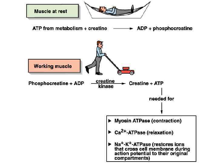
 ATP Use in the Resting Muscle Cell • ATP is necessary for cellular housekeeping duties. • ATP powers the combination of glucose monomers (which have been taken up from the blood stream) into the storage polymer glycogen. • ATP is used to create another energy storage compound called creatine phosphate or phosphocreatine: ATP + Creatine ADP + Creatine-Phosphate this rxn is catalyzed by the enzyme creatine kinase
 Working Muscle • As we begin to exercise, we almost immediately use our stored ATP. • For the next 15 seconds or so, we turn to the phosphagen system, a. k. a. , the energy stored in creatine-phosphate. Creatine-P + ADP Creatine Kinase Creatine + ATP – The ATP is then available to power contraction and relaxation: myosin ATPase, Ca 2+ ATPase in the SR membrane, and Na+/K+ ATPase in the sarcolemma. – The phosphagen system dominates in events such as the 100 m dash or lifting weights.
 Working Muscle • After the phosphagen system is depleted, the muscles must find another ATP source. • The process of anaerobic metabolism can maintain ATP supply for about 45 -60 s. • Anaerobic means “without air, ” and it is the breakdown of glucose without the presence of oxygen. – It usually takes a little time for the respiratory and cardiovascular systems to catch up with the muscles and supply O 2 for aerobic metabolism.
 Anaerobic Metabolism • Glucose is supplied by the breakdown of glycogen or via uptake from the bloodstream. • Glucose is broken down into 2 molecules of pyruvic acid, with the concomitant of 2 ATP and the conversion of 2 molecules of NAD+ into NADH. This process is known as glycolysis and it occurs in the sarcoplasm. – Unfortunately, w/o O 2, we cannot use the NADH in the ETC. – In order for more glycolysis to proceed, the muscle cell must regenerate the NAD+. It does this by coupling the conversion of pyruvic acid into lactic acid with the conversion of NADH into NAD+
 Anaerobic Metabolism • Lactic acid typically diffuses out of muscles into the blood stream and is taken to the liver, kidneys, or heart which can use it as an energy source. • Anaerobic metabolism is inefficient. Large amounts of glucose are used for very small ATP returns. Plus, lactic acid is a toxic end product whose presence contributes to muscle fatigue. • Anaerobic metabolism dominates in sports that requires bursts of speed and activity, e. g. , basketball.
 Aerobic Metabolism • Occurs when the respiratory and cardiovascular systems have “caught up with” the working muscles. – Prior to this, some aerobic respiration will occur thanks to the muscle protein, myoglobin, which binds and stores oxygen. • During rest and light to moderate exercise, aerobic metabolism contributes 95% of the necessary ATP. • Compounds which can be aerobically metabolized include: – Pyruvic acid (made via glycolysis), fatty acids, and amino acids.
 Aerobic Metabolism • It occurs in the mitochondria. • Pyruvic acid from glycolysis is the primary substrate. The cell also utilizes fatty acids and amino acids. • Aerobic respiration typically yields 36 ATP per molecule of glucose. Compare this to anaerobic metabolism.
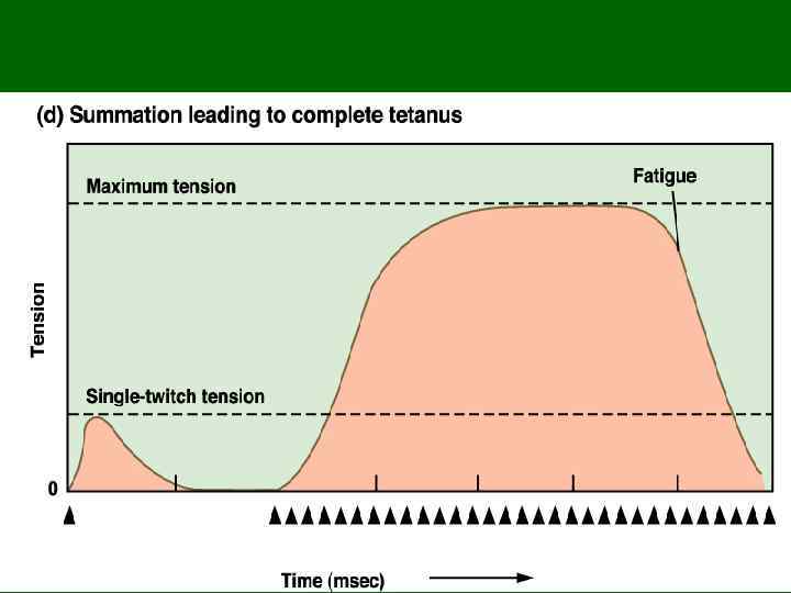
 Muscle Fatigue • Physiological inability to contract • Results primarily from a relative deficit of ATP. • Other contributing factors include the decrease in sarcoplasmic p. H (what causes this? ), increased sarcoplasmic [ADP], and ionic imbalances.
 Oxygen Debt • Refers to the fact that post-exercise breathing rate >>> resting breathing rate • This excess oxygen intake serves many tasks: – Replenish the oxygen stored by myoglobin and hemoglobin – Convert remaining lactic acid back into glucose – Used for aerobic metabolism to make ATP which is used to: • Replenish the phosphagen system • Replenish the glycogen stores • Power the Na+/K+ pump so as to restore resting ionic conditions within the cell.
 Whole Muscle Contraction • Why can you electrically stimulate a muscle to contract? (HINT: what kind of channels could an electric current open? ) • A sub-threshold stimulus would not cause contraction because no AP would be produced! • The response of a muscle to a single suprathreshold stimulus would be a twitch – the muscle quickly contracts and then relaxes. • Let’s take a look at a measurement of a neuron’s AP, a muscle fiber’s AP, and the tension developed by that muscle fiber.
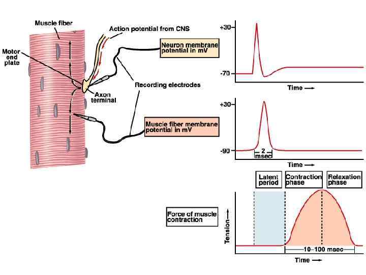
 Phases of the Muscle Twitch 1. Latent Period – Time btwn stimulus and generation of tension – Includes all time required for excitation, excitation-contraction coupling, and stretching of the series elastic components. 2. Contraction 3. Relaxation Now, let’s look at various types of muscle twitches
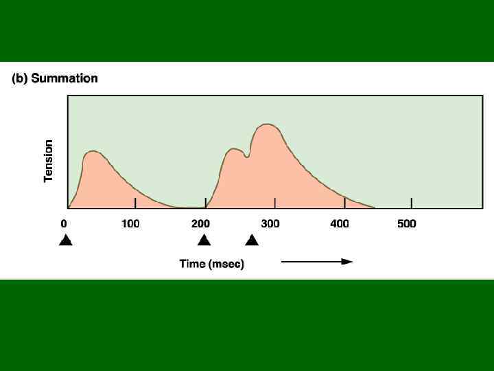
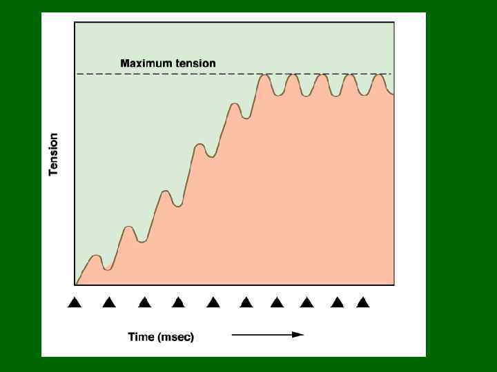
 The black arrows signify stimulation
 • A motor unit is defined as a somatic motor neuron and all the skeletal muscle fibers it innervates. • When this neuron is stimulated, all the muscle fibers it synapses upon will be stimulated and will contract as a unit • The # of muscle fibers per motor unit may be as high as several hundred or as few as four. – The smaller the motor unit, the finer and more delicate the movements. – Extraocular muscles typically have small motor units while the large postural muscles have large motor units Моторная единица Notice that the muscle fibers of a single unit are not clustered together but are spread out. What’s the advantage to this?
 Graded Responses • It should be obvious that you can contract a muscle at just about any rate and with any force you desire. • How does this fact concur with the quickness of a single muscle twitch. – We achieve smooth contractions of the whole muscle by varying the frequency of stimuli sent to the muscle fibers and by recruitment – varying the number and size of the motor units involved Thought problem: compare the act of picking up a pencil with the act of picking up a desk
 Internal vs. External Tension • When a skeletal muscle contracts, the myofibrils inside the muscle fibers generate internal tension. This internal tension is transferred to the series elastic components of the muscle – the fibers of the endomysium, perimysium, epimysium, and tendons. The tension of the SEC is known as external tension. • The SEC behaves like fat rubber bands. They stretch easily at first, but as they elongate they become stiffer and more effective at transferring the external tension to the resistance. – Attach a rubber band to a weight and then try to pick it up. What happens?
 Types of Contractions • Contractions can be: 1. Isometric • Iso= same, metr=measure 2. Isotonic • Iso=same, ton=tension
 Isotonic Contraction • Tension reaches a plateau and then the muscle shortens. Consider the following experiment: 1. A skeletal muscle 1 cm 2 in cross-sectional area can develop roughly 4 kg of force in complete tetanus. 2. If we hang a 3 kg weight from that muscle and stimulate it, the muscle will shorten. 3. Before the muscle can shorten, the cross-bridges must produce enough tension to overcome the resistance – in this case the 3 kg weight. Over this period, internal tension in the muscle fibers rises until the external tension in the tendon exceeds the amount of resistance. 4. As the muscle shortens, the internal and external tensions in the muscle remain constant at a value that just exceeds the resistance.
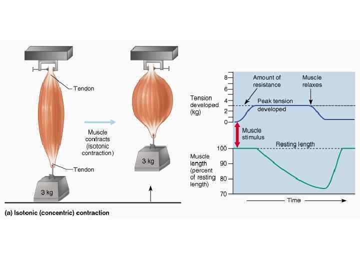
 Resistance and Speed of Contraction • There is an inverse relationship between the amount of resistance and the speed of contraction. • The heavier the load, the longer it takes for the movement to begin because muscle tension, which increases gradually, must exceed the resistance before shortening can occur – More cross-bridges must be formed, more fibers involved. This takes more time.
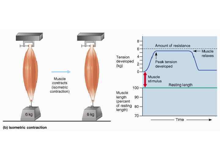
 Isometric Contractions • The muscle as a whole does not change length and the tension produced never exceeds the resistance. • Consider the following: – To the same muscle as before, we attach a 6 kg weight. – Although cross-bridges form and tension rises to peak values, the muscle cannot overcome the resistance of the weight and cannot shorten. – Although the muscle as a whole does not shorten, the individual fibers shorten until the tendons are taut and the external tension equals the internal tension. The muscle fibers cannot shorten further because the external tension does not exceed the resistance.
 Muscle Tone • Some of the motor units w/i particular muscle are always active, even when the muscle is not contracting. – Their contractions do not produce enough tension to cause movement, but they do tense and firm the muscle. – This resting tension in a skeletal muscle is called tone. – The identity of the motor units involved changes constantly. • Why do you suppose this is? • Resting muscle tone stabilizes the position of bones and joints.
 Muscle Fiber Types 2 main types: 1. Slow fibers 2. Fast fibers
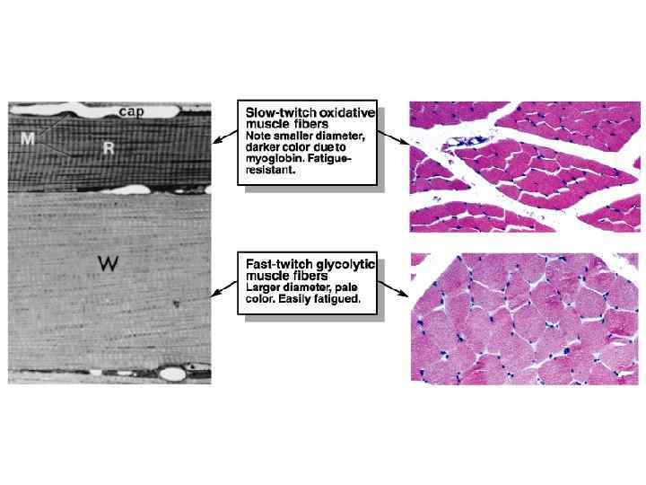
 Slow Fibers • Contract slowly because its myosin ATPases work slowly. • Depends on oxygen delivery and aerobic metabolism. • Is fatigue resistant and has high endurance. • Is thin in diameter – large amt of cytoplasm impedes O 2 and nutrient diffusion. • Cannot develop high tension – small diameter means few myofibrils. • Has rich capillary supply and lots of mitochondria. • Contains lots of the O 2 -storing protein, myoglobin which gives it a red color. • Uses lipids, carbs, and amino acids as substrates for it aerobic metabolism. • Best suited for endurance type activities. • A. k. a. red fibers, slow oxidative fibers, type I fibers.
 Fast Fibers • So named because they can contract in 0. 01 seconds or less after stimulation. • Fast fibers are large in diameter; they contain densely packed myofibrils, large glycogen reserves, and relatively few mitochondria. • Able to develop a great deal of tension b/c they contain a large number of sarcomeres. • Use ATP in massive amounts. Supported by anaerobic metabolism. Fatigue rapidly. • A. k. a. , fast fatigue (FF) fibers, fast glycolytic (FG) fibers, white fibers. • Best suited for short term, power activities.
 Thought questions: why do chickens have white breast meat and dark leg meat? What does this say about the activities of the associated muscles? Why do ducks have dark breast meat?
 Myasthenia Gravis • My=muscle, asthen=weakness, gravi=heavy • Autoimmune disease where antibodies attack the ACh receptors on neuromuscular junctions. • Results in progressive weakening of the skeletal muscles. Why? • Treated w/ anticholinesterases such as neostigmine or physostigmine. These decrease the activity of acteylcholinesterase. – Why would this help someone with myasthenia gravis?
 Muscular Dystrophy • Group of inherited muscle-destroying diseases that generally appear during childhood. • Dys=faulty; Troph=growth • Most common is Duchenne muscular dystrophy – DMD is caused by an abnormal X-linked recessive gene – Diseased muscle fibers lack the protein dystrophin which normally links the cytoskeleton to the ECM and stabilizes the sarcolemma – Age of onset is btwn 2 and 10. Muscle weakness progresses. Afflicted individuals usually die of respiratory failure, usually by age 25. Here is a slide of skeletal muscle from someone with DMD. Look how much connective tissue there is. Lots of adipose tissue too. Why do you think there’s so much?
 Other Important Terms • Flaccid paralysis – Weakness or loss of muscle tone typically due to injury or disease of motor neurons • Spastic paralysis – Sustained involuntary contraction of muscle(s) with associated loss of function • How do flaccid and spastic paralysis differ? • Spasm – A sudden, involuntary smooth or skeletal muscle twitch. Can be painful. Often caused by chemical imbalances.
 Other Important Terms • Cramp – A prolonged spasm that causes the muscle to become taut and painful. • Hypertrophy – Increase in size of a cell, tissue or an organ. • In muscles, hypertrophy of the organ is always due to cellular hypertrophy (increase in cell size) rather than cellular hyperplasia (increase in cell number) • Muscle hypertrophy occurs due to the synthesis of more myofibrils and synthesis of larger myofibrils.
 Other Important Terms • Atrophy – Reduction in size of a cell, tissue, or organ • In muscles, its often caused by disuse. Could a nerve injury result in disuse? Why might astronauts suffer muscle atrophy? • Fibrosis – Replacement of normal tissue with heavy fibrous connective tissue (scar tissue). How would fibrosis of skeletal muscles affect muscular strength? How would it affect muscle flexibility?
 Smooth Muscle • Involuntary, non-striated muscle tissue • Occurs within almost every organ, forming sheets, bundles, or sheaths around other tissues. • Cardiovascular system: – Smooth muscle in blood vessels regulates blood flow through vital organs. Smooth muscle also helps regulate blood pressure. • Digestive systems: – Rings of smooth muscle, called sphincters, regulate movement along internal passageways. – Smooth muscle lining the passageways alternates contraction and relaxation to propel matter through the alimentary canal.
 Smooth Muscle • Integumentary system: – Regulates blood flow to the superficial dermis – Allows for piloerection • Respiratory system – Alters the diameter of the airways and changes the resistance to airflow • Urinary system – Sphincters regulate the passage of urine – Smooth muscle contractions move urine into and out of the urinary bladder
 Smooth Muscle • Reproductive system – Males • Allows for movement of sperm along the male reproductive tract. • Allows for secretion of the non-cellular components of semen • Allows for erection and ejaculation – Females • Assists in the movement of the egg (and of sperm) through the female reproductive tract • Plays a large role in childbirth
 Smooth Muscle • Smooth muscle cells: – Are smaller: 5 -10 um in diameter and 30 -200 um in length – Are uninucleate: contain 1 centrally placed nucleus – Lack any visible striations – Lack T-tubules – Have a scanty sarcoplasmic reticulum • Smooth muscle tissue is innervated by the autonomic nervous system unlike skeletal muscle which is innervated by the somatic nervous system (over which you have control) • Only the endomysium is present. Nor perimysium or epimysium.
 Smooth Muscle Contraction • Myosin and actin are present and crossbridge formation powers contraction, but the thick and thin filaments do not have the strict repeating arrangement like that found in skeletal muscle. • There are no Z discs, instead thin filaments are attached to protein structures called dense bodies which attach to the sarcolemma.
 Smooth Muscle • Smooth muscle is always maintaining a normal level of activity – creating muscle tone. • Smooth muscle can respond to stimuli by altering this tone in either direction. – Smooth muscle can be inhibited and relax – Smooth muscle can be excited and contract • Possible stimuli include neurotransmitters, hormones, p. H, Pco 2, Po 2, metabolites (such as lactic acid, ADP), or even stretch.
 Smooth Muscle Contraction • Begins with the opening of membrane channels. Channels may be ligandgated (NTs, hormones, metabolites), voltage-gated, or mechanically-gated (stretch). • Channels will allow significant calcium entry from the ECF. Remember smooth muscle has little SR. • Calcium binds to a regulatory molecule called calmodulin and activates it. • Activated calmodulin activates an enzyme called Myosin Light Chain Kinase.
 Smooth Muscle Contraction • Activated MLCK will add a phosphate group to the myosin of the thick filament. This enables the myosin to interact with actin. – Tropomyosin is present but not blocking actin’s myosin binding sites – Troponin is not present • Contraction then ensues.
 Smooth muscle relaxation: Calcium is pumped out of the cell, which decreases the amount of active calmodulin which decreases the amount of active MLCK which decreases the number of crossbridges. Relaxation can occur subsequent to contraction or at any time if anything causes a decrease in the calcium permeability of the smooth muscle cell. Why are calcium channel blockers given to people with hypertension?
 Types of Smooth Muscle • Smooth muscle varies widely from organ to organ in terms of: – Fiber arrangement – Responsiveness to certain stimuli • How would the types of integral proteins that a smooth muscle cell contained contribute to this? • Broad types of smooth muscle: – Single unit (a. k. a. visceral) – Multi unit
 Single Unit Smooth Muscle • More common • Cells contract as a unit because they are all connected by gap junctions - protein complexes that span the PM’s of 2 cells allowing the passage of ions between them, i. e. , allowing the depolarization of one to cause the depolarization of another. • Some will contract rhythmically due to pacemaker cells that have a spontaneous rate of depolarization.
 Single Unit Smooth Muscle • Not directly innervated. Diffuse release of neurotransmitters at varicosities (swellings along an axon). • Responsive to variety of stimuli including stretch and concentration changes of various chemicals • Found in the walls of the digestive tract, urinary bladder, and other organs
 Multi-Unit Smooth Muscle • Innervated in motor units comparable to those of skeletal muscles • No gap junctions. Each fiber is independent of all the others. • Responsible to neural & hormonal controls • No pacemaker cells • Less common • Found in large airways to the lungs, large arteries, arrector pili, internal eye muscles (e. g. , the muscles that cause dilation of the pupil) • Why is good to have the digestive smooth muscle single unit and the internal eye muscles multi-unit?
 Cardiac Muscle • Striated, involuntary muscle • Found in walls of the heart • Consists of branching chains of stocky muscle cells. Unior binucleate. • Has sarcomeres & T-tubules • Cardiac muscle cells are joined by structures called intercalated discs – which consist of desmosomes and gap junctions. – Why do you suppose these are necessary? Notice the branching and the intercalated disc, indicated by the blue arrow.