
Muscle tissues Ass. Professor Goriachkina Valeria Lvovna


- Размер: 37.8 Mегабайта
- Количество слайдов: 33
Описание презентации Muscle tissues Ass. Professor Goriachkina Valeria Lvovna по слайдам
Muscle tissues Ass. Professor Goriachkina Valeria Lvovna
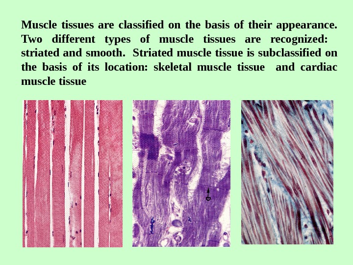
Muscle tissues are classified on the basis of their appearance. Two different types of muscle tissues are recognized: striated and smooth. Striated muscle tissue is subclassified on the basis of its location: skeletal muscle tissue and cardiac muscle tissue
General Features of Skeletal Muscle Tissue 1. It is form of striated muscle fibers. 2. Each muscle fiber contains many nuclei. They are located immediatly under the plasma membrane. 3. Each muscle fiber contains parallel myoffibrils (80% myofibrils in the fibre), which are trasversely striated. 4. The cytoplasm of the muscle fiber is known as Sarcoplasm (Greek sarkos — flesch) 5. The sarcoplasm contains glycogen, lipids and myoglobin (pigmental protein; it can take up, store, or give up, oxygen as needed). 6. The function of this tissue is controled voluntarily — it could be contracted or relaxe by will skeletal muscle came to be known also as voluntary muscle
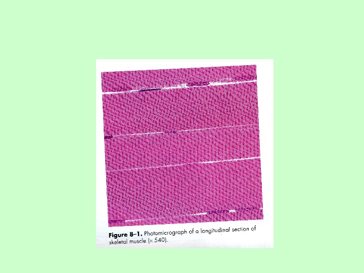
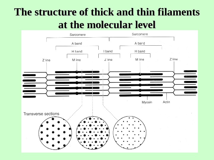
Skeletal muscle tissue Each myofibril has characteristic banding patterns (dark and light bands). When observed under polarized light, the dark-staining bands are birefringent (anisotropic), while the light-staining ones are isotropic. Accordingly, the dark bands are called A bands ( A for anisotropic) and the light ones, I bands (I for isotropic). Owing to these alternations of dark and light bands, transverse striations in a muscle fiber can be seen with light microscope. The arrangement of the contractile proteins within skeletal muscle fiber
The structure of thick and thin filaments at the molecular level
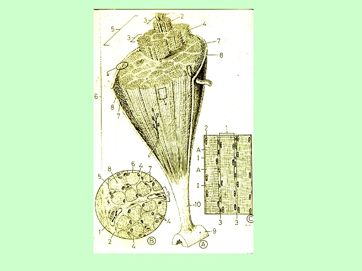
The structure of sarcomere
Sarcotubular system is composed of agranular (smooth) sarcoplasmic reticulum ( L-tubules ) and T-tubules
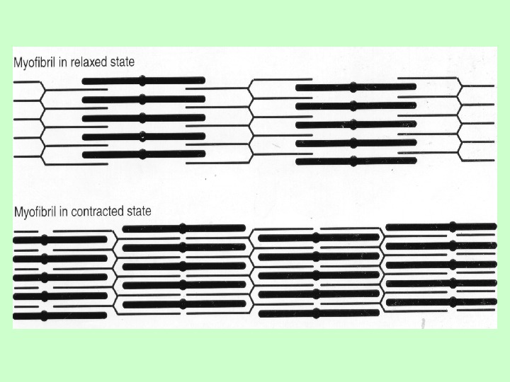
Contraction cycle of skeletal muscle
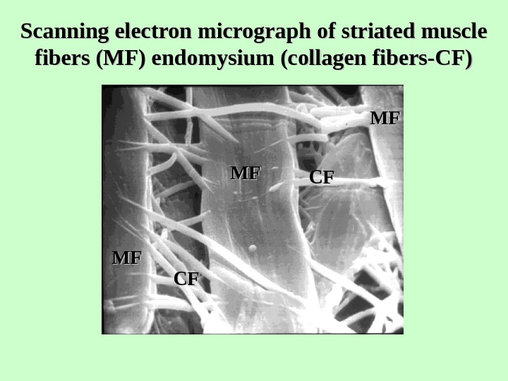
Scanning electron micrograph of striated muscle fibers (MF) endomysium (collagen fibers-СF) MFMF СFСFMFMF СFС
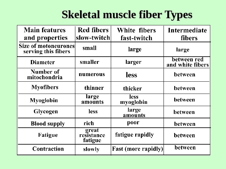
Skeletal muscle fiber Types less
Type of muscle fibres Aerobic (type I), anaerobic (type II) and intermediate fibres The activity of the specific mitochondrial enzyme succinate dehydrogenase ATP-ase activity
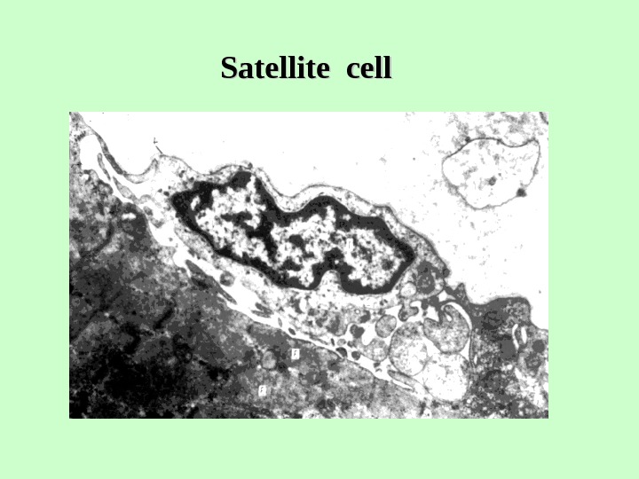
Histogenesis of Skeletal Muscle Fibers 1. From the myotomes arise myoblasts (two cell populations). 2. a) On one side presumptive myoblasts differentiate into true myoblasts. b) On other, presumptive myoblasts remain undifferentiated and give rise to satellite cells. 3. a) True myoblasts range in rows, fuse together and form myotube. b) Satellite cells adhere to myotube. 4. Myotube gradually differentiates into skeletal muscle fiber.
Satellite cell
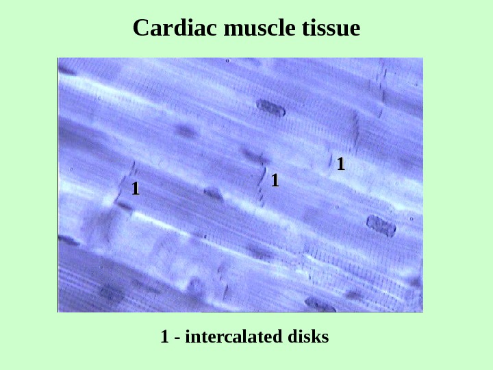
General Features of Cardiac Muscle Tissue 1. It is formed of striated muscle cells. 2. Each muscle cell contains one or two nuclei. They are located in the central part of the cell. 3. Each muscle cell contains parallel myofibrils (40% of the myofibrils in the cell), which are transversely striated. 4. Each cardiac muscle cell contains many (40% of the mitochondria in the cell). 5. Cardiac muscle cells are joined end — to — end by the intercalated discs (junctional complex of the two cell membranes of two adjacent cardiac muscle cells). 6. Cardiac muscle is involuntary striated muscle.
Cardiac muscle tissue 11 11 11 1 — intercalated disks
Cardiac muscle cells 3311 22 IDID 11 ID — Intercalated disk: 1 — desmosomes; 2 — fascia adherens; 3 — nexus.
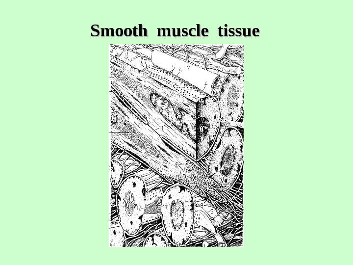
General Features of Smooth Muscle Tissue 1. It is formed of smooth muscle cells. 2. Each smooth muscle cell contains one rod-shape nucleus. It is located in the center of the cell. 3. Each smooth muscle cell contains myofibrils. They lack any cross striations. 4. Smooth muscle tissue is called involuntary muscle, because it is not controlled by the will.
Smooth muscle tissue
Muscular artery
Smooth muscle tissue
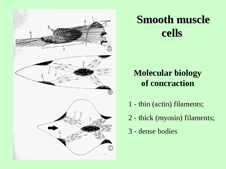
Smooth muscle cells Molecular biology of concraction 1 — thin (actin) filaments; 2 — thick (myosin) filaments; 3 — dense bodies