95b3b6f76f823a2a1126c6ef2761012d.ppt
- Количество слайдов: 84


Multisystem Disease 2 Lymphoma A Swartbooi Clinical Imaging Sciences 28 September 2012

Extranodal Lymphomatous infiltration of anatomic sites other than the lymph nodes n Primary and secondary involvement n Occur in both HD and NHL n – More common in NHL

Extranodal Lymphoma n Primary involvement – 30 -40% of cases – Lymph nodal involvement limited to regional nodes – Stage I or II disease – 1⁰ extranodal HD ext rare – disease involvement elsewhere for exclusion – Increased incidence noted as frequency of NHL is seen n Seen esp of CNS and Orbits

Extranodal Lymphoma n Primary involvement – Incidence of NHL n Age of pt (⇧ children) n Immune status (⇧ with ⇩ CD 4) – More aggressive subtype n Pathological subtype – Mantle cell lymphoma – Lymphoblastic lymphoma – Burkitt lymphoma – MALT lymphoma

Extranodal Lymphoma n Secondary involvement – Presence of widespread advanced disease elsewhere – Both HD and NHL n NHL more common – Adverse prognostic factor n Stage III or IV disease

Thorax n Pulmonary parenchyma – 2⁰ Involvement n HD more common 12% than NHL 4% n Intrathoracic LN n Extrathoracic LN – Direct ext of nodal disease n Paramediastinal / Perihilar lung involvement

Thorax
 mediastinal HD often confined")
Thorax n Pulmonary parenchyma – Recurrence after previously Rx (radiation) mediastinal HD often confined to lungs – In compar pt with NHL and pulmonary or pleural disease n No mediastinal nodes in 50% – As disease progress/relapse n Lung disease become common n Pt with HD 30 -40% have pulmon involvement

Thorax n Pulmonary parenchyma – 1⁰ Involvement n < 1% of lymhoma n Primary NHL – Usually low grade B-cell NHL (80 -85%) n Commonly single lung nodule n Multiple nodules or segm consolidation may occur n Pleural eff in 20% – High grade NHL (15 -20%) n Rapidly enlarging single or multiple nodules

Thorax n Pulmonary parenchyma – 1⁰ Involvement n Primary HD – Extremely rare – Single or multiple nodules – Upper lobe – Cavitate – Pleural eff in 20%

Thorax n Pleural disease – Pleural effusions – Usually accompanied by mediastinal lymphadenopathy – 10% of patients with NHL and 7% of patients with HD – Secondary to central lymphatic or venous obstruction – Rx of mediastinal disease – Pleural masses more common in relapse than presentation

Thorax

Thorax n Heart & Pericardium – Can occur with high-grade peripheral T-cell and large B-cell lymphomas – Direct spread rare n Except in AIDS related lymphoma (ARL) and posttransplant lymphoproliferative disorders (PTLD) – Pericardial effusions occur in 6% of patients with HD at presentation n Associated mass adjacent to heart

Thorax n Thymus – 1⁰ Thymic HD rare n Involvement noted in assoc with mediastinal nodes in 30 -50% n Often large B-cell lymphoma n Young woman (25 -40 yrs) n Typically rapidly growing with SVC obstr. (40%) n Thymus homogenous or heterogenous; may be nodular (lobulated) or smooth

Thorax n Thymus

Thorax n Chest wall – From direct extension n Ant Mediastinal nodes n Ax & Supra clav nodes – No bony destruction n Infection or Malignancy

Breast Widespread disease elsewhere n NHL of breast rare n – 2% lymphomas; 0. 5% breast malignancy n Bimodal – Pregnancy and lactation n High grade or burkitt n Billat with inflammatory picture

Breast

Breast n Bimodal – 50 yrs n Unilateral n Discrete often solitary mass n No archicteral distortion/skin thickening n No calcifications n 2⁰ involvement characterized by multiple nodules and enlarged LN’s

Abdomen n Hepatobiliary system – Liver involvement n 15 % in NHL – ⇧ in Paeds and with recurrence n 5% in HD n Primary hepatic disease rare (< 1%) – Increased in pt Hep. A or Hep. C + (25%) n Infiltration around portal tracts most common n Hepatomegaly suggest diffuse involvement

Abdomen n Hepatobiliary system – Liver involvement n 5 -10% focal infiltration – Resemble metastases n Hypoechoiec and well defined on US – GB n NHL ARL Rare, but ⇧ frequency in patients with

Abdomen n Spleen – Affected in 20% of patients with NHL – 30– 40% of patients with HD at presentation, considered a nodal organ – Presence of nodal disease above and below the diaphragm (stage III), but in a small proportion it is the sole focus of intra-abdominal disease. – Microscopic diffuse infiltration with only splenomegaly (not indicate involvement)

Abdomen n Spleen – 33% splenomeg – no infiltration – 33% normal – infiltration – Volume + indices not used – Focal lesions 10 -25% > 1 cm detected by cross sect imaging – 40% of NHL has involvement some time

Abdomen n Spleen – Imaging n Solitary, miliary, multiple masses n Ddx: infection, granulomatous disease n CT & US sensitive (us >3 mm) n MRI (not routine) n FDG–PET > CT or Scintigraphy – 1⁰ splenic lymphoma rare (1%)

Abdomen n Spleen

Abdomen n GIT – Most common site of 1⁰ EN NHL n 30 -45% of all n 10% adults + 30% children – 1⁰ HD of GIT rare n 2⁰ involvement common n Direct extension, multiple sites – 1⁰ lymphoma from lamina propria + submucosa
 – Sixth")
Abdomen n GIT – Frequently below the age of 10 years (BL) – Sixth decade (MALT type enteropathyassociated T-cell type) – Stomach 50%, Small bowel 35%, large bowel 15%

Abdomen n GIT – Stomach n 2– 5% of all gastric tumours n Submucosa, affecting the antrum more – Imaging n Multiple nodules, some with central ulceration, or a large, fungating lesion with or without ulceration, wall thickening, luminal narrowing, enlargement of the gastric folds (barium studies or endoscopically)

Abdomen n GIT – Imaging n Gastric wall thickening and accompanying nodal involvement, which is well shown on CT n Low grade – shallow ulceration n High grade – massive infiltration, masses

Abdomen n GIT – Small bowel n 50% of all primary tumours of the small bowel n Term ileum frequent, duodenal rare n Multifocal in 50% n Obstructive symptoms n Bowel wall thickening is well demonstrated on CT n With infiltration alternating areas of dilatation and constriction

Abdomen n GIT – Submucosal infiltration with multiple nodules or polyps of varying size result in intususception (most common cause < 6 yrs) – Barium studies typically show multiple polypoid filling defects, with or without central ulceration and irregular thickening of the valvulae.

Abdomen n GIT – Colon and rectum n Primary colonic lymphomas are usually of Burkitt or MALT subtypes n <0. 1% of all colonic neoplasms n caecum and rectum n diffuse or segmental distribution of small nodules 0. 2– 2. 0 cm in diameter n Mucosa intact n Polypoid mass uncommon, mimic ca unless term ileum involvement

Abdomen n GIT – Advanced disease, there may be marked thickening of the colonic or rectal folds resulting in focal strictures, fissures or ulcerative masses with fistula formation – Strictures longer than ca – Obstruc and rectal bleeding

Abdomen n GIT – Primary gastrointestinal lymphoma n An absence of superficial or intrathoracic lymph node enlargement n No involvement of the liver or spleen n A normal white cell count n No more than local regional lymph node enlargement

Abdomen n GIT

Abdomen n Pancreas – 1⁰ cases <1. 3% of pancreas tumours – 2% of NHL – Indistinguishable from primary adenocarcinoma on US, CT, or MRI – 2⁰ with disease elsewhere n Direct infiltration
 n 50% of")
Genito-urinary Tract Not commonly involved at the time of presentation (<5%) n 50% of patients will have involvement of some part of the genitourinary tract at autopsy n Testicle is the most commonly involved organ, followed by the kidney and the perirenal space n

Genito-urinary Tract Rarely are the bladder, prostate, uterus, vagina, or ovaries involved n 1⁰ cases rare, little lymphoid tissue n

Genito-urinary Tract n Kidneys – 90% of cases are in association with high-grade NHL (BL) – Often in recurrence, normal function – Multiple masses 60% n n n US hypoechoic Density reversal pattern on CT Solitary mass in 5 -15%, indistinguishable RCC NHL renal mass often no retroperit nodes Direct infil in 25% Vessel encasement, hilar extension

Genito-urinary Tract n Kidneys

Genito-urinary Tract n Testis – 5% of primary testicular tumours overall and 25– 50% of those in patients over 50 years – Commonest primary tumour over the age of 60 years – 1% of all patients of NHL, usually with DLBCL or BL – Association with lymphoma of Waldeyer's ring, the skin and central nervous system – Painless testicular swelling

Genito-urinary Tract n Testis – Ultrasonically, the lesions usually have a nonspecific appearance – Staging must always include ultrasonic evaluation of the contralateral testis and whole body CT

Adrenal Glands n n 25% of patients with NHL will develop secondary involvement of the adrenal gland Primary adrenal lymphoma is extremely rare, usually occurring in men over the age of 60. Secondary involvement detected in about 6% undergoing routine abdominal staging CT, in the presence of widespread retroperitoneal disease Indistinguishable from metastases

Central Nervous System n 1⁰ CNS Lymphoma – – – – Brain and spine only 3% of brain tumours 30% of NHL – immunodeficiency states n 6% AIDS pt get PCNSL Peaks 40 -60 yrs Lesions often frontal (personality) White matter close to CC (spread across) Isointense on CT&MRI, Hom enhancement

Central Nervous System n 2⁰ CNSL – 15% of NHL – In HD brain lesions often second dx – Often extra-axial and spinal spaces – MRI more sensitive (enhancing plaques) – Spinal presentation often from extension of prevertebral mass (dum-bell tumour)

Central Nervous System n CNSL

Central Nervous System n Orbital lymphoma – Pt’s 40 -70 yrs – Slow growing, infiltrating – NHL most common n 10 -15% of primary orbital tumour in adults n 4% primary EN NHL – 2⁰ cases 3. 5 – 5% of NHL & HD – Can involve any part, symptoms acc to site
 –")
Head & Neck n Waldeyer’s ring – Commonest site (1/3 of EN NHL) – Lymphoid tissue in nasopharynx, oropharynx, faucial palatine and lingual tonsils – Circumferential or multifocal involvement – 8% Paranasal sinus tumours are NHL – Assoc with GIT involvement – Most are DLBCL – MRI preferred imaging method

Head & Neck n Thyroid – NHL 2% malignant thyroid tumours – MALT type associated with Hashimoto’s – Can extend beyond gland – Hypoechoic on US – Low density on CT

Head & Neck n Salivary glands – All may be involved n Parotid most common – MALT most common – Single or multiple masses – High density on CT – Hypoechoic on US – Common in middle aged woman – Assoc with Sjogren’s disease

Musculoskeletal System Involvement of the bone, bone marrow and skeletal muscles can occur in both HD and NHL n NB sites of disease relapse n

Musculoskeletal System n Bone marrow – Lymphoma may arise within the marrow as true primary disease – Stage 1 E (present in 50 -80% low grade NHL) – Stage 4 if with widespread disease – In high grade NHL, marrow involvement is present in 20– 40% - poorer prognosis than liver and lung – BM biopsy NB in all staging – In HD, marrow involvement at presentation is rare (5 -15% during course)

Musculoskeletal System n Bone marrow – MRI – extremely sensitive in detecting bone marrow involvement n Low signal intensity on T 1 -weighted images and high signal on STIR sequences – FDG-PET is also very sensitive for bone marrow involvement at presentation

Musculoskeletal System n Bone – Primary lymphoma of bone is nearly all NHL (1% of NHL) - DLBCL – < 5% of bone malignancy – Appendicular skeleton in 1⁰ cases – eg. femur – Most cases secondary and HD (10 -25%) – Average at presentation is 24 years – Males>females – NHL lytic, HD sclerotic(skull, spine – ivory verteb)

Musculoskeletal System n Bone – Criteria for dx of primary lymphoma n Only a single bone is involved n There is unequivocal histological evidence of lymphoma n Other disease is limited to regional areas at the time of presentation n The primary tumour precedes metastases by at least 6 months

Musculoskeletal System n Bone – Imaging features of osseous lymphoma at conventional radiography, CT, and MR imaging are nonspecific – Radionuclide radiology has a sensitivity of close to 95% in the detection of bone involvement, far greater than plain radiography – FDG PET has proved to be more specific and sensitive than conventional bone scintigraphy in identifying osseous involvement by malignant lymphoma – MRI extraosseous and marrow involvement (1⁰ bone Lymphoma staging)

Musculoskeletal System n Bone

Musculoskeletal System n Bone

Musculoskeletal System n Cutaneous Lymphoma – Primary cutaneous lymphoma is the second most prominent group of NHLs, whereas primary or secondary skin involvement is very rare in patients with Hodgkin disease. – Almost 65% of primary cutaneous lymphomas are T-cell lymphomas, with the remainder being B-cell lymphomas – About 25% of primary cutaneous lymphomas demonstrate extracutaneous involvement at the time of diagnosis

Mucosa Associated Lymphoid Tissue Lymphoma n From mucosal sites that normally have no organized lymphoid tissue – Lymphoid tissue arise as a result of chronic inflammation or autoimmunity – Hashimoto's thyroiditis n 70 x increased risk for thyroid lymphoma – Sjögren's syndrome n 44 x increased risk for lymphoma – Helicobacter-induced chronic follicular gastritis

Mucosa Associated Lymphoid Tissue Lymphoma n n Often Adults - median age 60 BM involved in 20% Multiple sites in 10% Most common site – GIT (50%) n Stomach (85%) – Others n lung, head & neck, ocular adnexae, skin, and breast

Burkitt’s Lymphoma n n n Highly aggressive B-cell variant of NHL Associated with Epstein–Barr virus Extremely aggressive, rapidly growing but potentially curable 2– 3% of NHL 30– 50% of all childhood lymphoma

Burkitt’s Lymphoma 1. 2. Endemic form – Jaws and orbit are involved in 50% of cases – Ovaries, kidneys and breast may be involved Sporadic form – Predilection for the ileocaecal region (present with intussusception) – Also ovaries, kidneys and breasts – Retroperitoneal and paraspinal disease can cause paraplegia – Thoracic disease rare

Burkitt’s Lymphoma 3. n Immunodeficiency associated BL – Association with HIV infection – Initial manifestation of AIDS – EBV is identified in up to 40% of cases Extranodal disease is common and all three variants are at risk for CNS disease

Immunosuppression n Four WHO categories – Primary immunodeficiency – Infection with HIV – Immunosuppresion following transplant – Immunosupression for autoimmune disorders

Immunosuppression n Lymphoma and HIV – AIDS defining in 5% – Incidence of all subtypes of NHL is increased 60– 200 x – Incidence of HD increased up to 8 x – Tumours are aggresive, advanced – Involve GIT, CNS(multiple), Liver & BM – Multiple EN sites in 75%

Immunosuppression n Post transplant lymphoproliferative disorder – Low risk 1% ( renal and BM transplant ) – Highest 5% (heart-lung, liver-bowel) – Onset and site of EN disease correlate to transplant type and type of suppression n Eg. Cyclosporine affect GIT > CNS

Relapse n 10 -40% of pt’s with HD – Within 1 st 2 yrs 50% of pt’s with NHL n Follow up CT according to institution guideliness, pt’s symptoms n Functional imaging can detect relapse earlier than CT n

Post Treatment Evaluation n Imaging plays a critical role in monitoring response to treatment and in assessing the degree of response Lesions to be measured before, during and after treatment Well defined lesions either side of the diaphragm

Post Treatment Evaluation n Response criteria – HD n Complete response when no clinical or radiological evidence of disease n Complete remission – – – No clinical evidence Radiologcal abn not related to Rx Additional imaging or biopsy

Post Treatment Evaluation n Reponse criteria – NHL n Comple remission – No clinical or Rad evidence of disease – Nodes of >1. 5 cm now <1. 5 cm or 1 -1. 5 cm nodes <1 cm – Spleen normal sized with resolution of lesions – Marrow if involved, now cleared (BM biopsy proven)

Post Treatment Evaluation n Response Criteria – NHL n Complete remission – unconfirmed – Residual mass >1. 5 cm reduction of 75% in SPD n Partial response – > 50% reduction in SPD of all lesions – No progression n Progression – > 50% increase in SPD of mass – New lesion

Post Treatment Evaluation n CT – Imaging characteristics of extranodal involvement can be subtle or absent at conventional computed tomography – Response criteria n Remmision n Residual disease n Progression – Unable to diff Rx changes from residual on density – Limited sensitivity in detecting lymphomatous involvement of normal-sized lymph nodes, bone marrow, spleen, and extranodal tissues.

Post Treatment Evaluation n MRI – Differentiate active tumour from necrosis – Tumours often high signal intensity on T 2 n Residual or recurrent disease can be ID – Difficult in small lesions – False positive early with inflammation and necrosis

Post Treatment Evaluation n FDG-PET – Has facilitated the identification of affected extranodal sites, even when CT has demonstrated no lesions – Predict eventual outcome of NHL (early – after 1 -3 Cycles of Chemo) more accurately than PET – Ineffective Rx can be changed early – Sensitivity and specificity for the detection of residual disease after completion of first-line therapy are 84% and 90%, respectively, for Hodgkin disease, and 72% and 100%, respectively, for aggressive NH

Post Treatment Evaluation n FDG Uptake

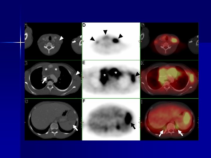
Post Treatment Evaluation n PET-CT – Certain PET/CT patterns are suggestive of extranodal disease and can help differentiate tumor from normal physiologic FDG activity, particularly in the mucosal tissues, bone marrow, and organs of the gastrointestinal tract – Standard imaging modality for initial staging, follow-up, and treatment response in some centre’s – NHL & HD upstaged in 31%

Post Treatment Evaluation n Radionuclide Imaging – Gallium-67 can be used n Taken up by tumour and not in fibrotic or necrotic tissue n ⇩ uptake – good response and the reverse n False +, in thymic hyperplasia, inflammation and benign uptake

PET-CT vs CE CT

Take Home Points n n n Most common extranodal sites of involvement are the stomach, spleen, Waldeyer ring, central nervous system, lung, bone, and skin Diagnosis of primary versus secondary extranodal lymphoma remains challenging FDG PET/CT is now the proposed imaging modality of choice for staging and follow-up in Hodgkin disease and most NHLs

Take Home Points n n It is important to determine whether extranodal involvement represents a primary manifestation or dissemination of systemic disease, which has a poorer prognosis At any stage during the course of the disease, the potential presence of extranodal involvement should be considered

References n n n Grainger & Allison – Diagnostic Radiology 5 th Ed; pages 1740 -1757 Paes, Kalkanis, Sideras, Serafini. FDG PET/CT of Extranodal Involvement in Non-Hodgkin Lymphoma and Hodgkin Disease. January 2010 Radio. Graphics, 30, 269 -291. Raanani P, Shasha Y, Perry C, et al. Is CT scan still necessary for staging in Hodgkin and non-Hodgkin lymphoma patients in the PET/CT era? Ann Oncol 2006; 17: 117– 122.

References n n Guermazi, Brice, Deb. Kerviler, Fermé. Extranodal Hodgkin Disease: Spectrum of Disease January 2001 Radio. Graphics, 21, 161 -179 Kwee TC, Kwee RM, Nievelstein RA. Imaging in staging of malignant lymphoma: a systematic review. Blood 2008; 111: 504– 516
95b3b6f76f823a2a1126c6ef2761012d.ppt