
Mucocutaneous Diseases Azmi Darwazeh BDS. ,


- Размер: 9 Mегабайта
- Количество слайдов: 64
Описание презентации Mucocutaneous Diseases Azmi Darwazeh BDS. , по слайдам
Mucocutaneous Diseases Azmi Darwazeh BDS. , MSc. , Ph. D. , FFD RCSI. Professor of Oral Medicine & Pathology Faculty of Dentistry Jordan University of Science & Technology
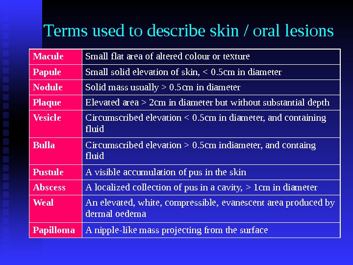
Terms used to describe skin / oral lesions Macule Small flat area of altered colour or texture Papule Small solid elevation of skin, 0. 5 cm in diameter Plaque Elevated area > 2 cm in diameter but without substantial depth Vesicle Circumscribed elevation 0. 5 cm indiameter, and containg fluid Pustule A visible accumulation of pus in the skin Abscess A localized collection of pus in a cavity, > 1 cm in diameter Weal An elevated, white, compressible, evanescent area produced by dermal oedema Papilloma A nipple-like mass projecting from the surface
Petechiae Pinhead-size macule of blood in the skin / mucosa Purpura A large macule or papule of blood Ecchymosis A large extravasation of blood into skin / mucosa Haematoma A swelling from gross bleeding Telangiectasia The visible dilatation of small blood vessels
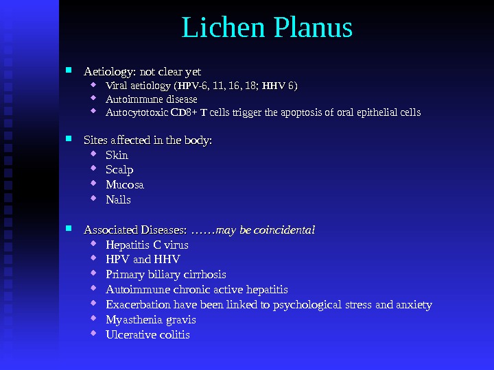
Lichen Planus Aetiology: not clear yet Viral aetiology (HPV-6, 11, 16, 18; HHV 6) Autoimmune disease Autocytotoxic CD 8+ T cells trigger the apoptosis of oral epithelial cells Sites affected in the body: Skin Scalp Mucosa Nails Associated Diseases: …… may be coincidental Hepatitis C virus HPV and HHV Primary biliary cirrhosis Autoimmune chronic active hepatitis Exacerbation have been linked to psychological stress and anxiety Myasthenia gravis Ulcerative colitis
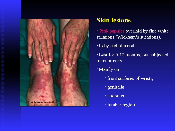
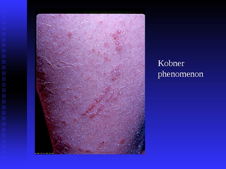
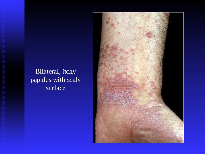
Skin lesions : • Pink papules overlaid by fine white striations (Wickham’s striations). • Itchy and bilateral • Last for 9 -12 months, but subjected to recurrency • Mainly on • front surfaces of wrists, • genitalia • abdomen • lumbar region
Kobner phenomenon
Bilateral, itchy papules with scaly surface
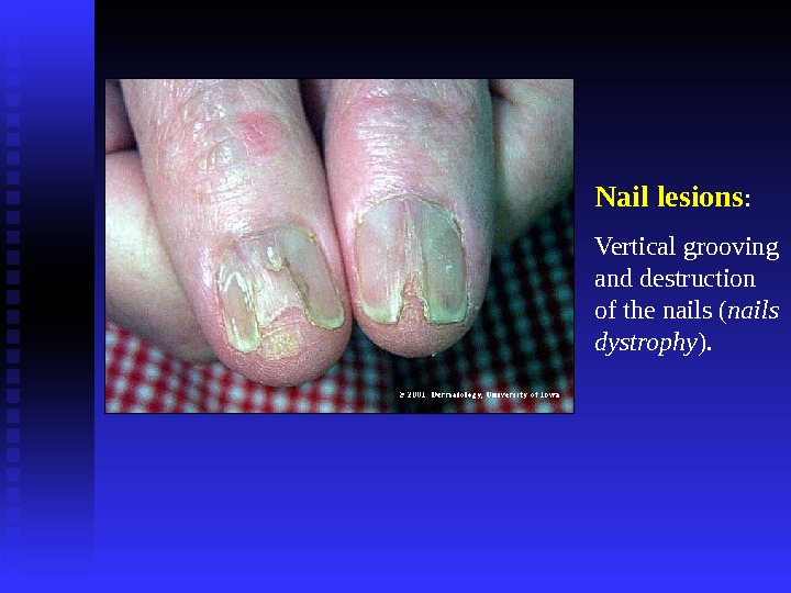
Nail lesions : Vertical grooving and destruction of the nails ( nails dystrophy ).
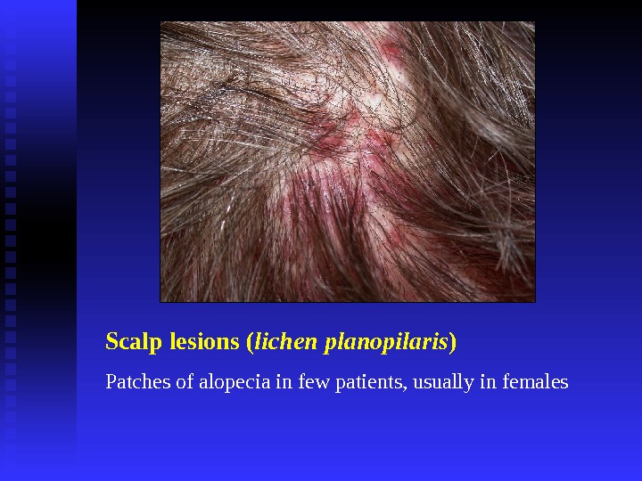
Scalp lesions ( lichen planopilaris ) Patches of alopecia in few patients, usually in females
Oral Lichen Planus Non-erosive Papular Linear Reticular Annular Plaque type Erosive Atrophic Bullous Ulcerative
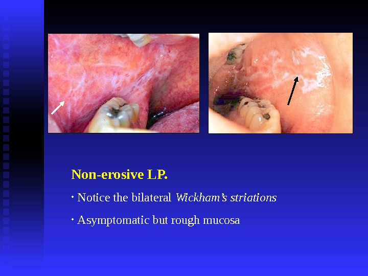
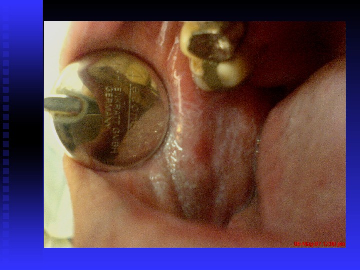
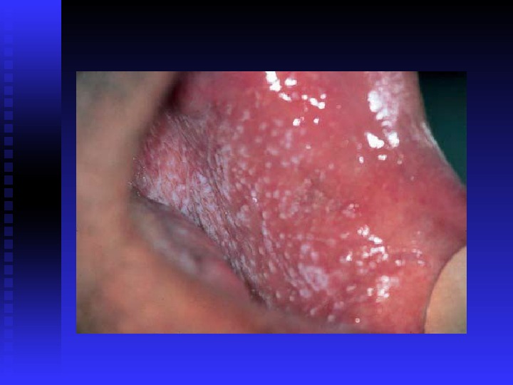
Non-erosive LP. • Notice the bilateral Wickham’s striations • Asymptomatic but rough mucosa
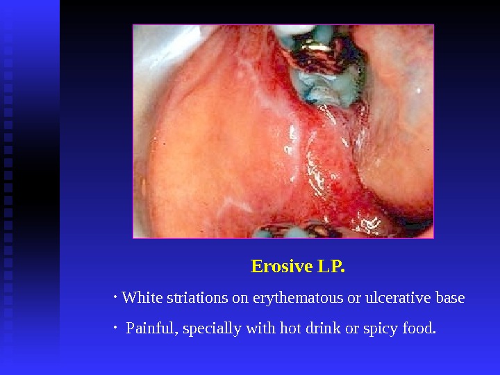
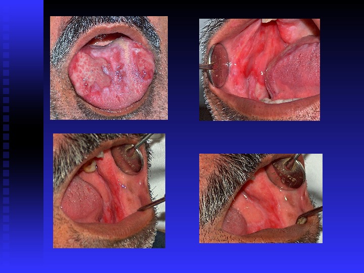
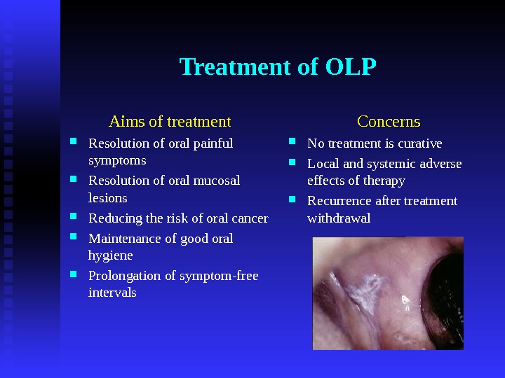
Erosive LP. • White striations on erythematous or ulcerative base • Painful, specially with hot drink or spicy food.
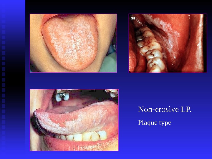
Non-erosive LP. Plaque type
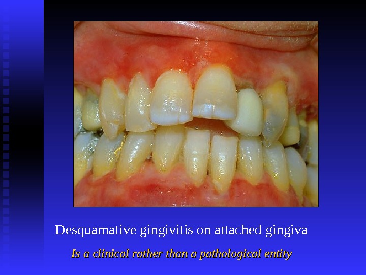
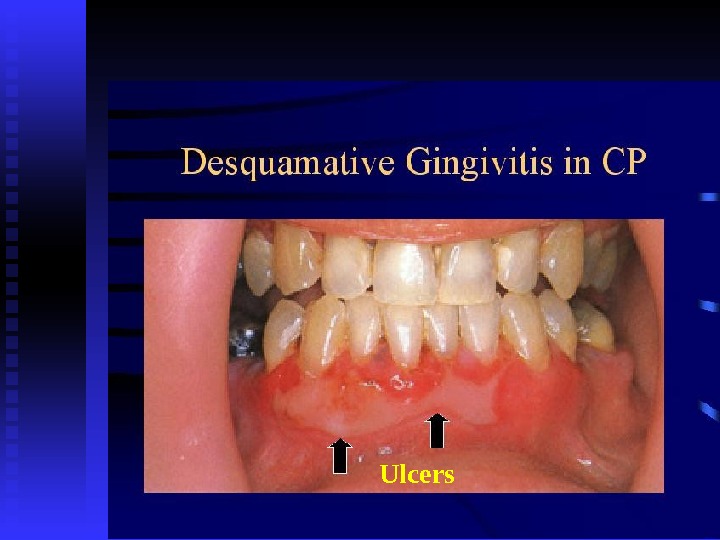
Desquamative gingivitis on attached gingiva Is a clinical rather than a pathological entity
Ulcers
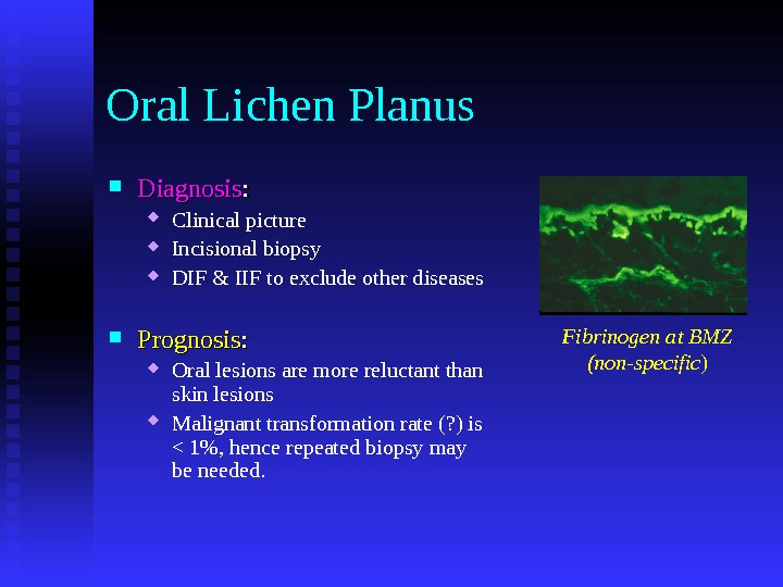
Oral Lichen Planus Diagnosis : : Clinical picture Incisional biopsy DIF & IIF to exclude other diseases Prognosis : : Oral lesions are more reluctant than skin lesions Malignant transformation rate (? ) is < 1%, hence repeated biopsy may be needed. Fibrinogen at BMZ (non-specific )
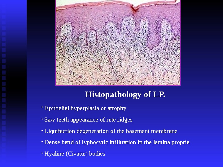
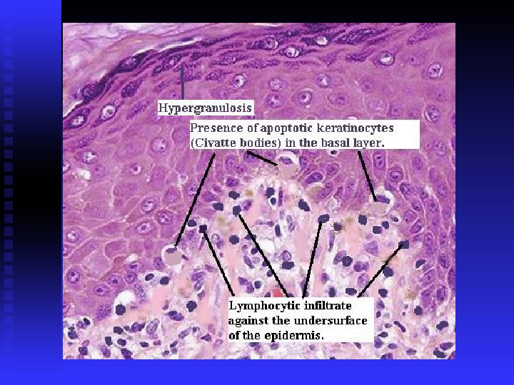
Histopathology of LP. • Epithelial hyperplasia or atrophy • Saw teeth appearance of rete ridges • Liquifaction degeneration of the basement membrane • Dense band of lyphocytic infiltration in the lamina propria • Hyaline (Civatte) bodies
Treatment of OLP Aims of treatment Resolution of oral painful symptoms Resolution of oral mucosal lesions Reducing the risk of oral cancer Maintenance of good oral hygiene Prolongation of symptom-free intervals Concerns No treatment is curative Local and systemic adverse effects of therapy Recurrence after treatment withdrawal
Treatment of OLP. Skin lesions Steroid cream Systemic steroids Oral lesions: Minor erosive type Remove trauma Antiseptic mouth wash Local steroids Betamethasone valerate aerosol, Beclomethasone dipropionate, 0. 1% triamcinolone acetonide in adhesoive paste, Beclometasone dipropionate 0. 05% Flucinonide ointment in an orabase paste 0. 025% clobetasol propionate Meconazole oral gel or chlorhexidine mouth wash
Treatment of OLP. Oral lesions: Major erosive type High concentration steroid mouth wash Triamcinolone intra-lesional injections (0. 2 to 0. 4 ml of 10 mg/m. L) Topical tacrolimus Systemic steroids: 40 -80 mg/day prednisolone for <2 weeks Azathioprine (50 to 100 mg/day) Levamisole (150 mg/day)
? Potentially malignant condition 0. 4%-2% per five years observation
OLP – Patient’s follow up Follow up: Every month till resolution of symptoms Every 6 -12 month afterward The risk of oral cancer in patients with OLP may be reduced by means of the following: Elimination of smoking and alcohol consumption Effective treatment of atrophic, erosive, and plaque oral lichen planus lesions Consumption of a nutritious diet including fresh fruit and vegetables Elimination of C albicans super-infection Regular clinical examination and repeat biopsy as required. Oral brush biopsy can be used to limit the number of scalpel biopsies
Lichenoid eruption The expression or unmasking of the lichen planus antigen may be induced by: Drugs (lichenoid drug reaction) Contact allergens in dental restorative materials or toothpastes (contact hypersensitivity reaction) Mechanical trauma (Koebner phenomenon) Viral infection Other unidentified agents
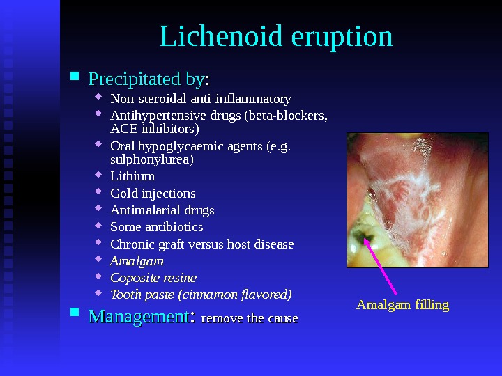
Lichenoid eruption Precipitated by : : Non-steroidal anti-inflammatory Antihypertensive drugs (beta-blockers, ACE inhibitors) Oral hypoglycaemic agents (e. g. sulphonylurea) Lithium Gold injections Antimalarial drugs Some antibiotics Chronic graft versus host disease Amalgam Coposite resine Tooth paste (cinnamon flavored) Management : : remove the cause Amalgam filling
Oral lichenoid lesions may be triggered by mechanical trauma ( Koebner phenomenon ) due to Calculus deposits Sharp teeth Rough surfaces of dental restorations or prostheses Cheek or tongue biting Oral surgical procedures
Pemphigus Auto-immune disease The patients usually between 40 -60 years old Racial difference in incidence Most of the patients have oral lesions which may be the initial presentation. The prognosis used to be very poor.
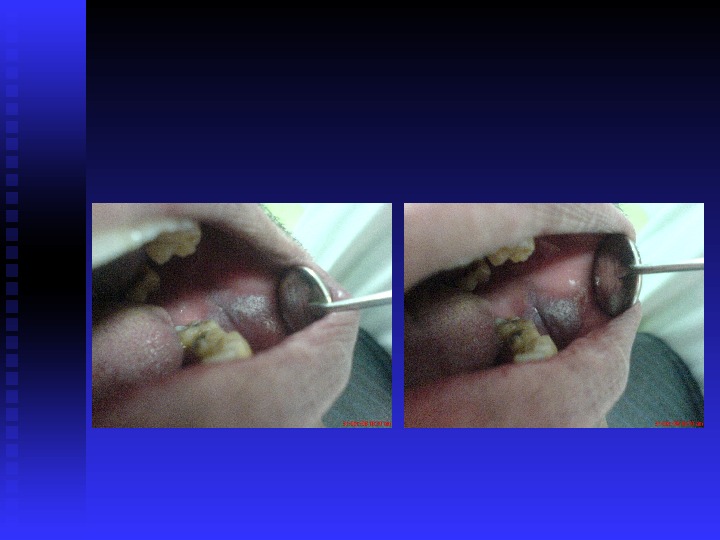
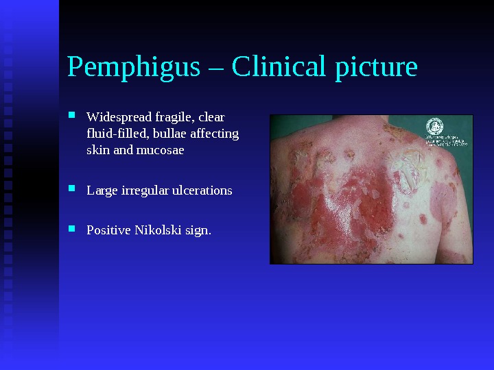
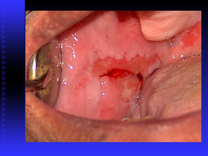
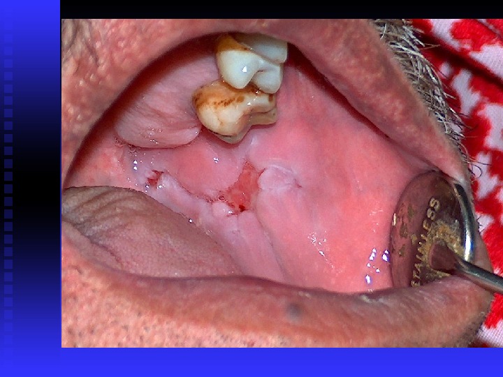
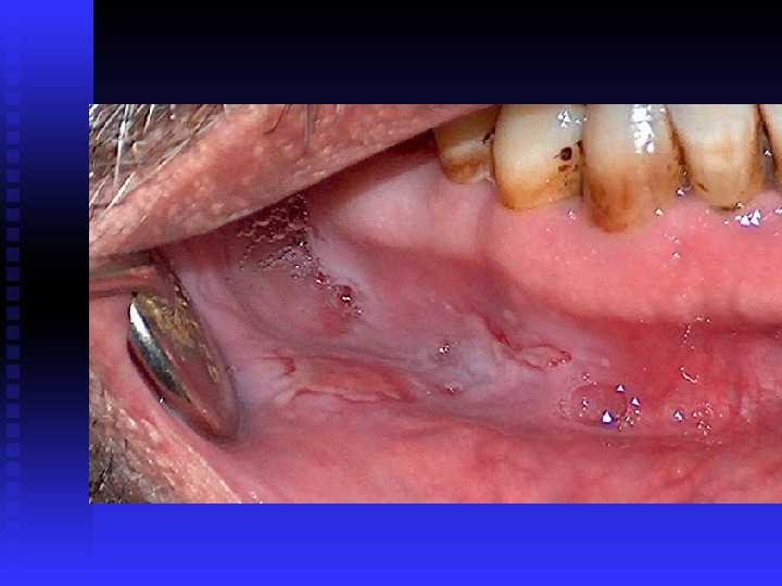
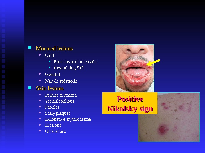
Pemphigus – Clinical picture Widespread fragile, clear fluid-filled, bullae affecting skin and mucosae Large irregular ulcerations Positive Nikolski sign.
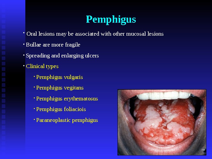
Pemphigus • Oral lesions may be associated with other mucosal lesions • Bullae are more fragile • Spreading and enlarging ulcers • Clinical types • Pemphigus vulgaris • Pemphigus vegitans • Pemphigus erythematosus • Pemphigus foliaciois • Paraneoplastic pemphigus
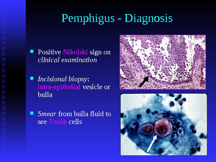
Pemphigus — Diagnosis Positive Nikolski sign on clinical examination Incisional biopsy : intra-epithelial vesicle or bulla Smear from bulla fluid to see Tzank cells
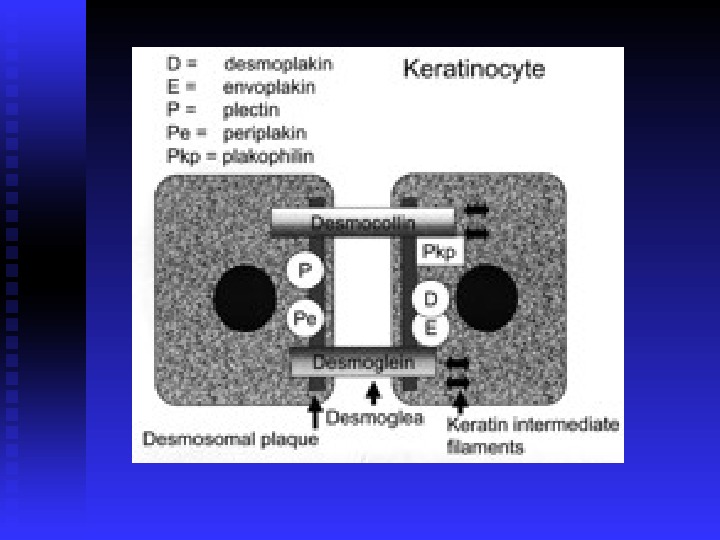
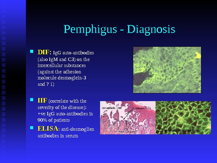
Pemphigus — Diagnosis DIFDIF : : Ig. G auto-antibodies (also Ig. M and C 3) on the intercellular substances (against the adhesion molecule desmoglein-3 and ? 1) IIF (correlate with the severity of the disease): +ve Ig. G auto-antibodies in 90% of patients ELISA : anti-desmoglien antibodies in serum
Direct & Indirect I
Pemphigus — Treatment Multidisplinary Initial treatment with high doses of steroid (100 mg prednisolone / day) Patients are maintained on steroids and/or steroid sparing drugs (e. g. azathioprine) High-concentration steroid mouth wash. Antifungal therapy may be needed. Dental considerations
Paraneoplastic pemphigus http: //emedicine. medscape. com/article/1064452 -overview Anhalt GJ, et al. Paraneoplastic pemphigus. An autoimmune mucocutaneous disease associated with neoplasia. N Engl J Med. Dec 20 1990 ; 323(25): 1729 -35. Autoimmune disease Anti-plakin antibodies 90% mortality rate Pemphigus + neoplasm commonly lymphoproliferative neoplasm (most commonly non-Hodgkin’s lymphoma) No race or gender predilection Age >60 yrs. The only type affects epithelia other than squamous !!!
Mucosal lesions Oral Erosions and mucositis Resembling SJS Genital Nasal: epistaxis Skin lesions Diffuse erythema Vesiculobullous Papules Scaly plaques Exfoliative erythroderma Erosions Ulcerations Positive Nikolsky sign
Pemphigoid Auto-immune disease No racial predominance Two basic clinical types: Bullous (generalized) pemhigoid Mucousal (cicatricial) pemhigoid
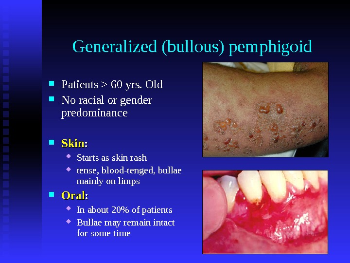
Generalized (bullous) pemphigoid Patients > 60 yrs. Old No racial or gender predominance Skin : : Starts as skin rash tense, blood-tenged, bullae mainly on limps Oral : : In about 20% of patients Bullae may remain intact for some time
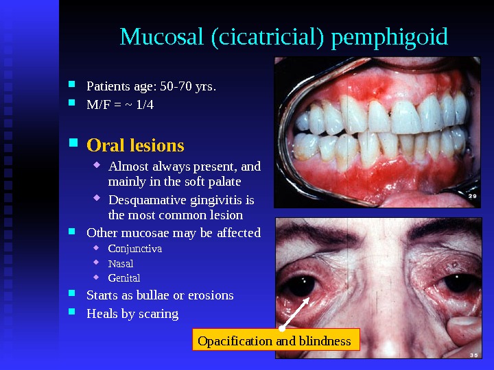
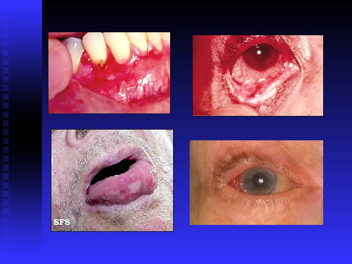
Mucosal (cicatricial) pemphigoid Patients age: 50 -70 yrs. M/F = ~ 1/4 Oral lesions Almost always present, and mainly in the soft palate Desquamative gingivitis is the most common lesion Other mucosae may be affected Conjunctiva Nasal Genital Starts as bullae or erosions Heals by scaring Opacification and blindness
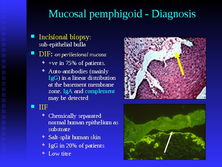
Mucosal pemphigoid — Diagnosis Incisional biopsy : sub-epithelial bulla DIF : on perilesional mucosa +ve in 75% of patients. Auto-antibodies (mainly Ig. G ) in a linear distribution at the basement membrane zone. Ig. A and complement may be detected IIF Chemically separated normal human epithelium as substrate Salt-split human skin Ig. G in 20% of patients Low titre
Pemphigoid – Treatment This disorder is extremely difficult to treat. Even with optimum control, blisters may continue to develop in some patients Generalized pemphigoid Systemic steroids and / or steroid sparing immunosuppressive agents. Topical steroid may be used for oral lesions. Mucosal pemphigoid Perforate the bullae if necessary Topical steroids: e. g. Beclamethasone spray Triamcinolone in orabase Antiseptic mouth wash. Multidisplinary management e. g. ophthalmologist, dermatologist and internest opinion is mandatory High-dose intravenous immune globulin in refractory cases
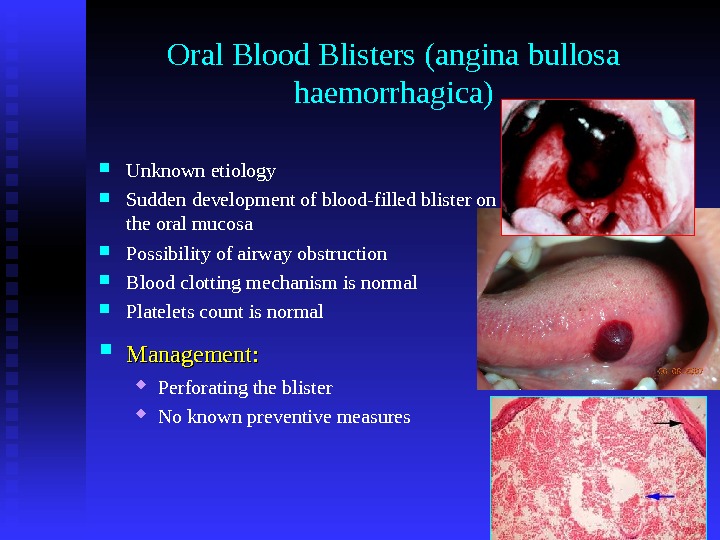
Oral Blood Blisters (angina bullosa haemorrhagica) Unknown etiology Sudden development of blood-filled blister on the oral mucosa Possibility of airway obstruction Blood clotting mechanism is normal Platelets count is normal Management: Perforating the blister No known preventive measures
Erythema multiforme Precipitating factors: Type IV hypersensitivity reaction Viral infections (e. g herpes simplex, mycoplasma) Bacterial infections Internal malignancy or its treatment with radiotherapy Pregnancy Drugs (e. g. sulphonamides, penicillins, phenylbutazone, barbiturates) Excessive exposure to UV light Unknown factors More in males More in young people
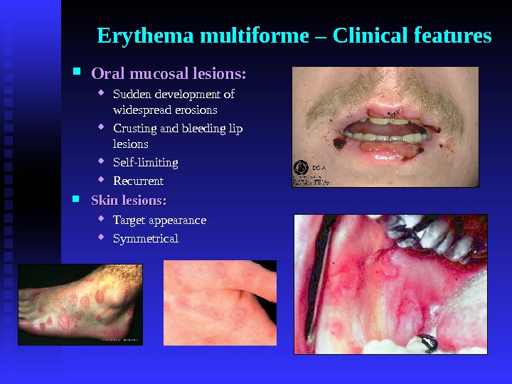
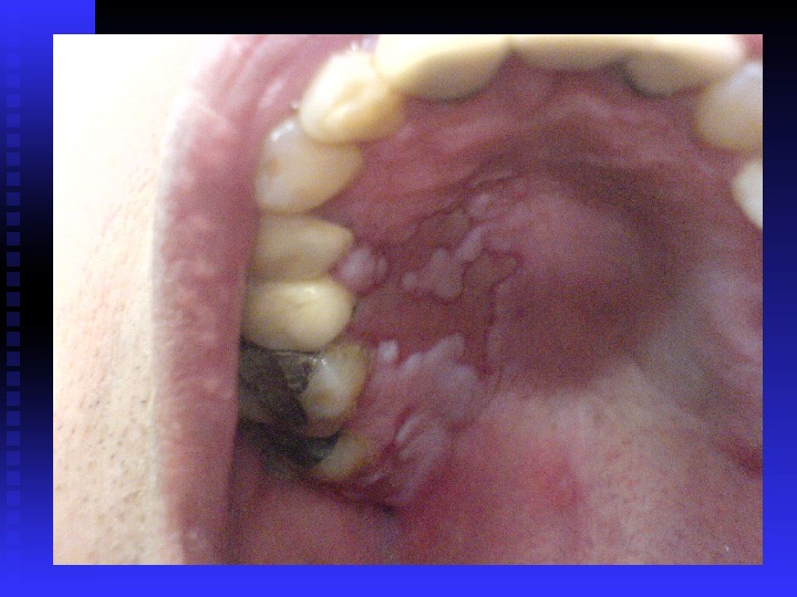
Erythema multiforme – Clinical features Oral mucosal lesions: Sudden development of widespread erosions Crusting and bleeding lip lesions Self-limiting Recurrent Skin lesions: Target appearance Symmetrical
EM General features : : • Cervical lymphadenitis • Pyrexia • Subside in 10 days • Subjected to recurrence • > in young patients
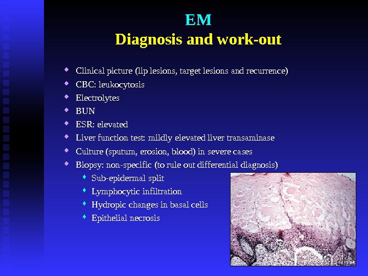
EM Diagnosis and work-out Clinical picture (lip lesions, target lesions and recurrence) CBC: leukocytosis Electrolytes BUN ESR: elevated Liver function test: mildly elevated liver transaminase Culture (sputum, erosion, blood) in severe cases Biopsy: non-specific (to rule out differential diagnosis) Sub-epidermal split Lymphocytic infiltration Hydropic changes in basal cells Epithelial necrosis
EM Treatment — Management Remove the cause if possible Treat infections Fluid intake and soft diet; possibly IV line is needed Oral antihistamines Antiseptic mouth wash Topical steroids ? ? Systemic steroids Acyclovir as a prophylaxis Consultations: Dermatologist Ophthalmologist Internal medicine specialist
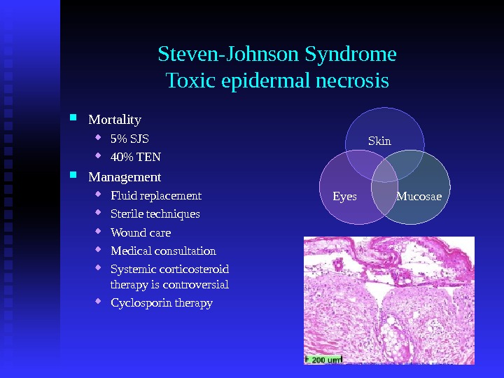
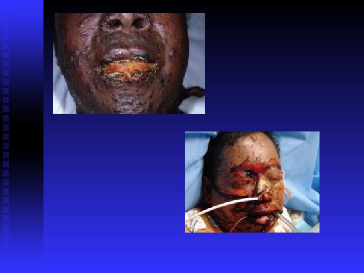
Steven-Johnson Syndrome Toxic epidermal necrosis Mortality 5% SJS 40% TEN Management Fluid replacement Sterile techniques Wound care Medical consultation Systemic corticosteroid therapy is controversial Cyclosporin therapy Skin Mucosae. Eyes
Lupus Erythematosus Autoimmune disease Two main clinical divisions: Discoid lupus erythematosus (DLE) Systemic lupus erythematosus (SLE) M: F = 9: 1 (18 -65 years; peak 25 -45 years) DLE & SLE may represent different ends of the spectrum of the disease activity SLE may be precipitated by some drugs e. g. Hydralazine (lupoid reaction). Oral mucosal lesions in 25 -75% of the cases
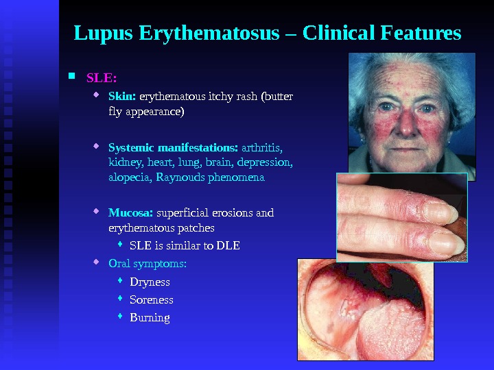
Lupus Erythematosus – Clinical Features SLE: Skin: erythematous itchy rash (butter fly appearance) Systemic manifestations: arthritis, kidney, heart, lung, brain, depression, alopecia, Raynouds phenomena Mucosa: superficial erosions and erythematous patches SLE is similar to DLE Oral symptoms: Dryness Soreness Burning
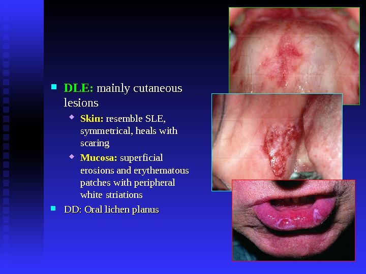
DLE: mainly cutaneous lesions Skin: resemble SLE, symmetrical, heals with scaring Mucosa: superficial erosions and erythematous patches with peripheral white striations DD: Oral lichen planus
Lupus Erythematosus Diagnosis: IMF: antinuclear antibodies (ANA) +ve in 90% of patients. DMF Biopsy and histopathological examination: resembles OLP
Prognosis No cure Renal disease is the main morbidity and mortality Thrombocytopenia and hemolytic anemia in 85% of patients Oral lesions are considered potentially malignant Treatment: antifungal agents may be required for oral lesions SLE: High doses systemic steroids + steroid sparing drugs. High concentration steroid mouth wash DLE: Topical steroids to reduce symptoms Antimalarial drug (chloroquine) may be useful (? Retinopathy) ? Potentially malignant condition
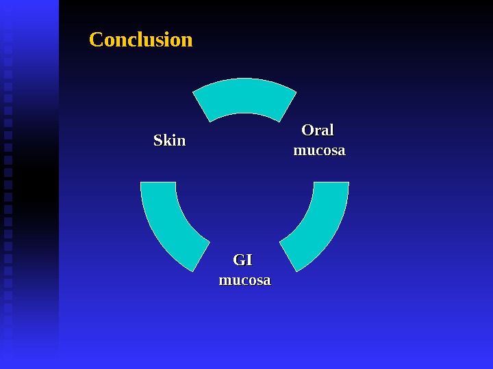
Oral mucosa GI GI mucosa. Skin. Conclusion