

eddd9211f8cc8647cf1f5e4242296e72.ppt
- Количество слайдов: 12
 Monochorionic diamniotic twin after intrauterine flushing of GCSF in natural IVF cycle. Case report. Konstantin Y. Boyarsky 1, 2, MD, Ph. D 1 IVF Clinics “GENESIS” 2 Department of Obstetrics and Gynecology, State Pediatric Medical Academy, St. Petersburg, Russia boyarsky@pochta. ru
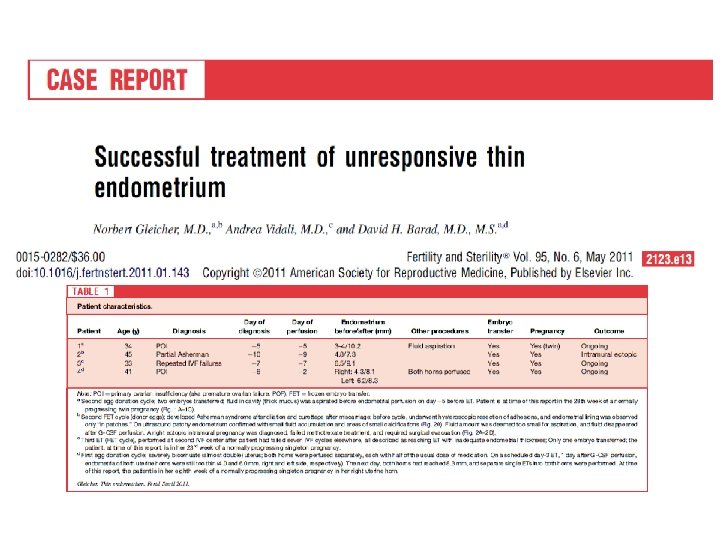
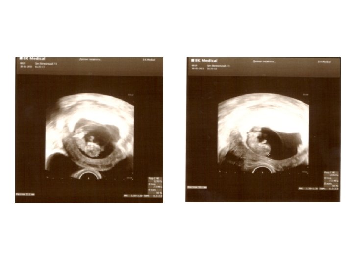
 Patient P. , 38 y. old, with tubal factor of infertility, had three tubal and ovarian surgery episodes and six IVF attempts in anamnesis. Previous IVF attempts had complications as poor response, thin endometrium and unexplained fluid accumulation in uterine cavity during ovarian stimulation. Two IVF attempts ended first trimester miscarriages with abnormal fetus karyotype. Ovarian reserve was low (serum AMH level <0. 5 ng/ml). The seventh IVF attempt was performed according to modified natural protocol. The antagonist daily injection with concomitant low FSH dose was administrated when leading follicle reached more than 15 mm in diameter. Three days later, when the follicle was 19 mm, ovulatory dose of 5000 IU HCG was administrated and 35 hours later follicle punction was performed. One mature COC was received. 24 hours after follicle punction, uterine cavity was flushed by 30 IU of recombinant G-CSF (Grasalva, Fillgrastim, TEVA, Israel) in one ml solution. 72 hours after follicle punction one 8 -cell embryo was transferred. Three weeks after ET monochorionic diamniotic twin was detected by ultrasound examination. Screening US scan on 12. 5 weeks of pregnancy showed absence of any chromosomal abnormality sings. Now, monozygotic female twin pregnancy reaches 20 weeks without any complications.
 GM-CSF (CSF-2) G-CSF (CSF-3) Localization in human genome 1 p 21 -p") M-CSF (CSF-1) GM-CSF (CSF-2) G-CSF (CSF-3) Localization in human genome 1 p 21 -p 13 5 q 31. 1 17 q 11. 2 -q 12 Molecular weight 60, 179 Da 16, 295 Da 22, 293 Da Molecular function Positive regulation of monocyte & macrophage differentiation, positive regulcellular protein metabolic process & multicellular organism growth, reproductive developmental process Negative regulation of cytolysis & apoptosis; epithelial fluid transport; positive regulation of cell proliferation; immune response; positive regulation of DNA replication; positive regulation of survival gene product activity Immune response granulocyte differentiation; cytokine and chemokine mediated signaling pathway; multicellular organismal development; positive regulation of cell proliferation; Name of commercial substance Lanimostim, Mirimostim (treatment of pancytopenia after anticancer chemotherapy) Molgramostin, Sargramostim (immunostimulation of white blood cells during cancer and sepsis treatment) Lenograstim, Pluripoietin (immunostimulation during Molecular structure Data from www. phosite. org cancer treatment)
M-CSF (CSF-1) GM-CSF (CSF-2) G-CSF (CSF-3) Localization in human genome 1 p 21 -p 13 5 q 31. 1 17 q 11. 2 -q 12 Molecular weight 60, 179 Da 16, 295 Da 22, 293 Da Molecular function Positive regulation of monocyte & macrophage differentiation, positive regulcellular protein metabolic process & multicellular organism growth, reproductive developmental process Negative regulation of cytolysis & apoptosis; epithelial fluid transport; positive regulation of cell proliferation; immune response; positive regulation of DNA replication; positive regulation of survival gene product activity Immune response granulocyte differentiation; cytokine and chemokine mediated signaling pathway; multicellular organismal development; positive regulation of cell proliferation; Name of commercial substance Lanimostim, Mirimostim (treatment of pancytopenia after anticancer chemotherapy) Molgramostin, Sargramostim (immunostimulation of white blood cells during cancer and sepsis treatment) Lenograstim, Pluripoietin (immunostimulation during Molecular structure Data from www. phosite. org cancer treatment)

 Molecular structure of CSF 1
 Molecular structure of CSF 2
 Molecular structure of CSF 3
 1 p 21") Name Localization in human genome CSF 1 MCSF| (COLONYSTIMULATING FACTOR, MACROPHAGESPECIFIC) 1 p 21 -p 13 CSF 2 GRANULOCYTEMACROPHAGE COLONYSTIMULATING FACTOR, GMCSF 5 q 31. 1 CSF 3 colony stimulating factor 3 (granulocyte) 17 q 11. 2 -q 12
Name Localization in human genome CSF 1 MCSF| (COLONYSTIMULATING FACTOR, MACROPHAGESPECIFIC) 1 p 21 -p 13 CSF 2 GRANULOCYTEMACROPHAGE COLONYSTIMULATING FACTOR, GMCSF 5 q 31. 1 CSF 3 colony stimulating factor 3 (granulocyte) 17 q 11. 2 -q 12
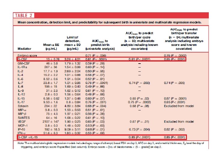
 CSF-2 (GM-CSF) CSF-3 (G-CSF) Ovary Regulates") Significance of CSFs in human reproduction CSF-1 (M-CSF) CSF-2 (GM-CSF) CSF-3 (G-CSF) Ovary Regulates steroidogenesis, enhances gonadotropin action on granulosa cells (1, 2) Enhances follicular development (7), histamine release during ovulation (6) Maintains number of primordial follicles(14), produced by granulosa cells in mature and ovulated follicles (15 -16) Endometrium M-CSF is produced by uterine glandular epithelial cells, involved in endometriosis pathogenesis (3) GM-CSF and GM-CSF-R are expressed with peak at midsecretory endometrium (8) G-CSF produces by luteal phase endometrium (17) Pregnancy and Placenta Plays a role in placental development (4) Growth factor for trophoblast and other placental cells (9), produced by trophoblast and u. NK cells (10), involved in pathogenesis of preeclampsia (11) Produces by trophoblast cells (17), affects decidual macrophages (18) Clinical use M-CSF can enhance oocytes number and PR in poor responders (5) Treatment of repeated implantation failure (12), GM-CSF production in endometrial co-culture associated with outcome (13), addition to culture (Embyo. Gene) FF level may be a predictor of PR in n. IVF cycles (19), S level may be a pedictor of pregnancy (20), treatment of thin endometrium (21), and RMs (22)
Significance of CSFs in human reproduction CSF-1 (M-CSF) CSF-2 (GM-CSF) CSF-3 (G-CSF) Ovary Regulates steroidogenesis, enhances gonadotropin action on granulosa cells (1, 2) Enhances follicular development (7), histamine release during ovulation (6) Maintains number of primordial follicles(14), produced by granulosa cells in mature and ovulated follicles (15 -16) Endometrium M-CSF is produced by uterine glandular epithelial cells, involved in endometriosis pathogenesis (3) GM-CSF and GM-CSF-R are expressed with peak at midsecretory endometrium (8) G-CSF produces by luteal phase endometrium (17) Pregnancy and Placenta Plays a role in placental development (4) Growth factor for trophoblast and other placental cells (9), produced by trophoblast and u. NK cells (10), involved in pathogenesis of preeclampsia (11) Produces by trophoblast cells (17), affects decidual macrophages (18) Clinical use M-CSF can enhance oocytes number and PR in poor responders (5) Treatment of repeated implantation failure (12), GM-CSF production in endometrial co-culture associated with outcome (13), addition to culture (Embyo. Gene) FF level may be a predictor of PR in n. IVF cycles (19), S level may be a pedictor of pregnancy (20), treatment of thin endometrium (21), and RMs (22)