Описание презентации Minimally Classic CNV Red-free Photograph Image courtesy по слайдам
 Minimally Classic CNV
 Red-free Photograph Image courtesy of J Slakter
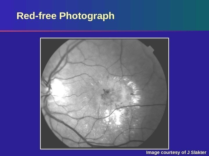
 Red-free Photograph Digital red-free photograph shows: – Lipid, blood and fluid in exudative AM
 Early-phase Fluorescein Angiogram Image courtesy of J Slakter
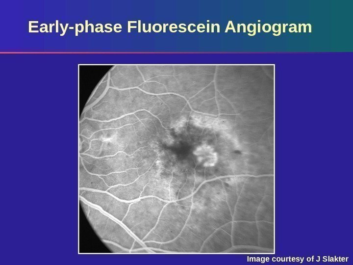
 Early-phase Fluorescein Angiogram Early phase: – Well-defined classic CNV centrally – Stippled mild hyperfluorescence from occult CNV surrounding classic CNV – Blockage from blood
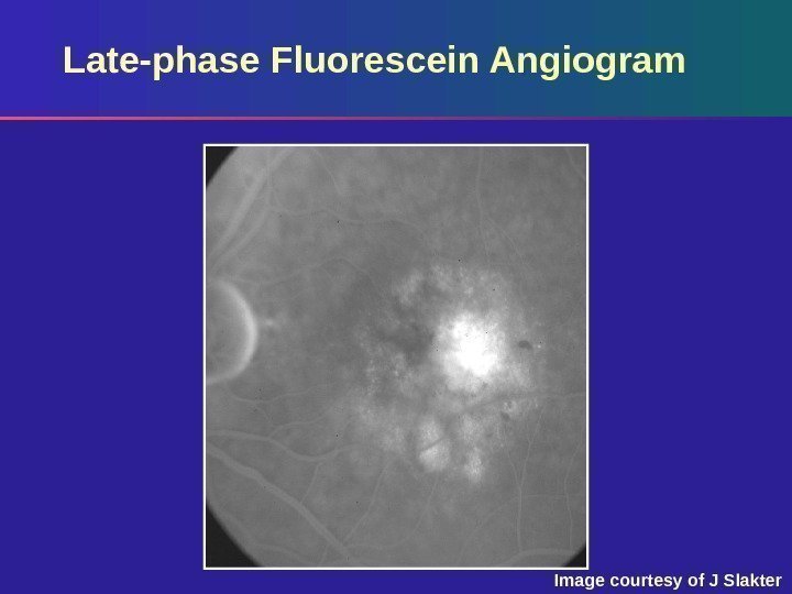 Late-phase Fluorescein Angiogram Image courtesy of J Slakter
 Late-phase Fluorescein Angiogram Late phase: – Leakage from the central classic CNV – Surrounding area of stippled fluorescence from occult CNV
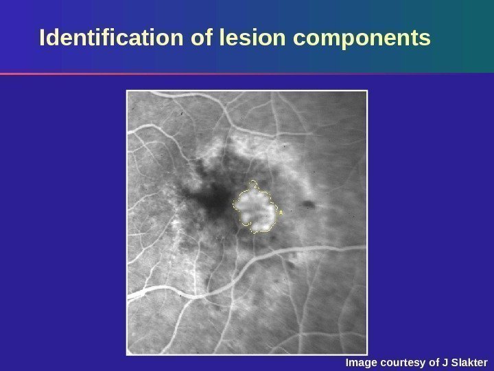 Identification of lesion components Image courtesy of J Slakter
 Digital tracing of the classic component of the CNV – Classic CNV is traced in the early phase when it is best delineated. Identification of lesion components
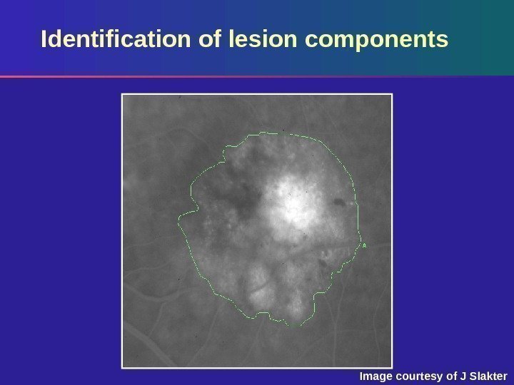 Image courtesy of J Slakter. Identification of lesion components
 Digital tracing of the occult component – Occult component is measured in the late phase where the entire extent of involvement can be seen. Identification of lesion components
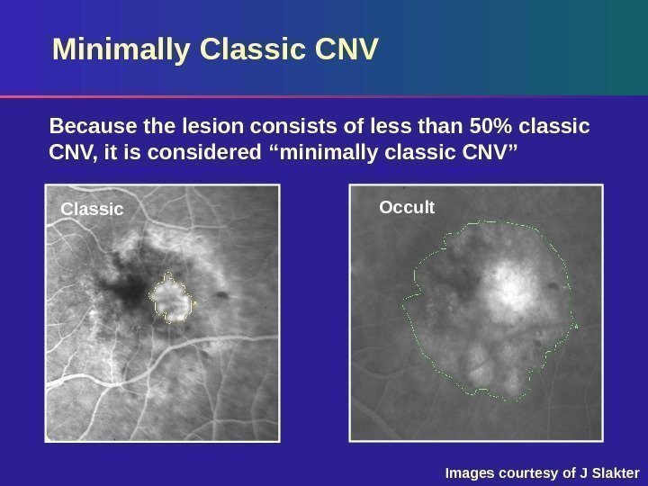 Minimally Classic CNV Classic Occult. Because the lesion consists of less than 50% classic CNV, it is considered “minimally classic CNV” Images courtesy of J Slakter


