microscopy_1.pptx
- Количество слайдов: 34


Микроскопия Лекция 1 А. П. Савицкий Институт биохимии им. А. Н. Баха РАН Московский Государственный университет им. М. В. Ломоносова


Molecular biology DNA Genomics Proteins Proteomics Cell biology Cells Cytomics Histology Physiology Medicine Tissue Animal Human Phenomic Clinic Information level

ГЕНОМИКА-ПРОТЕОМИКА-ФУНКЦИЯ

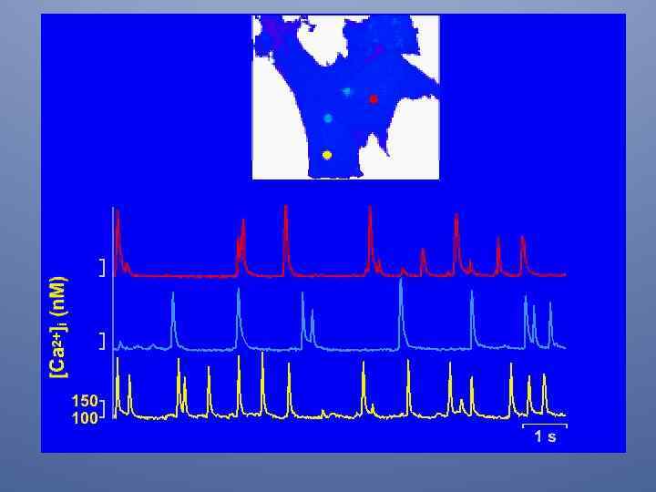
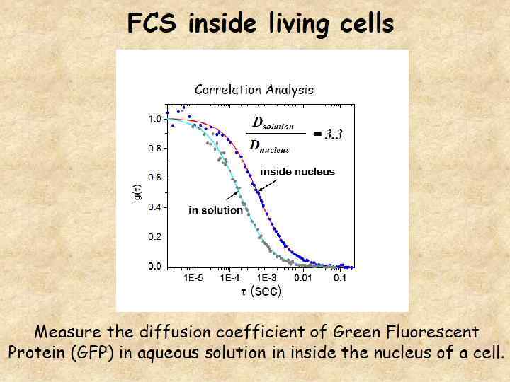
Live cell imaging and systems biology Much of the experimental data used to construct mathematical models of molecular networks are derived from in vitro measurements. However, there is increasing evidence that in vitro measurements fail to capture both the complexity and the individuality found in single, living cells. These limitations can be overcome by live cell microscopy which is evolving to enable in vivo biochemistry. live cell microscopy illustrate how a number of different imaging approaches could be applied to analyze a specific molecular network. incorporation of such quantitative live cell imaging methods is critical for the progress of systems biology.
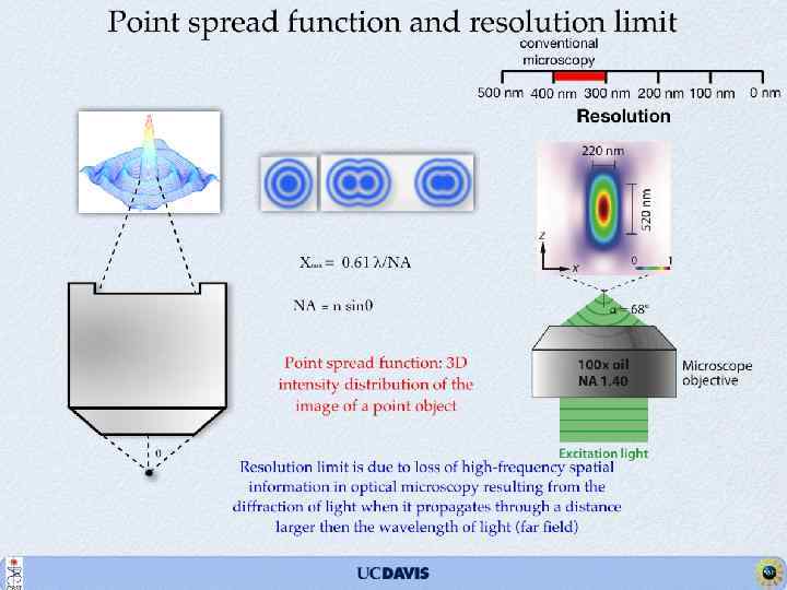

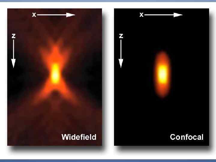
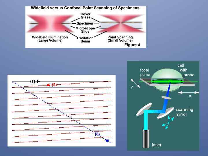
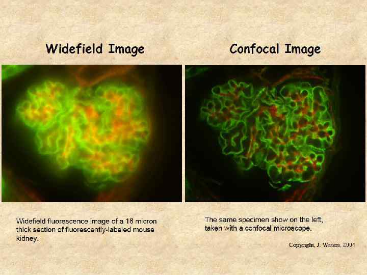
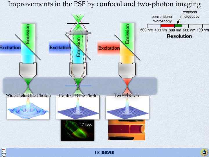
Confocal Principle ILLUMINATION DETECTOR PINHOLE Illumination and detection through the pinhole microscope CONFOCAL PLANE cell

Принцип пошагового сканирования
 3 -D (x-y,")
Multi-D Imaging Multi-W 2. 5 -D Over Time 2 -D (x-y) 3 -D (x-y, z) 3. 5 -D 4 -D (x-y, z, t) 5, 6 -D (x-y, z, t, W)

Флуоресценция родамина 6 G в этиленгликоле Рамановский спектр этиленгликоля

Интенсивность рамановского рассеяния воды по отношению к флуоресценции 1 молекулы родамина 6 G

Оптическая геометрия для детекции одиночных молекул

Принцип TIRF измерений

microscopy_1.pptx