

5f79e38c563c78e347dc16162588d1e6.ppt
- Количество слайдов: 61
 Microvascular Decompression Surgery for Trigeminal Neuralgia Pearls and Pitfalls Aaron Cohen-Gadol, MD, MSc
 Disclosures • Aaron A. Cohen-Gadol • Research Grants from Anspach and Zeiss

 Harvey Cushing
 “TN/MVD – Desirable Practice” • TN is relatively easy to diagnose • MVD is one of the most satisfying operations in neurosurgery • Prescient/pleasing anatomy • Relatively short procedure • Microsurgical environment • Patients do well and are thankful • TN is “one of the worst pains humans have been afflicted with” • Minimal side-effects
 TN Pain • • • Isolated to the 3 divisions of the nerve Convulsive, not crossing midline Has oral and skin triggers Normal neurologic exam Responds to Tegretol & antiepileptic drugs The character of the pain may change with the use of neuropathic pain medications and have more of a constant character • Detailed history is important • Patients with predominantly constant or burning pain or facial numbness are not good candidates
 Classification of TN TN, type 1 TN, type 2 TNP, neuropathic TDP, deafferentation pain of spontaneous onset with > 50% limited to duration of an episode of pain of spontaneous onset with > 50% as constant unintentional injury to trigeminal system from trauma, oral surgery, ENT surgery, root injury from posterior fossa or skull base surgery, stroke, etc. pain in a region of trigeminal numbness resulting from intentional injury to trigeminal system from neurectomy, gangliolysis, rhizotomy, nucleotomy, tractotomy, or other denervating procedures STN, symptomatic pain resulting from multiple sclerosis PHN, postherpetic pain resulting from trigeminal Herpes zoster outbreak ATN, atypical pain predominantly having a psychological rather than a physiological origin Burchiel, Neurosurg Focus 18: 1 -3, 2005
 • Ablative •") Treatment • Medication • Surgical • Physiologic • Microvascular decompression (MVD) • Ablative • • Radiosurgery Percutaneous stereotactic rhizotomy (PSR) Glycerol injection Balloon compression
Treatment • Medication • Surgical • Physiologic • Microvascular decompression (MVD) • Ablative • • Radiosurgery Percutaneous stereotactic rhizotomy (PSR) Glycerol injection Balloon compression
 Pathogenesis and Treatment • • No matter what the responsible etiology MVD - Effective and durable palliative option Percutaneous procedures - Less invasive Posterior fossa exploration offers the only chance for a non-destructive procedure and a more durable result
 Imaging for MVD • Brain MRI or CT scan • Exclude a structural pathology such as a meningioma, acoustic neuroma or an epidermoid tumor • High resolution MRI: Negative • Posterior fossa exploration is reasonable • We have routinely offered MVD to the patients who did not harbor an “MRI evident” vascular loop and have found compressive arterial loops during their posterior fossa exploratory surgery
 MRI findings
 Indications for MVD • • Failure of medical or percutaneous therapy Patient preference Location of pain in V 1 or multiple divisions Physiologic young and can tolerate procedure • • No perfect procedure, no cure Be caring, listen and stay optimistic Cure a few, comfort all! Dysesthesia is not responsive to more procedures
 Positioning
 Incision
 Microvascular Decompression Surgery
 Operating room set-up
 Patient positioning
 Head clamp positioning
 Positioning Video
 Incision
 Linear incision
 Curvilinear incision
 Surgical corridors
 Burr hole placement
 Craniotomy
 Mastoid bone drilling
 Bone removal over the dural sinuses
 Waxing the mastoid air cells and dural opening
 Exposure
 Petrous-tentorial junction
 Opening arachnoid membranes
 “Watching” for vessels
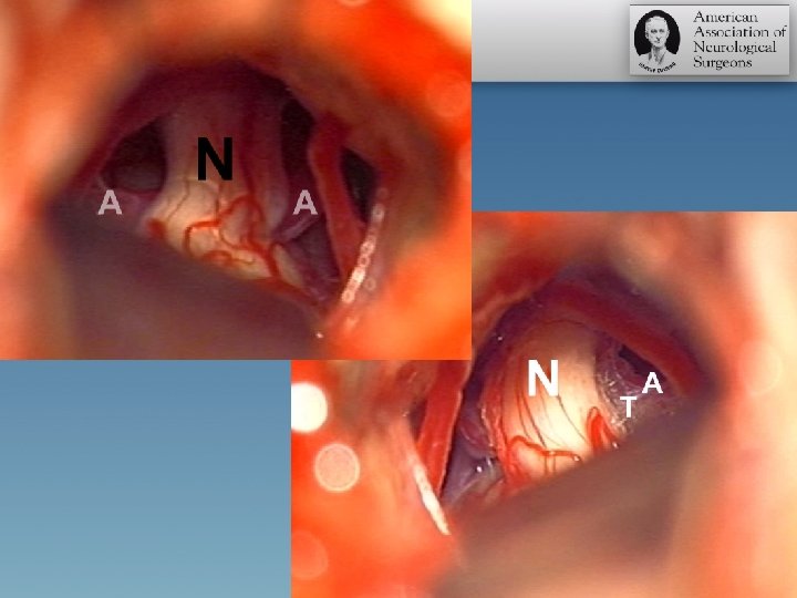
 Variations of nerve-vessels
 Implant insertion
 Nuance of technique
 Complete decompression

 Venous “conflict”
 Ectatic basilar artery
 Surgical Videos
 PEARLS and PITFALLS
 Pearls • Control bleeding from the sinus with “ bone wax” and avoid aggressive packing • Control bleeding from SPV with gelfoam powder • Neurovascular conflict at the root entry zone of the nerve • Detailed and careful inspection of the space around the root entry zone (360) • Gentle handling of the nerve for a thorough inspection • The manipulation (“gentle” rhizotomy) of the nerve is responsible for some of the pain control afforded by MVD operations
 Pearls • Arterial compression by the superior cerebellar artery along the medial axilla of the root entry zone is one of most common overlooked sites of compression • Covered by motor rootlets or a vein • Small space and surrounding vessels • Inexperience in mobilizing plays a role • Discovery of discoloration along the root entry zone and the nerve
 Hiding artery
 should be") Pearls • A possibility of multiple offending vessels (arterial and/or venous loops) should be excluded with careful inspection • DO NOT GIVE UP TOO EALRY • DO NOT PERSEVERE TOO LATE • Perforators deserve respect
Pearls • A possibility of multiple offending vessels (arterial and/or venous loops) should be excluded with careful inspection • DO NOT GIVE UP TOO EALRY • DO NOT PERSEVERE TOO LATE • Perforators deserve respect
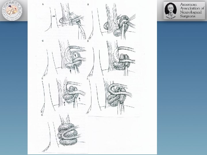
 Pearls • For a large artery embedded in the axilla • Work superior and inferior to the nerve to partially mobilize the artery • Small pieces of shredded Teflon may be inserted from the inferior aspect of the nerve and pushed superiorly in a semi-blinded fashion • Identification of the Teflon patch superior to the nerve confirms adequate mobilization of the nerve • Endoscopic techniques?
 Pitfalls • Avoid cerebellar retraction – Drain CSF • Watch for the veins • Sacrificing any vein: Superior petrosal vein? • Arteries hide within arachnoid membranes • Open arachnoid membranes widely • Watch for the tip of your scissors • “Does not look right? ” Please stop! • Inadequate exposure leads to inadequate decompression • Place your retractor WISELY • “I do not want to retract the brainstem”
 Pitfalls • No compression or venous compression – controversial? • Rhizotomy • Forceps “squeeze” • Gentle bipolar coagulation of the root entry zone • We have avoided partial root transaction due to the potential risk of disabling anesthesia De La Rosa
 Pitfalls • Aggressive manipulation of the nerve should be avoided • Overzealous use of Teflon should be avoided • Teflon granuloma has been reported as a cause of pain recurrence • Decompression of the wrong nerve (VII/VII complex) has been reported • Irrigate to assure no implant displacement • Wax in! Wax out!
 Decompression
 “Teflon granuloma conflict”
 Final thoughts • • Intraop monitoring for uncomplicated cases? Perioperative steroids Tapering preoperative meds CSF leak (wound versus nose) Pain recurrence Redo operation Atypical cases?