Лекция_12_метаботропные_рецепторы_част.pptx
- Количество слайдов: 110


Метаботропные рецепторы
")
Родопсин-подобные метаботропные рецепторы (выделены синим)

Ацетилхолиновые метаботропные рецепторы Агонисты: Ац. Х, карбахол, мускарин Антагонисты: атропин, скополамин, галламин м. Ац. ХР локализованы - в мембранах различных тканей (железы, сосуды, гладкие мышцы и др. ), которые иннервируются постганглионарными волокнами вегетативной НС (парасимпатический отдел); - в пресинаптических мембранах постганглионарных нейронов симпатической НС (торможение со стороны постганглионарных нейронов парасимпатической НС ); - в пресинаптических мембранах мотонейронов в нервно-мышечных синапсах; - в ЦНС в пост- и пресинаптических мембранах.

Ацетилхолиновые метаботропные рецепторы 55– 70 k. Da Аmino acid sequence and transmembrane domain structure of the human M 1 muscarinic receptor Amino acids that are identical among the m 1, m 2, m 3 and m 4 receptors are dark orange. The shaded cloud represents the approximate region that determines receptor–G-protein coupling. Arrows denote amino acids important for specifying G protein coupling. Amino acids predicted to be involved in agonist or antagonist binding are denoted by white letters.
 рецепторы")
Ацетилхолиновые метаботропные (мускариновые) рецепторы
")
Агонисты (выделены)

Мускариновые рецепторы в парасимпатической системе

Ацетилхолиновые метаботропные рецепторы
")
Ацетилхолиновые метаботропные рецепторы Выделено 5 подтипов мускариновых рецепторов - М(1 -5)
")
Ацетилхолиновые метаботропные рецепторы Выделено 5 подтипов мускариновых рецепторов - м. Ац. ХР(1 -5)
 в вегетативных ганглиях и")
Примеры эффектов мускариновых рецепторов М 1 (М 3, М 5) в вегетативных ганглиях и в желудке через Gq-белки активируют фосфолипазу С, в результате образуются ИФ 3 и ДАГ. ИФ 3 повышает концентрацию Са 2+ в цитозоле, который вместе с ДАГ активирует протенкиназу С. М 2 (М 4) в ЦНС и сердце активируют Gi-белки и подавляют аденилатциклазу, уменьшая концентрацию ц. АМФ, Go-белки (βγ-димер) увеличивают К+- и снижают Са 2+-проводимость

Примеры эффектов мускариновых рецепторов M 1 receptor This receptor is found mediating slow EPSP at the ganglion in the postganglionic nerve, is common in exocrine glands and in the CNS. It is predominantly found bound to G proteins of class Gq which use upregulation of phospholipase C and therefore inositol trisphosphate and intracellular calcium as a signalling pathway. However, Gi (causing a downstream decrease in c. AMP) and Gs (causing an increase in c. AMP) have also been shown to be involved in interactions in certain tissues. M 2 receptor The M 2 muscarinic receptors are located in the heart, where they act to slow the heart rate down to normal sinus rhythm after stimulatory actions of the parasympathetic nervous system, by slowing the speed of depolarization. They also reduce contractile forces of the atrial cardiac muscle, and reduce conduction velocity of the atrioventricular node (AV node). It also serves to slightly decrease the contractile forces of the ventricular muscle. M 2 muscarinic receptors act via a Gi type receptor, which causes a decrease in c. AMP in the cell, generally leading to inhibitory-type effects. Effects include formation of IP 3 and DAG.

Примеры эффектов мускариновых рецепторов M 3 receptor The M 3 muscarinic receptors are located at many places in the body. They are located in the smooth muscles of the blood vessels, as well as in the lungs. Because the M 3 receptor is Gq -coupled and mediates an increase in intracellular calcium, it typically causes constriction of smooth muscle, such as that observed during bronchoconstriction. However, with respect to vasculature, activation of M 3 on vascular endothelial cells causes increased synthesis of nitric oxide which diffuses to adjacent vascular smooth muscle cells and causes their relaxation thereby explaining the paradoxical effect of parasympathomimetics on vascular tone and bronchiolar tone. Indeed, direct stimulation of vascular smooth muscle M 3 mediates vasconstriction in pathologies whereby the vascular endothelium is disrupted. The M 3 receptors are also located in many glands which help to stimulate secretion in salivary glands and other glands of the body. Like the M 1 muscarinic receptor, M 3 receptors are G proteins of class Gq which upregulate phospholipase C and therefore inositol trisphosphate and intracellular calcium as a signalling pathway.

Примеры эффектов мускариновых рецепторов M 4 receptors are found in the CNS. Receptors work via Gi receptors to decrease c. AMP in the cell and thus produce generally inhibitory effects. M 5 receptor Location of M 5 receptors is not well known. Like the M 1 and M 3 muscarinic receptor, M 5 receptors are coupled with G proteins of class Gq which upregulate phospholipase C and therefore inositol trisphosphate and intracellular calcium as a signalling pathway.

Общая схема каскадов, инициируемых мускариновыми рецепторами

Каскады, инициируемые мускариновыми рецепторами М 1, М 3 и М 5 структурно похожи, активируют фосфолипазу С (Gq) М 1 также активирует гуанилатциклазу (Gs) и ингибирует аденилатциклазу (Gi) М 2 и М 4 структурно похожи, ингибируют (Gi) аденилатциклазу открывают К+-каналы, закрывают Са 2+-каналы (Go).

GIRK - G protein-coupled inwardly-rectifying potassium channel

Muscarinic cholinergic receptors can be subdivided based upon their G-protein– coupling characteristics and effector mechanisms M 1, M 3 and M 5 m. ACh. Rs preferentially couple to G-proteins of the Gq/G 11 family, whereas M 2 and M 4 receptors typically activate G-proteins of the Gi/Go family. Agonist occupancy of the two groups of m. ACh. Rs results in the activation of different downstream effector proteins, as indicated, although some effectors (e. g. , mitogen-activated protein kinase) (MAPK) are activated by both groups of receptors. Note that the effects of m. ACh. R activation are mediated by both the α and βγ subunits of the G-proteins. An increase or decrease in the activity of the effector mechanism is indicated by the direction of the arrow. GIRK, G-protein–activated inwardly rectifying K+ channel; PLCβ, phosphoinositide-specific phospholipase C.

Ацетилхолиновые метаботропные рецепторы: эффекты активации The M 1, M 3 and M 5 m. ACh. Rs preferentially couple to G-proteins of the Gq/11 family, which, via either α or βγ subunits, can increase the activity of phosphoinositide-specific phospholipase C (PLC) with the attendant formation of inosito-1, 4, 5 -trisphosphate and diacylglycerol. These second messengers are responsible for the mobilization of intracellular Ca 2+ and activation of protein kinase C (PKC) and subsequently, that of mitogen-activated protein kinase (MAPK). M 1 receptors have also been shown to inhibit a voltage-sensitive current known as M-current (“M” for muscarinic). m. ACh. R–mediated inhibition of K+ efflux through the M-channels results in the slow depolarization of the cell and a facilitation of repetitive cell firing.
 (PKC –")
Каскад m 1, вызывающий снижение К +-тока из-за блокады К +-проводимости (IM-ток) (PKC – активируется DAG и Ca 2+)

m 1: Снижение К+-проводимости происходит в результате фосфорилирования субъединиц канала с участием PKC
 и,")
m 1: Снижение К+-проводимости приводит к снижению выходящего К+-тока (сравни а и в) и, как следствие, повышение возбудимости клетки, в результате чего возникает спайковый разряд. Oxo-M - muscarinic agonist oxotremorine-methiodide Выходящий ток Спайковая активность
 специального красителя при его связывании с")
Активация М 1 -рецептора сопровождается снижением флуоресценции (светимости) специального красителя при его связывании с ИФ 3. Снижение флуоресценции цитоплазмы происходит из-за увеличения концентрации ИФ 3 и сопровождается снижением К +-тока.

Ацетилхолиновые метаботропные рецепторы: эффекты активации One of the major consequences of the activation of either M 2 or M 4 receptors is the negative regulation of adenylyl cyclase activity, an effect mediated by the release of the αi subunit from pertussis–sensitive Gi. The reduction in cyclic AMP production results in a decrease in the activity of protein kinase A. M 2 and M 4 m. ACh. Rs can also cause a rapid activation of G-protein-coupled, inwardly rectifying K+-channels (GIRKs). However, activation of these channels, which results in membrane hyperpolarization, is a result of the direct interaction of the βγ subunits with the channel itself; no second messenger formation is required. M 2 and M 4 receptors can also negatively modulate Ca 2+ currents whereas they activate MAPK.
")
GIRK - G protein-coupled inwardly-rectifying potassium channel активируют К+-каналы внутреннего выпрямления (К+-ток внутрь клетки) Generalized diagram of G protein-gated ion channel: (A) Typically, the activated effector protein begins a signaling cascade which leads to the eventual opening of the ion channel. (B) The GTP-bound α-subunit in some cases can directly activate the ion channel. (C) In other cases, the activated βγ-complex of the G protein may interact with the ion channel.
")
GIRK - G protein-coupled inwardly-rectifying potassium channel (усиление входящего К+-тока)

Протон-активируемые метаботропные рецепторы из класса родопсин-подобных рецепторов
 Signaling mechanisms of proton-sensing GPCRs: - OGR 1 is coupled")
Протон-активируемые метаботропные рецепторы (каскады) Signaling mechanisms of proton-sensing GPCRs: - OGR 1 is coupled with Gq/11 proteins and phospholipase C (PLC)/Ca 2+ signaling pathways - - TDAG 8 and GPR 4 are coupled with the Gs proteins and adenylyl cyclase (AC)/c. AMP pathways in native cells - the proton-sensing role of G 2 A is in question.

Протон-активируемые метаботропные рецепторы (каскады, предполагаемая роль OGR 1 в секреции инсулина и его синтезе в β-клетках)

http: //www. nature. com/nature/journal/v 414/n 6865/fig_ta b/414788 a_F 1. html Glucose is transported into the β-cell by the glucose transporter 2 isoform (GLUT 2). By catalysing the transfer of phosphate from ATP to glucose to form glucose-6 -phosphate, glucokinase (MODY 2) functions as the glucose sensor of the β-cell. The generation of ATP by glycolysis and the Krebs cycle leads to closure of the ATPsensitive K+ channel — a hetero-octamer comprised of four subunits of the sulphonylurea 1 receptor (SUR 1) and four subunits of the inwardly rectifying K+ channel Kir 6. 2 (ref. 59. Mutations in these proteins are associated with familial persistent hyperinsulinaemia hypoglycaemia of infancy 59. The closing of the ATP-sensitive K+ channel leads to depolarization of the plasma membrane and influx of extracellular calcium. Together with calcium mobilized from intracellular stores, this leads to fusion of insulin-containing secretory granules with the plasma membrane and the release of insulin into the circulation. The pancreatic β-cells have insulin receptors and there is evidence for an autocrine action of insulin on β-cell function, including transcription of the glucokinase and insulin genes. The MODY-associated transcription factors HNF-4α (MODY 1), HNF-1α (MODY 3), HNF-1β (MODY 5), IPF 1 (MODY 4) and Neuro. D 1 (MODY 6) regulate the transcription of insulin and other β-cell genes. Mutations in islet-1 (Isl-1) may also lead to β-cell dysfunction. Protein kinase Bα may be important in determining β-cell mass.
")
Глютаматные метаботропные рецепторы (выделены оранжевым)

Глютаматные метаботропные рецепторы
, Са 2+-чувствительные рецепторы")
Глютаматные метаботропные рецепторы включают восемь типов метаботропных рецепторов (m. Glu. R), Са 2+-чувствительные рецепторы и ГАМКВ рецепторы. Характеризуются длинными N- и С-терминалями. Лиганд-связывающий участок у m. Glu. R локализован на N-терминалях двух субъединиц рецептора, которые связаны между собой дисульфидным мостиком. Два цистеиновых остатка на внеклеточных петлях образуют дисульфидный мостик. Уникальной особенностью этого семейства рецепторов является короткая и высоко консервативная внутриклеточная петля ТМ 5 -ТМ 6. Агонист

Метаботропные глутаматные рецепторы The m. Glu. Rs perform a variety of functions in the central and peripheral nervous systems: for example, they are involved in learning, memory, anxiety, and the perception of pain. They are found in pre- and postsynaptic neurons in synapses of the hippocampus, cerebellum, and the cerebral cortex, as well as other parts of the brain and in peripheral tissues. Подразделяются на три группы в соответствии со структурой и физиологической активностью. Внутри групп гомология составляет 60 -70%, между группами – около 40%.

Каскады групп I и II глутаматных метаботропных рецепторов

Метаботропные глутаматные рецепторы Group I The m. Glu. Rs in group I, including m. Glu. R 1 and m. Glu. R 5, are stimulated most strongly by the excitatory amino acid analog L-quisqualic acid. Stimulating the receptors causes the associated enzyme phospholipase C to hydrolyze phosphoinositide phospholipids in the cell's plasma membrane. This leads to the formation of inositol 1, 4, 5 -trisphosphate (IP 3) and diacyl glycerol. Due to its hydrophilic character IP 3 can travel to the endoplasmic reticulum where it induces, via fixation on its receptor, the opening of calcium channels increasing in this way the cytosolic calcium concentrations. The lipophilic diacylglycerol remains in the membrane acting as a cofactor for the activation of protein kinase C. These receptors are also associated with Na+- and K+-channels. Their action can be excitatory, increasing conductance, causing more glutamate to be released from the presynaptic cell, but they also increase inhibitory postsynaptic potentials, or IPSPs. They can also inhibit glutamate release and can modulate voltage-dependent calcium channels. Group I m. Glu. Rs, but not other groups, are activated by 3, 5 -dihydroxyphenylglycine (DHPG), a fact which is useful to experimenters because it allows them to isolate and identify them.

Метаботропные глутаматные рецепторы Group II & Group III The receptors in group II, including m. Glu. Rs 2 and 3, and group III, including m. Glu. Rs 4, 6, 7, and 8, (with some exceptions) prevent the formation of cyclic adenosine monophosphate, or c. AMP, by activating a G protein that inhibits the enzyme adenylyl cyclase, which forms c. AMP from ATP. These receptors are involved in presynaptic inhibition, and do not appear to affect postsynaptic membrane potential by themselves. Receptors in groups II and III reduce the activity of postsynaptic potentials, both excitatory and inhibitory, in the cortex. The chemicals 2 -(2, 3 -dicarboxycyclopropyl)glycine (DCG-IV) and eglumegad activate only group II m. Glu. Rs, while 2 -amino-4 -phosphonobutyrate (L-AP 4) activates only group III m. Glu. Rs. Several subtype-selective positive allosteric modulators have also now been developed which activate only the m. Glu 2 subtype, such as Biphenylindanone A. LY-341, 495 is a drug which acts as a selective antagonist blocking both of the group II metabotropic glutamate receptors, m. Glu. R 2 and m. Glu. R 3.

Вкусовые рецепторы T 1 R - обеспечивает вкус «сладкого» T 2 R/TRB - обеспечивает вкус «горького» taste-m. Glu. R 4 - обеспечивает вкус «umami» ( «чистой воды» )
 Evidence also")
m. Glu. R обеспечивают рецепцию вкуса аминокислот (в т. ч. глутамата, Umami) Evidence also suggests that m. GLu. Rs or taste specific variants thereof contribute to umami taste. Contacts of umami tasting molecules with the specific taste receptor cells trigger signal transduction reactions leading to receptor potentials, i. e. , electrical excitation - через активацию TRPM 5 -каналов. Семейство TRP-каналов насчитывает 33 разновидности, разделенные на 8 подсемейств. TRPM 5 - (transient receptor potential melastatin) каналы относятся к потенциалзависимым каналам и активируются Са 2+. TRPM 4 and TRPM 5 form Ca 2+-activated Na+ channels, impermeable for Ca 2+. !!! на рис. ошибка

Вкусовые рецепторы A model for the major signaling mechanisms for the transduction of sweet, bitter and umami stimuli. The individual steps are detailed in the text. Note that stimuli of each of these taste qualities interact with GPCRs: bitter stimuli with T 2 Rs, and sweet and umami stimuli with T 1 Rs. αGustducin has been implicated in the transduction of all three types of stimuli, but other αsubunits likely also couple to T 1 Rs or T 2 Rs in some TRC populations. PLC-β 2 and the Ca 2+activated TRP channel subunit TRPM 5 are essential for normal sweet, bitter and umami taste. The role of IP 3 and the IP 3 R in the stimulus-dependent increase in intracellular Ca 2+ as depicted are speculative.
 channel")
Transient receptor potential (TRP) channel

Bitter, sweet & umami taste transduction

Вкусовые рецепторы вкусовой почки желудочно-кишечного тракта выделяется глюкагоноподобный пептид

Пример действия m. Glu. R 6 в биполярных клетках сетчатки

Темновой ток в фоторецепторах сетчатки - деполяризация

Темновой ток в фоторецепторах сетчатки на свету устраняется

Сигнал от фоторецепторов на On- и OFF-биполярных клетках сетчатки
")
m. Glu. R 6 в биполярных клетках. Раньше полагали так: В темноте (при деполяризации) фоторецепторы выделяют Glu и активируют m. Glu. R 6 -рецепторы (R), которые (Giбелки) активируют фермент фосфодиэстеразу (PDE). Фосфодиэстераза снижает уровень ц. ГМФ, что приводит к уменьшению ц. ГМФ-зависимого Na+/Са 2+-тока в ONбиполярах. На свету (при гиперполяризации) фоторецепторы перестают выделять Glu (фосфодиэстераза не активируется), и ц. ГМФ-зависимый Na+/Са 2+-ток восстанавливается, что приводит к деполяризации ONбиполяров.

m. Glu. R 6 в биполярных клетках. Раньше полагали так: m. Glu. R 4 m. Glu. R 6 Gi/G 0, ↓c. GMP m. Glu. R 7 Gi/G 0 m. Glu. R 8 Group III Gi/G 0 В темноте (при деполяризации) фоторецепторы выделяют Glu и активируют m. Glu. R 6 -рецепторы (R), которые (Gi-белки) активируют фермент фосфодиэстеразу (PDE). Фосфодиэстераза снижает уровень ц. ГМФ, что приводит к уменьшению ц. ГМФ-зависимого Na+/Са 2+-тока в ON-биполярах (как и фоторецепторах).
 фоторецепторы")
m. Glu. R 6 в биполярных клетках. Современные представления: В темноте (при деполяризации) фоторецепторы выделяют Glu и активируют m. Glu. R 6 рецепторы, которые через Go-белки активируют неизвестный каскад вторичных посредников, которые деактивируют TRPM 1 -каналы. При их активации в ON-биполярах возникают катионные токи. Для этого необходимо присутствие в ON-биполярах белка nyctalopin (NYX), поскольку в его отсутствие активации не происходит. Shen Y, Heimel JA, Kamermans M, et al. 2009. A transient receptor potential-like channel mediates synaptic transmission in rod bipolar cells. J Neurosci 29: 6088– 93. ? ? Koike C, Sanuki R, Miyata K, et al. 2007. The functional analysis of TRPM 1 in retinal bipolar cells. Neurosci Res 58 S: S 41.
 фоторецепторы")
m. Glu. R 6 в биполярных клетках. Современные представления: В темноте (при деполяризации) фоторецепторы выделяют Glu и активируют m. Glu. R 6 рецепторы, которые через Go-белки активируют неизвестный каскад вторичных посредников, которые деактивируют TRPM 1 -каналы. При их активации в ON-биполярах возникают катионные токи. Для этого необходимо присутствие в ON-биполярах белка nyctalopin (NYX), поскольку в его отсутствие активации не происходит. Shen Y, Heimel JA, Kamermans M, et al. 2009. A transient receptor potential-like channel mediates synaptic transmission in rod bipolar cells. J Neurosci 29: 6088– 93. Koike C, Sanuki R, Miyata K, et al. 2007. The functional analysis of TRPM 1 in retinal bipolar cells. Neurosci Res 58 S: S 41.

m. Glu. R 6 в биполярных клетках. Роль TRPM 1 -каналов В отсутствие TRPM 1 -каналов (у мышей-нокаутов) на свет ON-биполяры не активируются (нет входящих токов) В отсутствие TRPM 1 -каналов на свет OFF-биполяры тормозятся (выходящие токи) реакции палочковых ON-биполяров колбочковых ON-биполяров

Метаботропные ГАМКВ рецепторы Широко распространены в ЦНС и вегетативной НС. Тормозное действие осуществляется через Gi/o-белки: - i-субъединица ингибирует аденилатциклазу; - / -димер (Go) напрямую активирует К+-каналы; - / -димер (Go) напрямую ингибирует Са 2+-каналы. ГАМКB рецептор гетеродимер и образован двумя субъединицами GABABR 1 и GABABR 2. Агонист: (R)-baclofen Антагонист: phaclofen
 напрямую")
Тормозное действие осуществляется через Gi/o-белки: - i-субъединица ингибирует аденилатциклазу; - / -димер (Go) напрямую активирует К+-каналы; - / -димер (Go) напрямую ингибирует Са 2+-каналы. ГАМКB рецептор гетеродимер и образован двумя субъединицами GABABR 1 и GABABR 2. ГАМК связывается только с GABABR 1 -субъединицей GABABR 2 обеспечивает лишь аллостерическую модуляцию Сигнализация осуществляется GABABR 2 -субъединицей через Gi/о-белки

Внеклеточно Са 2+-активируемые метаботропные рецепторы
")
Внеклеточно Са 2+-активируемые метаботропные рецепторы (модель димерной формы)
")
Внеклеточно Са 2+-активируемые метаботропные рецепторы (каскады, активируются множеством лигандов)

Рецепторы катехоламинов

Адренергические рецепторы

Адренергические рецепторы
 Receptor type Agonist potency order Selected action of agonist Mechanism")
Адренергические рецепторы ( -тип) Receptor type Agonist potency order Selected action of agonist Mechanism Agonists Antagonists α 1: A, B, D noradrenaline≥ smooth muscle adrenaline >> contraction isoprenaline (Alpha blockers) noradrenaline phenoxybenz Gq: phospholipase C phenylephrin amine (PLC) activated, IP 3 e phentolamine and calcium up methoxamine prazosin Cirazoline tamsulosin terazosin α 2: A, B, C adrenaline > noradrenaline >> isoprenaline Gi: adenylate cyclase inactivated, c. AMP down ↑К+, ↓Са 2+ smooth muscle contraction clonidine lofexidine xylazine Tizanidine Guanfacine (Alpha blockers) yohimbine
 Selected action of agonist Recept or type Agonist potency order")
Адренергические рецепторы ( -тип) Selected action of agonist Recept or type Agonist potency order β 1 isoprenaline > heart noradrenaline > muscle adrenaline contraction Mechanism Agonists Antagonists Gs: adenylate cyclase activated, c. AMP up noradrenaline isoprenaline dobutamine β 2 isoprenaline > adrenaline > noradrenaline smooth muscle relaxation Gs: adenylate cyclase activated, c. AMP up (Short/long) salbutamol bitolterol mesylate (Beta blockers) formoterol butoxamine isoproterenol propranolol levalbuterol metaproterenol salmeterol terbutaline ritodrine β 3 isoprenaline > Enhance noradrenaline = lipolysis adrenaline Gs: adenylate cyclase activated, c. AMP up L-796568 CL 316, 243 LY 368842 Ro 40 -2148 (Beta blockers) metoprolol atenolol (Beta blockers)]

Каскады адренергических рецепторов Gq Gi Gs

Каскады адренергических рецепторов Adrenaline or noradrenaline are receptor ligands to either α 1, α 2 or βadrenergic receptors. α 1 couples to Gq, which results in incerased intracellular Ca 2+ which results in e. g. smooth muscle contraction. α 2, on the other hand, couples to Gi, which causes a decrease of c. AMP activity, resulting in e. g. smooth muscle contraction. β receptors couple to Gs, and increases c. AMP activity, resulting in e. g. heart muscle contraction, smooth muscle relaxation and glycogenolysis

Адренергические рецепторы группа α 1 сопряжена с Gq-белком activates phospholipase C, leading to increased Ca 2+ release and protein kinase C activation in the cell

Адренергические рецепторы группа α 2 сопряжена с Gi/Go-белками inhibit adenylyl cyclase and stimulate phospholipase A 2 activities activation of α 2 -adrenergic receptors leads to release of Gβγ resulting in activation of K+ channels and inhibition of Ca 2+ channels.

Адренергические рецепторы группа β сопряжена с Gs-белком activate adenylyl cyclase activity

Activate adenylyl cyclase activity

Классические и неклассические каскады β-рецепторов

Классические и неклассические каскады β-рецепторов

Дофаминовые рецепторы
 D 1")
Дофаминовые рецепторы: каскады подразделяют на два семейства: D 1 -like family (excitatory) D 1 D 5 D 2 -like family (inhibitory) D 2 D 3 D 4

Дофаминовые рецепторы D 1 -семейства Activation of D 1 -like family receptors (D 1 и D 5) is coupled to the G protein Gαs, which subsequently activates adenylate cyclase, increasing the intracellular concentration of c. AMP.

Дофаминовые рецепторы D 2 -семейства D 2 -like activation is coupled to the G protein Gαi, which subsequently increases phosphodiesterase activity. Phosphodiesterases break down c. AMP, producing an inhibitory effect in neurons. D 2 -like activation is coupled also to the G protein Go, которые активируют К+каналы и инактивируют Са 2+-каналы, обеспечивая тормозные процессы.

Дофаминовые рецепторы D 2 -семейства D 2 -like activation is coupled to the G protein Gαi, which subsequently increases phosphodiesterase activity. Phosphodiesterases break down c. AMP, producing an inhibitory effect in neurons. D 2 There is a short version of D 2 (D 2 Sh) and a long version of D 2 (D 2 Lh): • The D 2 Sh are pre-synaptic situated, having modulatory functions (called autoreceptor, they regulate the neurotransmission by feed-back mechanisms, i. e. , synthesis, storage and release of dopamine into the synaptic cleft). • The D 2 Lh may have the classic function of a post-synaptic receptor, i. e. , keep going on the neurotransmission (excitatory or inhibitory) once blocked by a receptor antagonist or stimulated by the endogenous neurotransmitter itself or a synthetic full or partial agonist.

Дофаминовые рецепторы D 2 -семейства D 3 Maximum expression of dopamine D 3 receptors is noted in the islands of Calleja and nucleus accumbens. D 4 The D 4 receptor has the following variants D 4. 2, D 4. 3 a, D 4. 3 b, D 4. 4 a, D 4. 4 b, D 4. 4 c, D 4. 4 d, D 4. 4 e, D 4. 5 a, D 4. 5 b, D 4. 6 a, D 4. 6 b, D 4. 7 a, D 4. 7 b, D 4. 7 c, D 4. 7 d, D 4. 8, D 4. 10. These variants differ in a variable number tandem repeat domain present within the coding sequence of exon 3. Some of these alleles are associated with greater incidence of certain diseases. For example, the D 4. 7 alleles have an established association with attention-deficit hyperactivity disorder.

Дофаминовые рецепторы: общая характеристика

Дофаминовые рецепторы: функции

Серотониновые рецепторы

Серотониновые рецепторы
 Mammalian 5 -HT receptor subtypes and their signal transduction pathways. The")
Серотониновые рецепторы (каскады) Mammalian 5 -HT receptor subtypes and their signal transduction pathways. The dotted lines show the proposed new signal cascade. AC, adenylyl cyclase; c. AMP, cyclic adenosine monophosphate; GIRK, G protein-gated inwardly rectifying K+ channel; ADPR, ADP-ribosyl cyclase; c. ADPR, cyclic adenosine diphosphate ribose; PLC, phospholipase C. ADPR c. ADPR
 5 -HTR signaling pathways and effectors. Blue 5 -HTR signal transduction")
Серотониновые рецепторы (каскады) 5 -HTR signaling pathways and effectors. Blue 5 -HTR signal transduction in neurons, gray signaling linkages only in transfected cell lines/ PL phospholipase, ERK extracellular signal-regulated kinase, PK protein kinase, IP 3 inositol triphosphate, DAG diacylglycerol, MAPK mitogen-activated protein kinase, NOS nitric oxide synthase, AHP after-hyperpolarization, JAK Janus kinase, STAT transcription pathway, ih hyperpolarization-activated current, Epa activated exchange factor.
")
Серотониновые рецепторы (каскады)
")
Серотониновые рецепторы (каскады)

Серотониновые рецепторы: функции

Гистаминовые рецепторы

Каскады гистаминовых рецепторов

Гистаминовые рецепторы Receptor H 1 Mechanism Gq Function ileum contraction modulate circadian cycle systemic vasodilatation bronchoconstriction (asthma) speed up sinus rhythm Stimulation of gastric acid secretion Smooth muscle relaxation Inhibit antibody synthesis, T-cell proliferation and cytokine production H 2 Gs ↑ Ca 2+ H 3 Gi/o Neurotransmitter in CNS Presynaptic autoreceptors H 4 Gi/o mediate mast cell chemotaxis. [2] Antagonists H 1 antihistamines loratadine cetirizine ranitidine cimetidine

Гистаминовые рецепторы H 1 рецептор через Gq-белки активирует фосфолипазу С, вызывая синтез ИФ 3. Это приводит к уменьшению К+-проводимости и увеличению тетродотоксиннечувствительной Na+-проводимости и, соответственно, к деполяризации нейронов. H 2 рецептор через Gs-белки активирует аденилатциклазу, вызывая увеличение Са 2+-тока, что в конечном итоге приводит к возбудительным эффектам во внутренних органах (желудочно-кишечном тракте, в кровеносных и лимфатических сосудах). H 3 рецептор является ауторецептором и через Gi/o-белки напрямую снижает Са 2+-проводимость, тем самым уменьшая выделение гистамина из пресинаптических окончаний (отрицательная обратная связь). H 3 рецептор также описан как постсинаптический рецептор в стриатуме и коре мозга. H 4 инициирует хемотаксис тучных клеток и не задействован в цепях нейронной сигнализации.

Пуриновые метаботропные рецепторы: АТФ Метаботропные Р-рецепторы – P 2 Y, P 2 T и P 2 U – встречаются в основном за пределами ЦНС, однако непосредственный эффект АТФ обнаружен в нейронах. Семейство P 2 Y включает 12 метаботропных рецепторов, локализованных в постсинаптических мембранах. Осуществляют свои эффекты главным образом через Gq-белки (активация фосфолипазы С), реже через Gi- и Gs-белки, соответственно, ингибируя и активируя аденилатциклазу.

Пуриновые метаботропные рецепторы: АТФ Protein Gene Coupling Nucleotide P 2 RY 1 Gq/11 ADP P 2 RY 2 Gq/11 ATP, UTP P 2 RY 4 Gi and Gq/11 UTP P 2 RY 5 P 2 RY 6 P 2 RY 8 orphan receptor P 2 RY 9 / GPR 23 Lysophosphatidic acid P 2 RY 10 orphan receptor P 2 RY 11 Gs and Gq/11 ATP P 2 RY 12 Gi ADP P 2 RY 13 Gi ADP P 2 RY 14 Gq/11 UDP-glucose Lysophosphatidic acid[2] Gq/11 UDP

Пуриновые метаботропные рецепторы: АТФ

Аденозиновые рецепторы

Аденозиновые рецепторы Receptor Mechanism Effects Agonists N 6 -Cyclopentyladenosine CCPA 2'-Me. CCPA GR 79236 Antagonists Gi/o -> c. AMP↑/↓ Inhibition: ↓ vesicle release ↓ NMDA receptor activity Gq -> ? decrease heart rate A 2 a Gs -> c. AMP↑ coronary artery vasodilatation CGS 21680 ATL-146 e caffeine theophylline KW 6002 SCH-58261 A 2 b Gq -> PLC -> IP 3↑, DAG↑ bronchospasm 5'-N-ethylcarboxamidoadenosine theophylline Gq -> PLC? cardioprotective in cardiac ischemia inhibition of neutrophil degranulation Cl-IB-MECA MRS 3558 MRS 1191 MRS 1523 MRE 3008 F 20 A 1 A 3 SDZ WAG caffeine theophylline DPCPX CPT CPX CCPA
 аденозин")
Пуриновые метаботропные рецепторы: аденозиновые Через пресинаптические А 1 -рецепторы (при сопряжении с Go-белками) аденозин может уменьшать синаптическое выделение ряда медиаторов, например, ГАМК, что приводит к уменьшению торможения в постсинаптических нейронах. А 1 -рецепторы ингибируют аденилатциклазу (при сопряжении с Gi-белками), а также активируют фосфолипазу С (через Gq-белки). Активируя А 2 а-рецепторы, аденозин через Gs-белки активирует аденилатциклазу. А 2 в-рецепторы через Gq-белки активируют фосфолипазу С. В результате синтеза липидов в нейронах активируются Са 2+-зависимые К+каналы, что приводит к усилению следовой гиперполяризации и значительному тормозному эффекту на центральные нейроны. Рецепторы А 3 содержатся в нервной ткани в очень малом количестве, их функция в механизмах межнейронной сигнализации мало изучена. Предположительно они активируют фосфолипазу С.

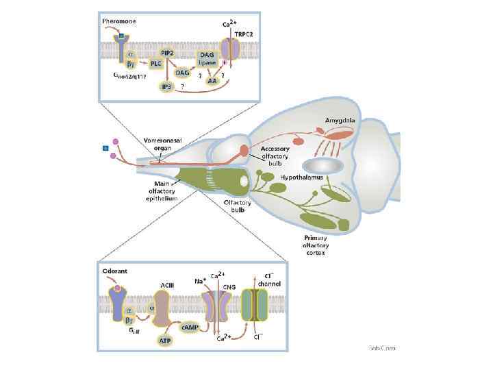
Обонятельные рецепторы

trace-amine-associated receptors

Обонятельные рецепторы A model for the transduction of odors in canonical OSNs The individual steps are detailed in the text. Note that several feedback loops modulate the odor response, including inhibition of the CNG channel by Ca 2+ (purple balls) that permeate the channel, and a Ca 2+/calmodulin (Ca. M)-mediated desensitization of the CNG channel that underlies rapid odor adaptation. Several other mechanisms have also been described, including phosphodiesterase-mediated hydrolysis of the second messenger c. AMP and phosphorylation of the OR by various kinases.

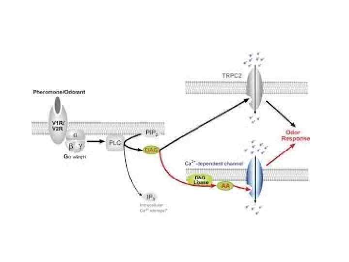
Обонятельные рецепторы A model for chemosensory transduction in vomeronasal sensory neurons The individual steps are detailed in the text. In contrast to the transduction cascade in OSNs, the mechanism of vomeronasal transduction is less well characterized. Vomeronasal sensory neurons express V 1 R, V 2 R or FPR receptors and either Gαi or Gαo. The TRPC 2 channel subunit is expressed in all VSNs, and may be part of a multimeric channel complex. Ca 2+ ions are represented as purple balls; Na+ ions as blue balls. VR, vomeronasal receptor (V 1 R, V 2 R or FPR); PIP 2, phosphatidylinositol 4, 5 -bisphosphate; IP 3, inositol 1, 4, 5 -trisphosphate. DAG, diacylglycerol.
-рецепторы сопряжены с Gi/o-белками, которые обеспечивают закрытие")
Опиоидные рецепторы μ-, δ-, κ- и ORL(opioid receptor-like)-рецепторы сопряжены с Gi/o-белками, которые обеспечивают закрытие Са 2+-каналов (κ-рецепторы) и открытие К+каналов (μ-, δ- и ORL-рецепторы). В зависимости от локализации рецепторов это приводит к уменьшению высвобождения медиатора и снижению возбудимости нейронов.

Опиоидные рецепторы
 = pain-inhibiting neurotransmitters produced by reticular formation in")
Inhibition of pain: - endorphins (enkephalins) = pain-inhibiting neurotransmitters produced by reticular formation in brain - descending fibers synapse (1) at the spinal cord dorsal horn release endorphins into synapse between sensory neurons (2) and ascending pain neurons (3) - endorphins have specific receptor sites on postsynaptic neurons - inhibitory action > opening of K+-channels > closing Ca 2+-channels hyperpolarizing post-synaptic membrane act as pain killers by inhibiting pain signals along ascending pain neurons 1 2 3
, нейрокинин А (NKA) и нейрокинин В (NKB). Афинность лигандов")
Тахикинины Включают вещество Р (SP), нейрокинин А (NKA) и нейрокинин В (NKB). Афинность лигандов (в порядке уменьшения) к рецепторам: Общий механизм сопряжен с Gq-белками, каскады фосфолипазы С (ИФ 3/ДАГ). Эффект заключается в медленной деполяризации через закрытие К+-каналов.

Функциональная роль некоторых пептидов
 2 -арахидонил-глицерол (2 -arachidonylglycerol, 2 -AG) Синтезируются в результате повышения внутриклеточной")
Эндоканнабиноиды Анандамид (anandamide) 2 -арахидонил-глицерол (2 -arachidonylglycerol, 2 -AG) Синтезируются в результате повышения внутриклеточной концентрации Са 2+. Механизм высвобождения из клеток неизвестен. Предполагается, что они диффундируют через клеточную мембрану и достигают соседних клеток. Через свои рецепторы эндоканнабиноиды уменьшают выделение ГАМК из тормозных терминалей, предположительно действуя на потенциалзависимые Са 2+- и/или К+-каналы пресинаптической мембраны.
.")
Рецепторы эндоканнабиноидов Идентифицировано два эндоканнабиноидных рецептора – СВ 1 и СВ 2 (44% гомологии). СВ 1 сопряжен с Gi/o-белками (реже с Gs). Каскад с Gi/o-белками приводит к ингибированию аденилатциклазы и открытию Kir-каналов. При активации G / -белков пресинаптическими СВ 1 блокируются Са 2+-каналы.
.")
Рецепторы эндоканнабиноидов Идентифицировано два эндоканнабиноидных рецептора – СВ 1 и СВ 2 (44% гомологии). До недавнего времени считалось, что СВ 2 распространены только на периферии. Однако сейчас СВ 2 описаны и в мозге. СВ 2 сопряжен с Gi/o-белками, но эффекты не включают открытие K+каналов и блокаду Са 2+-каналов.

Рецепторы эндоканнабиноидов Пресинаптические СВ 1 снижают высвобождение глютамата, Ац. Х и ГАМК через активацию G / -димера, который блокирует Са 2+-каналы и активирует K+-каналы. Синтез эндоканнабиноидов запускается при увеличении внутриклеточного Са 2+ и (или) активации липидных каскадов. Эндоканнабиноиды транспортируются (механизм неизвестен) из постсинаптической клетки и связываются с пресинаптическими рецепторами – т. н. эндоканнабиноидная ретроградная регуляция выделения медиатора.
Лекция_12_метаботропные_рецепторы_част.pptx