
LUNG Diseases Function of the Respiratory System


- Размер: 25.7 Mегабайта
- Количество слайдов: 76
Описание презентации LUNG Diseases Function of the Respiratory System по слайдам
LUNG Diseases
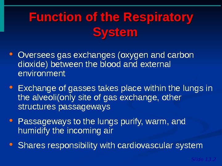
Function of the Respiratory System Slide 13. 2 Oversees gas exchanges (oxygen and carbon dioxide) between the blood and external environment Exchange of gasses takes place within the lungs in the alveoli(only site of gas exchange, other structures passageways Passageways to the lungs purify, warm, and humidify the incoming air Shares responsibility with cardiovascular system
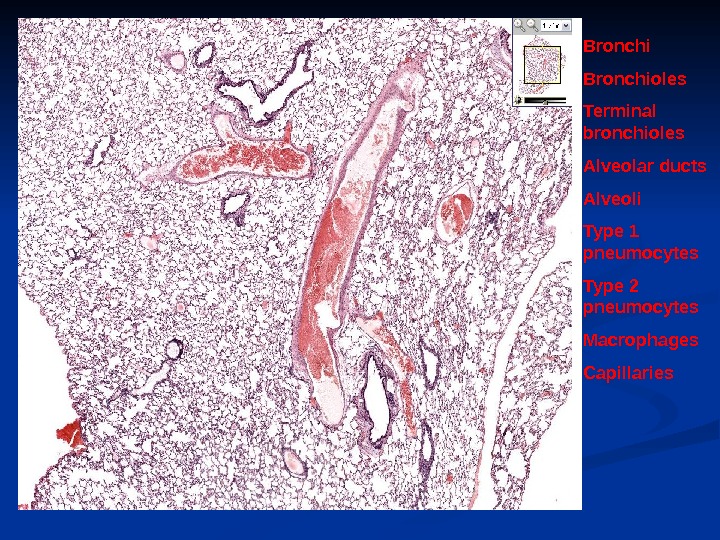
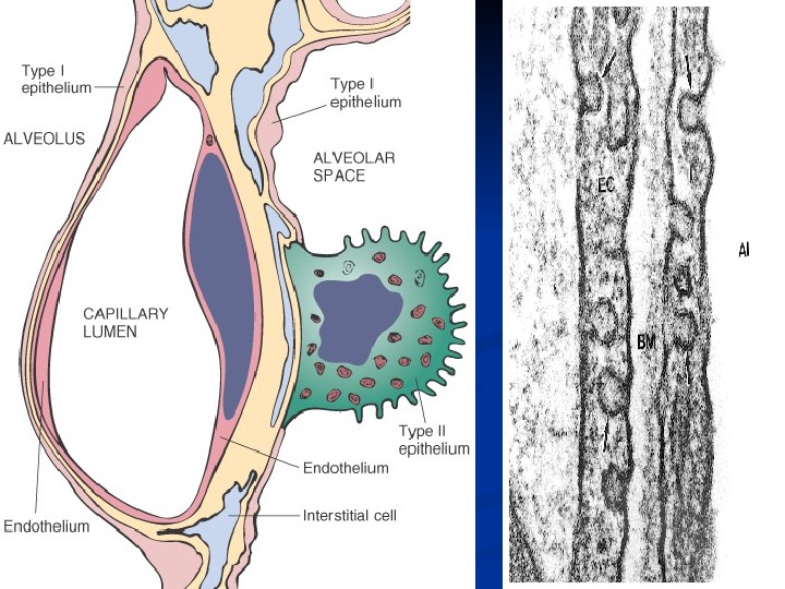
Bronchioles Terminal bronchioles Alveolar ducts Alveoli Type 1 pneumocytes Type 2 pneumocytes Macrophages Capillaries
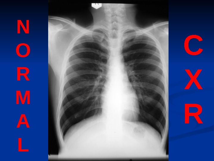
N O R M A L C X R
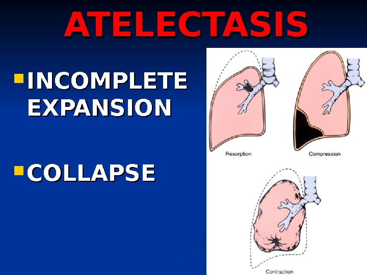
ATELECTASIS INCOMPLETE EXPANSION COLLAPS
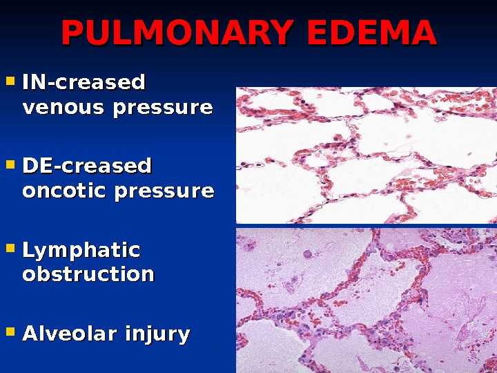
PULMONARY EDEMA IN-creased venous pressure DE-creased oncotic pressure Lymphatic obstruction Alveolar injury
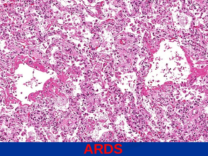
ARDS
ACUTE INTERSTITIAL PNEUMONIA Think of it as ARDS with NO known etiology!
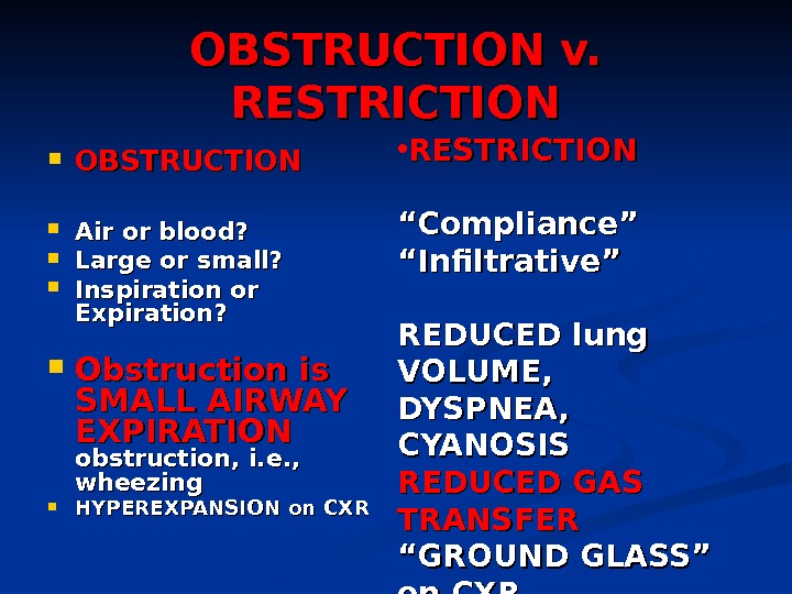
OBSTRUCTION v. RESTRICTION OBSTRUCTION Air or blood? Large or small? Inspiration or Expiration? Obstruction is SMALL AIRWAY EXPIRATION obstruction, i. e. , wheezing HYPEREXPANSION on CXR • RESTRICTION ““ Compliance” ““ Infiltrative” REDUCED lung VOLUME, DYSPNEA, CYANOSIS REDUCED GAS TRANSFER ““ GROUND GLASS” on CXR
OBSTRUCTION (c. OPD) EMPHYSEMA (almost always chronic) CHRONIC BRONCHITIS emphysema ASTHMA BRONCHIECTASIS
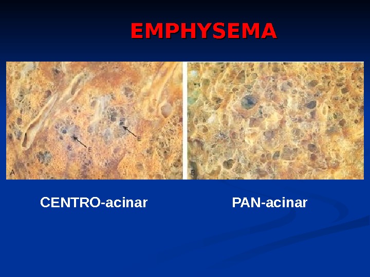
EMPHYSEMA COPD, or “END-STAGE” lung disease Centri-acinar, Pan-acinar, Paraseptal, Irregular Like cirrhosis, thought of as END-STAGE of multiple chronic small airway obstructive etiologies NON-specific IN-creased crepitance, BULLAE (BLEBS) Clinically linked to recurrent pneumonias, and progressive failure
CENTRO-acinar PAN-acinar. EMPHYSEM
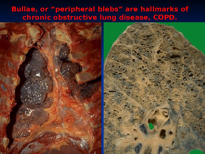
Bullae, or “peripheral blebs” are hallmarks of chronic obstructive lung disease, COPD.
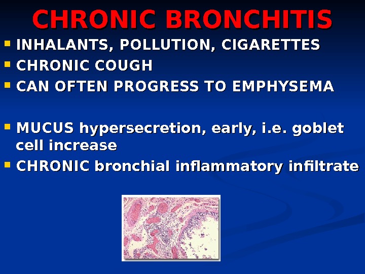
CHRONIC BRONCHITIS INHALANTS, POLLUTION, CIGARETTES CHRONIC COUGH CAN OFTEN PROGRESS TO EMPHYSEMA MUCUS hypersecretion, early, i. e. goblet cell increase CHRONIC bronchial inflammatory infiltrate
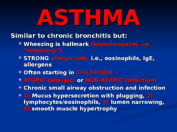
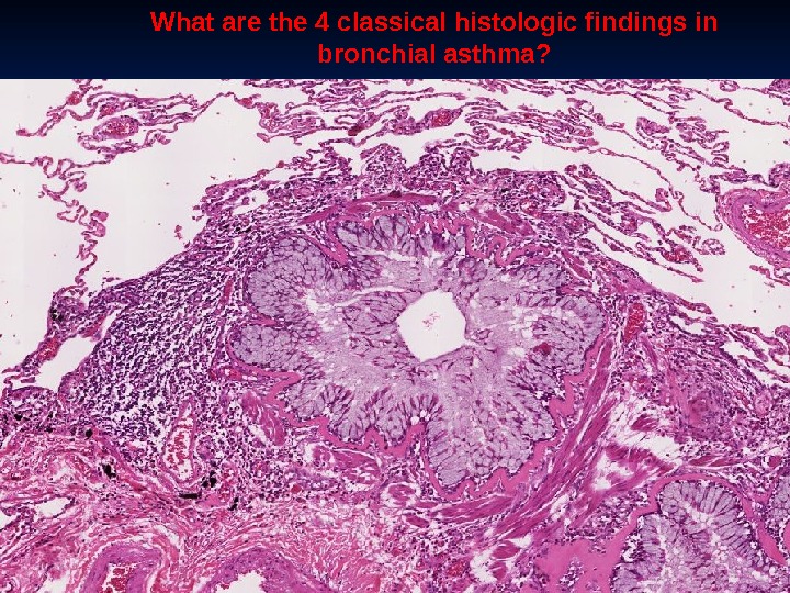
ASTHMA Similar to chronic bronchitis but: Wheezing is hallmark (( bronchospasm, i. e. “wheezing” )) STRONG allergic role , , i. e. , eosinophils, Ig. E, allergens Often starting in CHILDHOOD ATOPIC (allergic) oror NON-ATOPIC (infection) Chronic small airway obstruction and infection 1)1) Mucus hypersecretion with plugging, 2)2) lymphocytes/eosinophils, 3)3) lumen narrowing, 4)4) smooth muscle hypertrophy
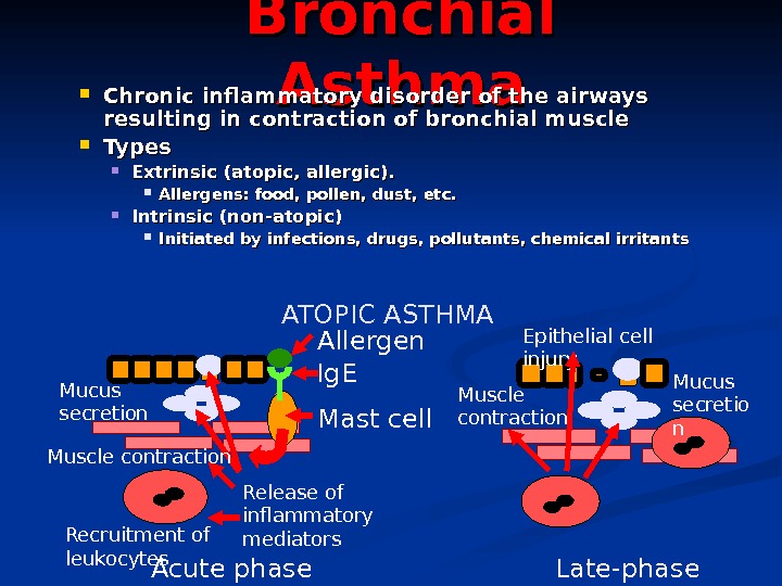
Bronchial Asthma Chronic inflammatory disorder of the airways resulting in contraction of bronchial muscle Types Extrinsic (atopic, allergic). Allergens: food, pollen, dust, etc. Intrinsic (non-atopic) Initiated by infections, drugs, pollutants, chemical irritants Release of inflammatory mediators Ig. E Mast cell. Allergen Muscle contraction Mucus secretion Recruitment of leukocytes Epithelial cell injury Muscle contraction Mucus secretio n Acute phase Late-phase. ATOPIC ASTHM
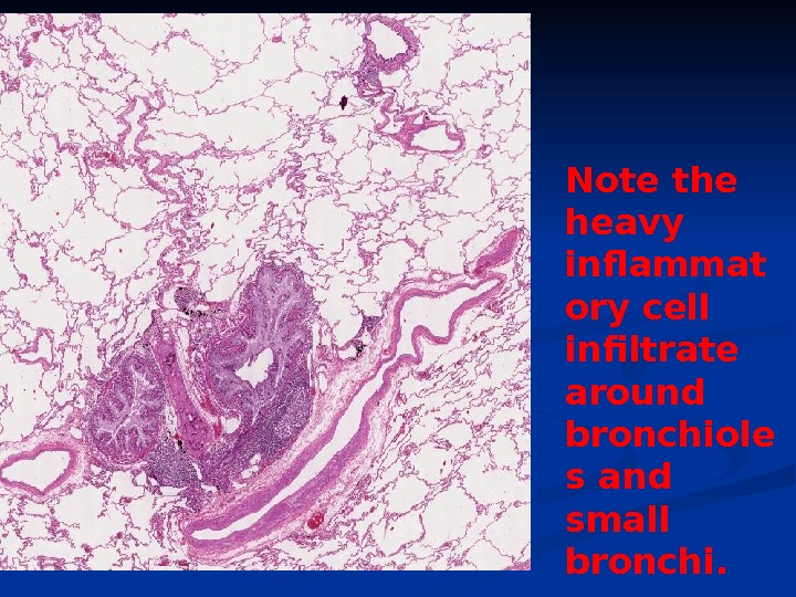
Note the heavy inflammat ory cell infiltrate around bronchiole s and small bronchi.
What are the 4 classical histologic findings in bronchial asthma?
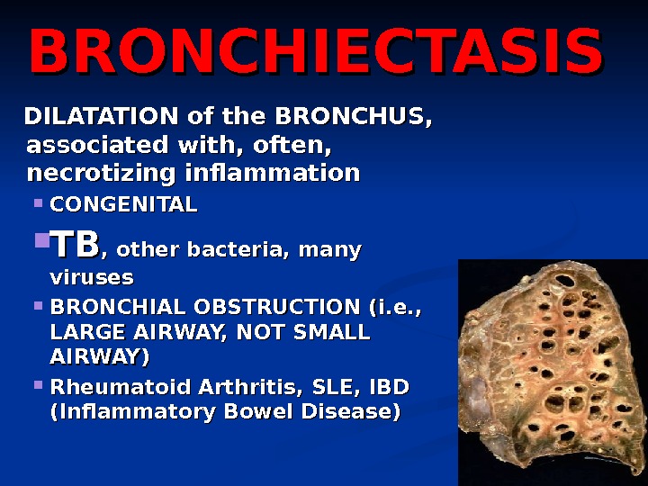
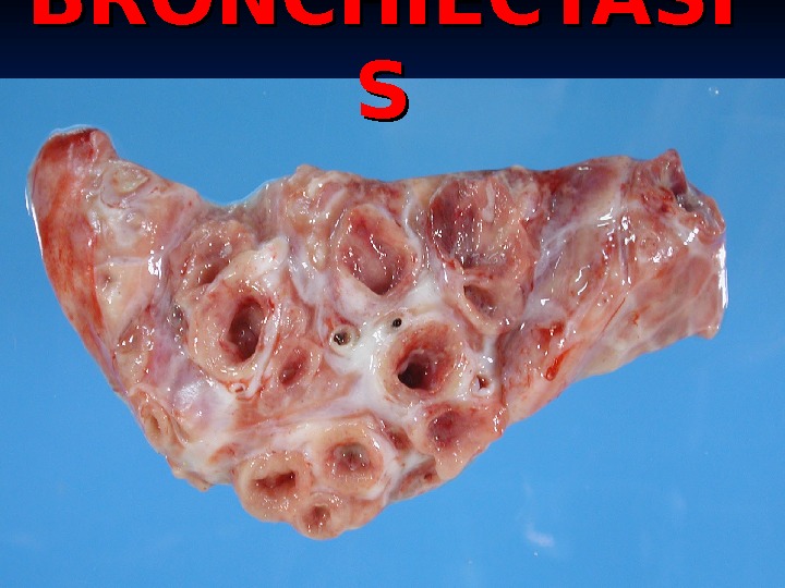
BRONCHIECTASIS DILATATION of the BRONCHUS, associated with, often, necrotizing inflammation CONGENITAL TBTB , other bacteria, many viruses BRONCHIAL OBSTRUCTION (i. e. , LARGE AIRWAY, NOT SMALL AIRWAY) Rheumatoid Arthritis, SLE, IBD (Inflammatory Bowel Disease)
BRONCHIECTASI SS
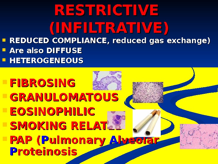
RESTRICTIVE (INFILTRATIVE) REDUCED COMPLIANCE, reduced gas exchange) Are also DIFFUSE HETEROGENEOUS FIBROSING GRANULOMATOUS EOSINOPHILIC SMOKING RELATED PAP ( PP ulmonary AA lveolar PP roteinosis
FIBROSING ““ IDIOPATHIC” PULMONARY FIBROSIS (IPF) NONSPECIFIC INTERSTITIAL FIBROSIS ““ CRYPTOGENIC” ORGANIZING PNEUMONIA ““ COLLAGEN” VASCULAR DISEASES PNEUMOCONIOSES DRUG REACTIONS RADIATION CHANGES
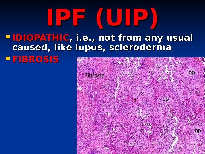
IPF (UIP) IDIOPATHIC , i. e. , not from any usual caused, like lupus, scleroderma FIBROSIS
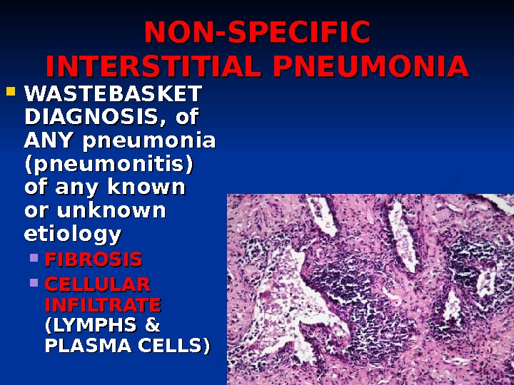
NON-SPECIFIC INTERSTITIAL PNEUMONIA WASTEBASKET DIAGNOSIS, of ANY pneumonia (pneumonitis) of any known or unknown etiology FIBROSIS CELLULAR INFILTRATE (LYMPHS & PLASMA CELLS)
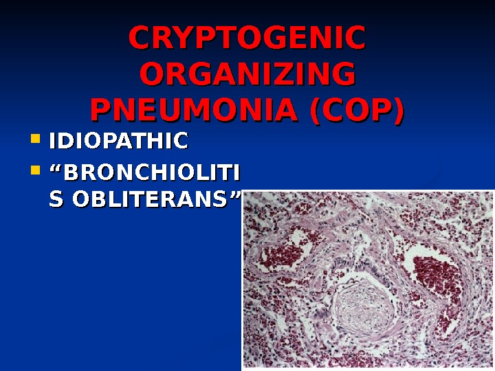
CRYPTOGENIC ORGANIZING PNEUMONIA (COP) IDIOPATHIC ““ BRONCHIOLITI S OBLITERANS”
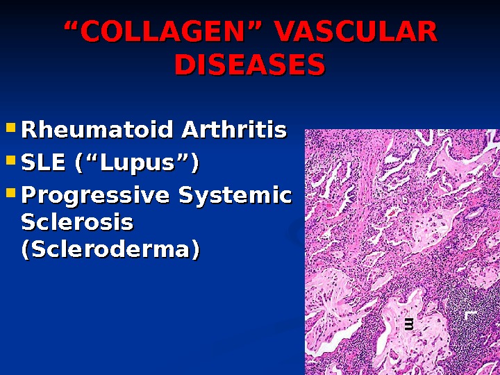
““ COLLAGEN” VASCULAR DISEASES Rheumatoid Arthritis SLE (“Lupus”) Progressive Systemic Sclerosis (Scleroderma)
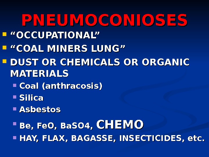
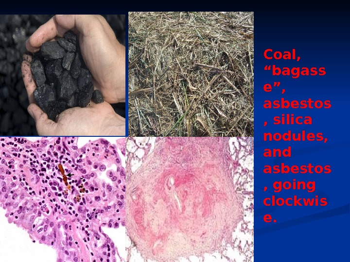
PNEUMOCONIOSES ““ OCCUPATIONAL” ““ COAL MINERS LUNG” DUST OR CHEMICALS OR ORGANIC MATERIALS Coal (anthracosis) Silica Asbestos Be, Fe. O, Ba. SO 4, CHEMO HAY, FLAX, BAGASSE, INSECTICIDES, etc.
Coal, “bagass e”, asbestos , silica nodules, and asbestos , going clockwis e.
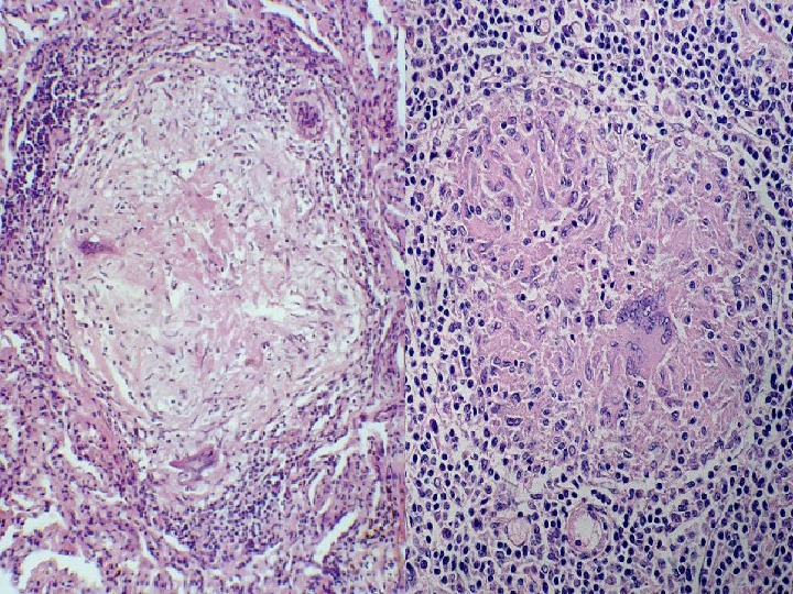
GRANULOMATOUS SARCOIDOSIS , , i. e. , NON-caseating granulomas (IDIOPATHIC) HYPERSENSITIVITY (DUSTS, bacteria, fungi, Farmer’s Lung, Pigeon Breeder’s Lung)
SARCOIDOSIS Mainly LUNG, but eye, skin or ANYWHERE UNKNOWN ETIOLOGY IMMUNE, GENETIC factors F>>M B>>W YOUNG ADULT BLACK WOMEN
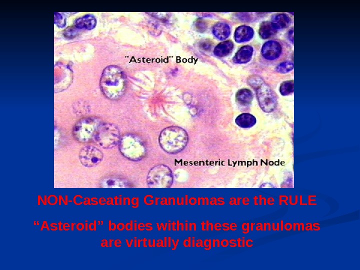
NON-Caseating Granulomas are the RULE “ Asteroid” bodies within these granulomas are virtually diagnostic
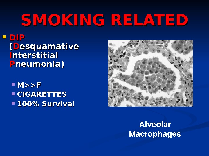
SMOKING RELATED DIPDIP (( DD esquamative II nterstitial PP neumonia) M>>F CIGARETTES 100% Survival Alveolar Macrophages
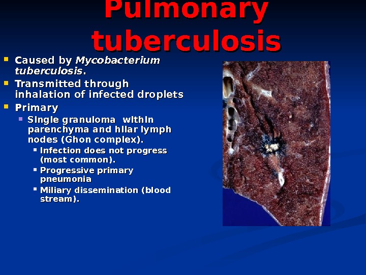
Pulmonary tuberculosis Caused by Mycobacterium tuberculosis. . Transmitted through inhalation of infected droplets Primary Single granuloma within parenchyma and hilar lymph nodes (Ghon complex). Infection does not progress (most common). Progressive primary pneumonia Miliary dissemination (blood stream).
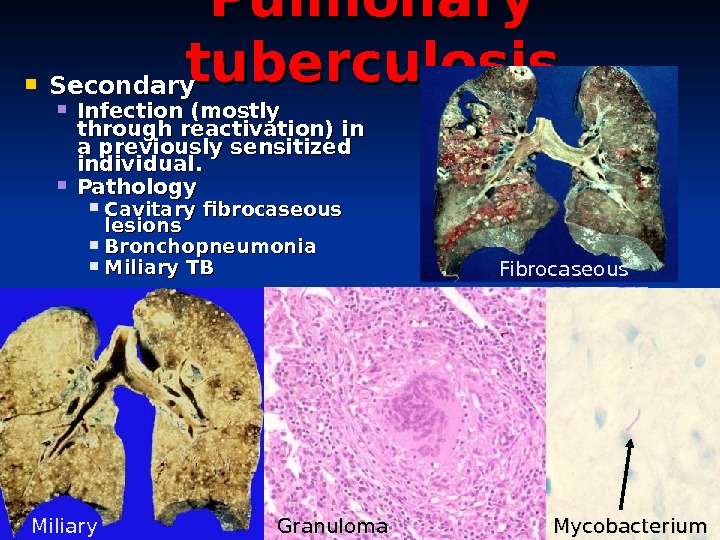
Pulmonary tuberculosis Secondary Infection (mostly through reactivation) in a previously sensitized individual. Pathology Cavitary fibrocaseous lesions Bronchopneumonia Miliary TB Miliary Granuloma Fibrocaseous Mycobacterium
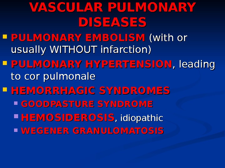
VASCULAR PULMONARY DISEASES PULMONARY EMBOLISM (with or usually WITHOUT infarction) PULMONARY HYPERTENSION , leading to cor pulmonale HEMORRHAGIC SYNDROMES GOODPASTURE SYNDROME HEMOSIDEROSIS , idiopathic WEGENER GRANULOMATOSIS
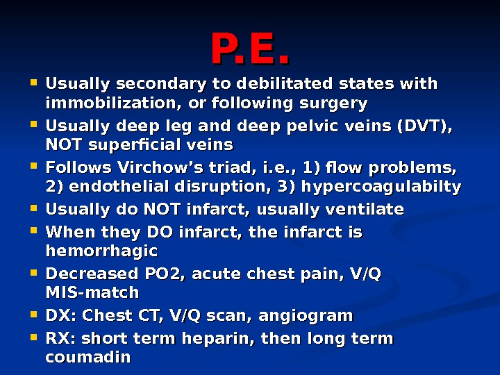
P. E. Usually secondary to debilitated states with immobilization, or following surgery Usually deep leg and deep pelvic veins (DVT), NOT superficial veins Follows Virchow’s triad, i. e. , 1) flow problems, 2) endothelial disruption, 3) hypercoagulabilty Usually do NOT infarct, usually ventilate When they DO infarct, the infarct is hemorrhagic Decreased PO 2, acute chest pain, V/Q MIS-match DX: Chest CT, V/Q scan, angiogram RX: short term heparin, then long term coumadin
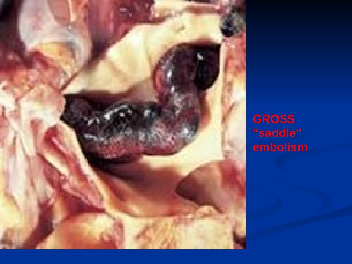
GROSS “saddle” embolism
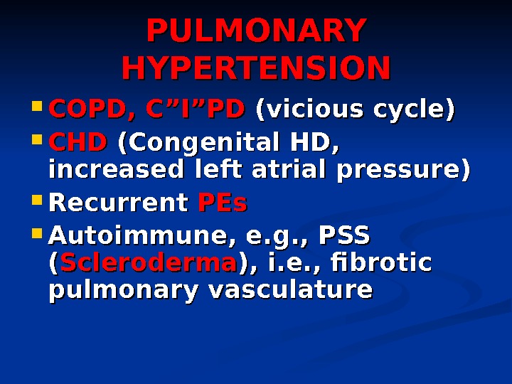
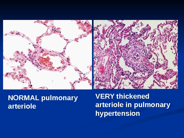
PULMONARY HYPERTENSION COPD, C”I”PD (vicious cycle) CHDCHD (Congenital HD, increased left atrial pressure) Recurrent PEs Autoimmune, e. g. , PSS (( Scleroderma ), i. e. , fibrotic pulmonary vasculature
VERY thickened arteriole in pulmonary hypertension. NORMAL pulmonary arteriole
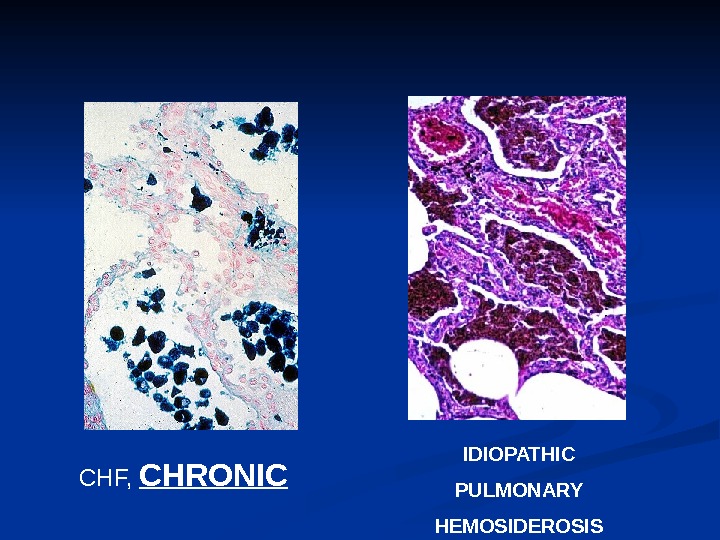
CHF, CHRONIC IDIOPATHIC PULMONARY HEMOSIDEROSIS
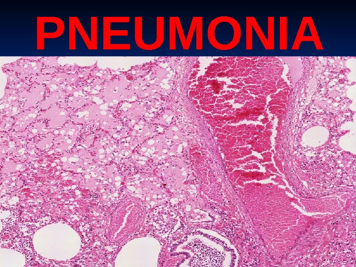
PNEUMONI
PULMONARY INFECTIONS COMM UNITY-ACQUIRED BACTERIAL ACUTE PNEUMONIAS Streptococcus Pneumoniae Haemophilus Influenzae Moraxella Catarrhalis Staphylococcus Aureus Klebsiella Pneumoniae Pseudomonas Aeruginosa Legionella Pneumophila COMMUNITY-ACQUIRED ATYPICAL (VIRAL AND MYCOPLASMAL) PNEUMONIAS Morphology. Clinical Course. Influenza Infections Severe Acute Respiratory Syndrome (SARS) NOSOCOMIAL PNEUMONIA ASPIRATION PNEUMONIA LUNG ABSCESS Etiology and Pathogenesis. CHRONIC PNEUMONIA Histoplasmosis, Morphology Blastomycosis, Morphology Coccidioidomycosis, Morphology PNEUMONIA IN THE IMMUNOCOMPROMISED HOST PULMONARY DISEASE IN HUMAN IMMUNODEFICIENCY VIRUS INFECTION
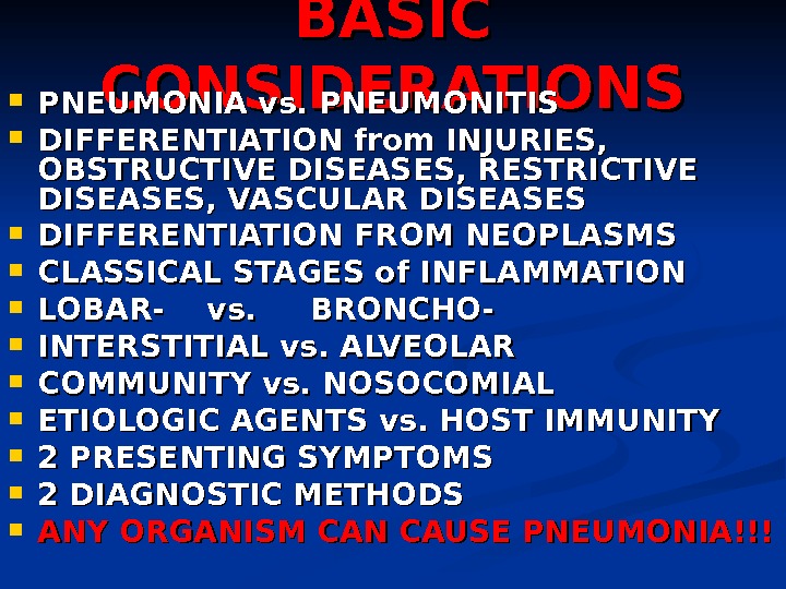
BASIC CONSIDERATIONS PNEUMONIA vs. PNEUMONITIS DIFFERENTIATION from INJURIES, OBSTRUCTIVE DISEASES, RESTRICTIVE DISEASES, VASCULAR DISEASES DIFFERENTIATION FROM NEOPLASMS CLASSICAL STAGES of INFLAMMATION LOBAR- vs. BRONCHO- INTERSTITIAL vs. ALVEOLAR COMMUNITY vs. NOSOCOMIAL ETIOLOGIC AGENTS vs. HOST IMMUNITY 2 PRESENTING SYMPTOMS 2 DIAGNOSTIC METHODS ANY ORGANISM CAN CAUSE PNEUMONIA!!!
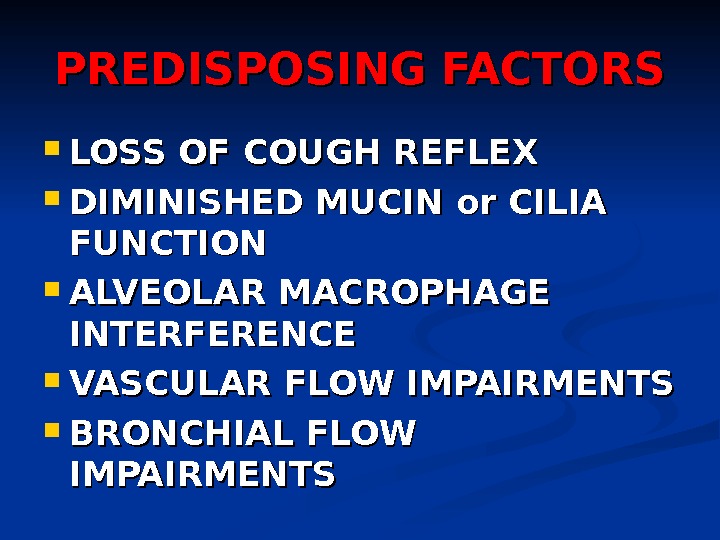
PREDISPOSING FACTORS LOSS OF COUGH REFLEX DIMINISHED MUCIN or CILIA FUNCTION ALVEOLAR MACROPHAGE INTERFERENCE VASCULAR FLOW IMPAIRMENTS BRONCHIAL FLOW IMPAIRMENTS
Although pneumonia is one of the most common causes of death, it usually does NOT occur in healthy people spontaneously
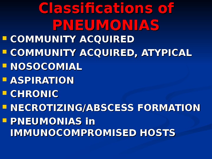
Classifications of PNEUMONIAS COMMUNITY ACQUIRED, ATYPICAL NOSOCOMIAL ASPIRATION CHRONIC NECROTIZING/ABSCESS FORMATION PNEUMONIAS in IMMUNOCOMPROMISED HOSTS
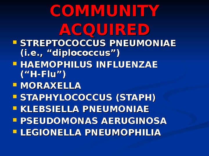
COMMUNITY ACQUIRED STREPTOCOCCUS PNEUMONIAE (i. e. , “diplococcus”) HAEMOPHILUS INFLUENZAE (“H-Flu”) MORAXELLA STAPHYLOCOCCUS (STAPH) KLEBSIELLA PNEUMONIAE PSEUDOMONAS AERUGINOSA LEGIONELLA PNEUMOPHILI
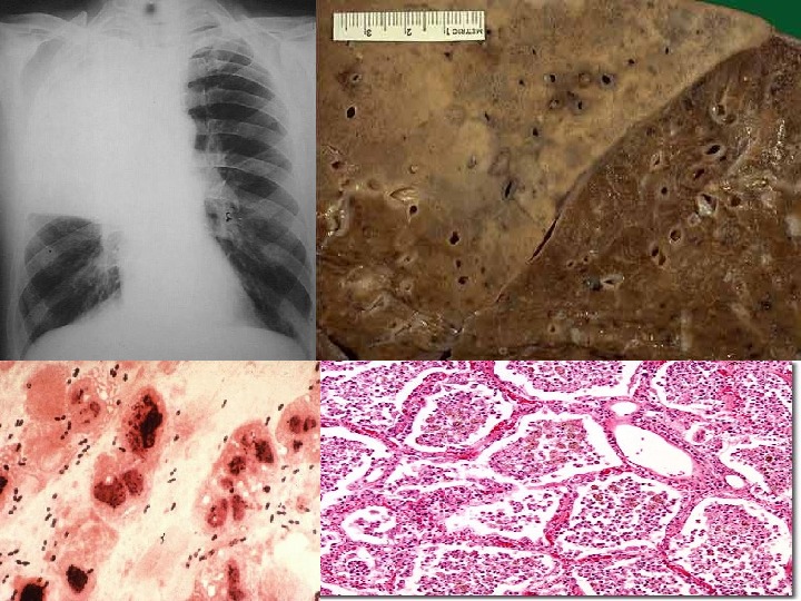
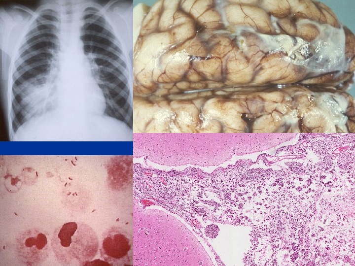
STREPTOCOCCUS The classic LOBAR pneumonia Normal flora in 20% of adults Only 20% of victims have ++ blood cultures ““ Penicillins” are often 100% curative Vaccines are often 100% preventive
MORPHOLOGY ACUTE ORGANIZING CHRONIC FIBROSIS vs. FULL RESOLUTION ““ HEPATIZATION ”, RED vs. GREY CONSOLIDATION ““ INFILTRATE ”, XRAY vs. HISTOPATH Loss of “ CREPITANCE ””
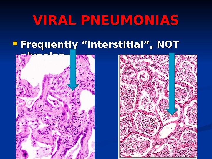
VIRAL PNEUMONIAS Frequently “interstitial”, NOT alveolar
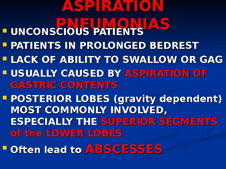
ASPIRATION PNEUMONIAS UNCONSCIOUS PATIENTS IN PROLONGED BEDREST LACK OF ABILITY TO SWALLOW OR GAG USUALLY CAUSED BY ASPIRATION OF GASTRIC CONTENTS POSTERIOR LOBES (gravity dependent) MOST COMMONLY INVOLVED, ESPECIALLY THE SUPERIOR SEGMENTS of the LOWER LOBES Often lead to ABSCESSES
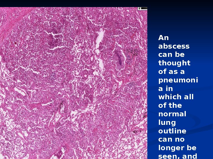
LUNG ABSCESSES ASPIRATION SEPTIC EMBOLIZATION NEOPLASIA From NEIGHBORING structures: ESOPHAGUS SPINE PLEURA DIAPHRAGM ANY pneumonia which is severe and destructive, and UN-treated enough
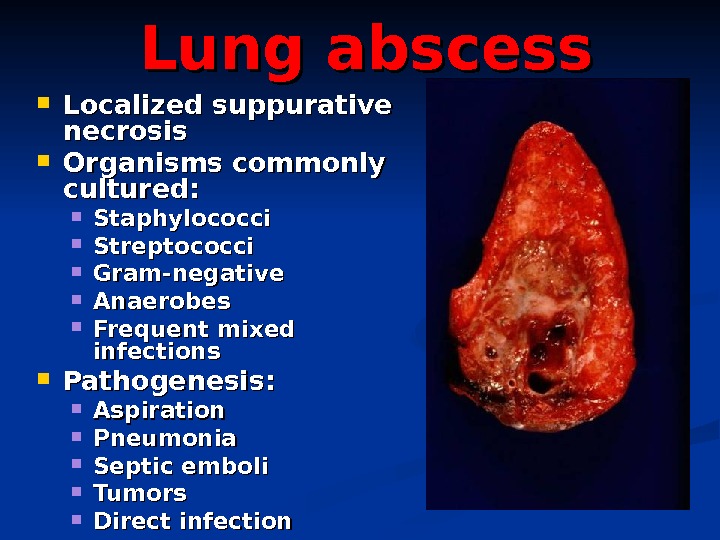
Lung abscess Localized suppurative necrosis Organisms commonly cultured: Staphylococci Streptococci Gram-negative Anaerobes Frequent mixed infections Pathogenesis: Aspiration Pneumonia Septic emboli Tumors Direct infection
An abscess can be thought of as a pneumoni a in which all of the normal lung outline can no longer be seen, and there is 100% pus.
CHRONIC Pneumonias USUALLY NOT persistences of the community or nosocomial bacterial infections, but CAN BE, at least histologically Often SYNONYMOUS with the 4 classic fungal or granulomatous pulmonary infections, i. e. , TB, Histo-, Blasto-, Coccidio- If you see pulmonary granulomas, think of a CHRONIC process, often years
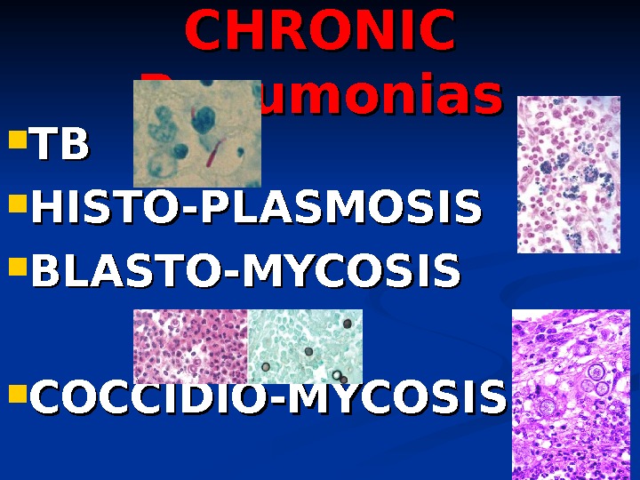
CHRONIC Pneumonias TBTB HISTO-PLASMOSIS BLASTO-MYCOSIS COCCIDIO-MYCOSIS
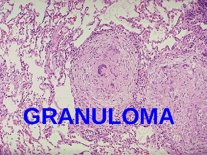
GRANULOM
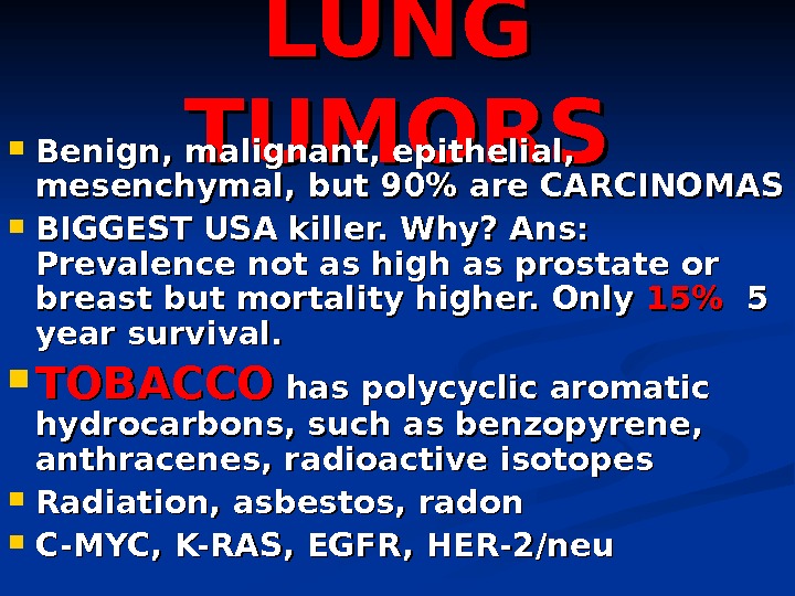
LUNG TUMORS Benign, malignant, epithelial, mesenchymal, but 90% are CARCINOMAS BIGGEST USA killer. Why? Ans: Prevalence not as high as prostate or breast but mortality higher. Only 15%15% 5 5 year survival. TOBACCO has polycyclic aromatic hydrocarbons, such as benzopyrene, anthracenes, radioactive isotopes Radiation, asbestos, radon C-MYC, K-RAS, EGFR, HER-2/neu
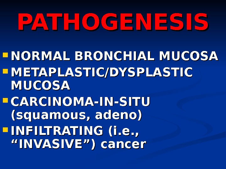
PATHOGENESIS NORMAL BRONCHIAL MUCOSA METAPLASTIC/DYSPLASTIC MUCOSA CARCINOMA-IN-SITU (squamous, adeno) INFILTRATING (i. e. , “INVASIVE”) cancer
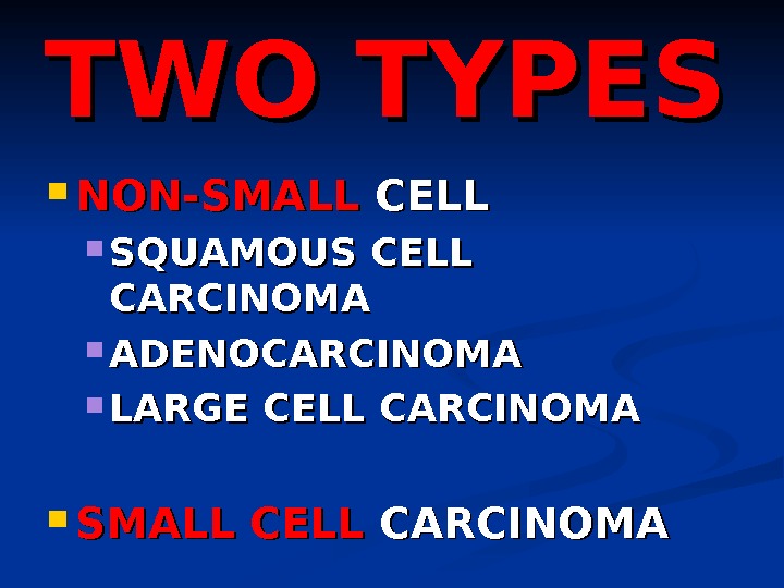
TWO TYPES NON-SMALL CELL SQUAMOUS CELL CARCINOMA ADENOCARCINOMA LARGE CELL CARCINOMA SMALL CELL CARCINOM
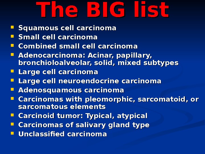
The BIG list Squamous cell carcinoma Small cell carcinoma Combined small cell carcinoma Adenocarcinoma: Acinar, papillary, bronchioloalveolar, solid, mixed subtypes Large cell carcinoma Large cell neuroendocrine carcinoma Adenosquamous carcinoma Carcinomas with pleomorphic, sarcomatoid, or sarcomatous elements Carcinoid tumor: Typical, atypical Carcinomas of salivary gland type Unclassified carcinoma
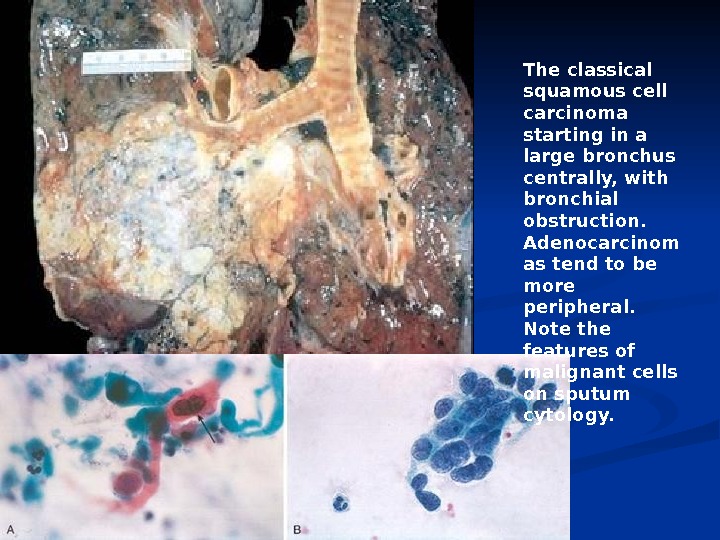
The classical squamous cell carcinoma starting in a large bronchus centrally, with bronchial obstruction. Adenocarcinom as tend to be more peripheral. Note the features of malignant cells on sputum cytology.
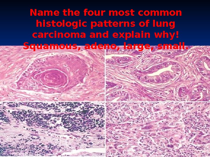
Name the four most common histologic patterns of lung carcinoma and explain why! Squamous, adeno, large, small.
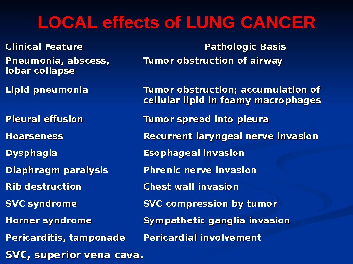
Clinical Feature Pathologic Basis Pneumonia, abscess, lobar collapse Tumor obstruction of airway Lipid pneumonia Tumor obstruction; accumulation of cellular lipid in foamy macrophages Pleural effusion Tumor spread into pleura Hoarseness Recurrent laryngeal nerve invasion Dysphagia Esophageal invasion Diaphragm paralysis Phrenic nerve invasion Rib destruction Chest wall invasion SVC syndrome SVC compression by tumor Horner syndrome Sympathetic ganglia invasion Pericarditis, tamponade Pericardial involvement SVC, superior vena cava. LOCAL effects of LUNG CANCER
METASTATIC TUMORS LUNG is the MOST COMMON site for all metastatic tumors, regardless of site of origin It is the site of FIRST CHOICE for metastatic sarcomas for purely anatomic reasons!
PLEURA PLEURITIS PNEUMOTHORAX EFFUSIONS HYDROTHORAX HEMOTHORAX CHYLOTHORAX MESOTHELIOMAS
PLEURITIS Usual bacteria, viruses, etc. Infarcts Lung abscesses, empyema TBTB ““ Collagen” diseases, e. g. , RA, SLESLE Uremia Metastatic
PNEUMOTHORAX SPONTANEOUS, TRAUMATIC, THERAPEUTIC OPEN or CLOSED ““ TENSION” pneumothorax, “valvular” effect ““ Bleb” rupture Perforating injuries Post needle biopsy
EFFUSIONS TRANSUDATE (HYDROTHORAX) EXUDATE (HYDROTHORAX) BLOOD (HEMOTHORAX) LYMPH (CHYLOTHORAX)
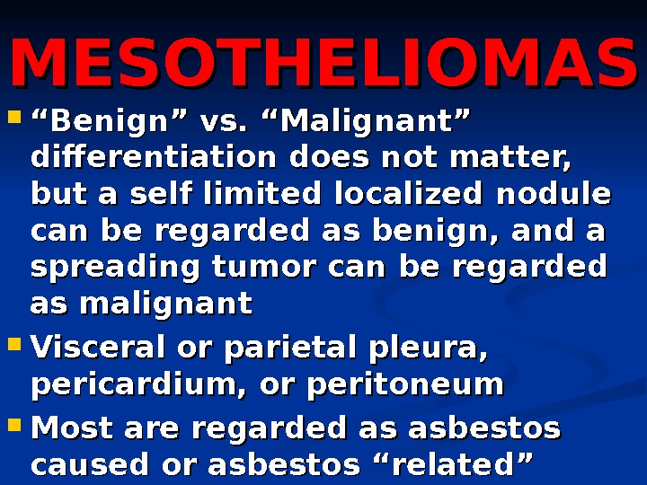
MESOTHELIOMAS ““ Benign” vs. “Malignant” differentiation does not matter, but a self limited localized nodule can be regarded as benign, and a spreading tumor can be regarded as malignant Visceral or parietal pleura, pericardium, or peritoneum Most are regarded as asbestos caused or asbestos “related”
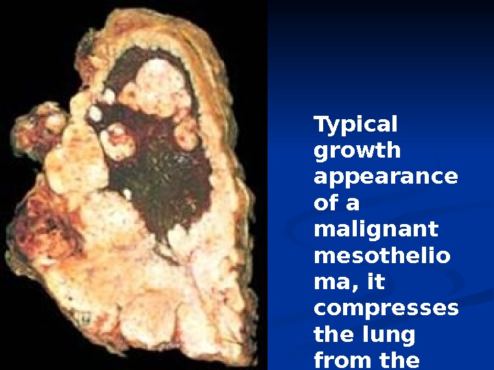
Typical growth appearance of a malignant mesothelio ma, it compresses the lung from the OUTSIDE.
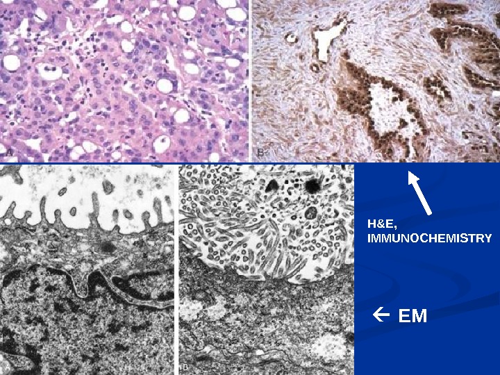
EMH&E, IMMUNOCHEMISTRY