5bb09663894624698c389b4a1661b791.ppt
- Количество слайдов: 87


Low Back Pain Prof. Dr. Hidayet Sarı l Cerrahpaşa Medical School l Physical Medicine and Rehabilitation Department l

Lumbosacral Pain l l l 60 -90% life time incidence 5% annual incidence Peak in 40’s 12 -26% in children and adolescents cost in US upwards of 100 billion per year

Lumbosacral Pain l l l 15 -25% of workman’s comp= LBP 30 -40% of workman’s comp payments Return to work rates l l l 50% if disabled for 6 months 25% if disabled 1 year 0% if disabled > 2 years

Lumbosacral Pain l 90% resolve in 6 -12 weeks l l l Croft et al (1998) found that 90% did not seek care after three months 40 -80% in 1 week 75% sciatica clear in 1 -6 months l 70 -90% recur

Diagnosis: Low Back Pain ? l A physiologic cause of back pain can not be definitively determined in 85% of patients

Anatomy l Vertebra l l Body, anteriorly l Functions to Support weight Vertebral arch, posteriorly l Formed by two pedicles and two laminae l Functions to protect neural structures
 l Attached anteriorly to body l Continuous")
Vertebral Arch Pedicles (Latin for Little Feet) l Attached anteriorly to body l Continuous posteriorly with laminae l Intervertebral foramen l. Superior vertebral notch l. Inferior vertebral notch l Laminae (Latin for Thin Plates) l Meet posteriorly to form spinous process l

Facet Joint l l l Formed by articulation of inferior and superior processes of subsequent vertebrae Orientation in lumbar spine is toward sagittal plane, allowing flexion and extension but limiting rotation of the lumbar vertebrae Helps to prevent anterior movement of superior vertebra on inferior vertebra Articular surfaces are made up of non-innervated articular cartilage Capsule and synovial membrane are innervated with pain receptors

Ligaments l l l Anterior longitudinal ligament Posterior longitudinal ligament Interspinous ligament Supraspinous ligament Ligamentum flavum

Intervertebral Disc l l l Most common site of back pain Normally comprises ~ 25% of length of spine Consists of a central nucleus pulposus l l l Reticulated and collagenous substance Composed of ~ 88% water Annulus fibrosus l l Consists of concentric lamellae of fibrocartilage fibers arranged obliquely With each layer, they are arranged in opposite directions

Muscles l l l l Psoas Major/minor Quadratus lumborum Intertransversalis Interspinals Multifidus Longissimus thoracis Iliocostalis lumborum Erector spinae

Differential Diagnosis l l MSLBP/Mechanical/. . . Osteoarthritis Facet/disk/SI Facet Syndrome Diskitis l Fracture – l l l Stress Compression Other Spinal Stenosis Tumor Discogenic
")
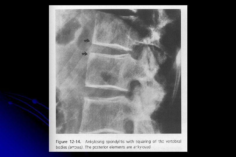
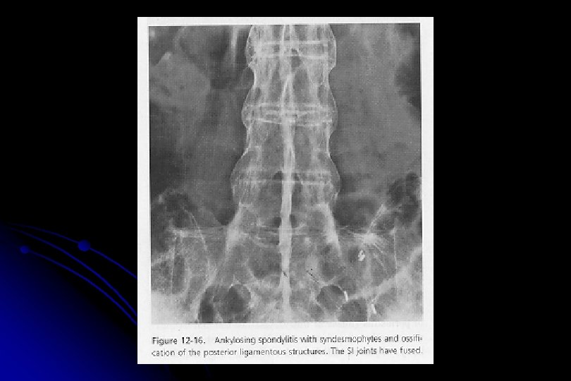
Differential Diagnosis l Non-back pain l l retroperitoneal process (Pancreatic, Renal, Duodenal, Gyn, Prostate) AAA Zoster Diabetic radiculopathy l l SI joint Rheumatologic disorders l l Reiters Ankylosing Spondylitis

Differential Diagnosis

Common Causes of Low Back Pain Muscular spasm, strain l Ligament sprain l Spondylosis l Herniated nucleus pulposus l Facet joint dysfunction l Spondylo-lysis or -listhesis l Seronegative spondyloarthropathies l

Clearing up the terms l Spondylosis l Degenerative joint disease affecting the vertebrae and intervertebral disc l Spondylolysis l Fracture l in pars interarticularis Spondylolisthesis l Displacement of one vertebra on another

Spondylo-lysis and -listhesis

Spondilo-lysthesis

Facet joint pain

Ankylosing spondylitis

History

History l Three major concerns: l Is there evidence of systemic disease l Is there evidence of neurological disease l Is there social or psychological stress which is contributing? l Exclude serious underlying pathology, such as infection, malignancy or cauda equina syndrome

Red Flags l General l> l l l Rest +/- l l Cancer 50 l History of Cancer l Weight loss l Unrelenting night pain Infection l l l IVDU Steroid use Fever Fracture l 1 month l> l l Age > 70 Steroid use Trauma hx Bladder dysfunction Osteoporosis Cauda Equina Syndrome l Saddle anesthesia l Bowel/bladder dysfunction l l l Loss of sphincter tone Rapid progression Unilat or bilat major motor weakness

Yellow Flags l l Belief that back pain is harmful or severely disabling Fear-avoidance behavior and reduced activity level Social withdrawal and low mood Expectation that passive treatments will help

Back Pain Risk Factors l l l Caucasian Western states Smoker Increasing age up to 55 Prolonged driving of vehicle Hard physical labor l vibration or repetitive lift > 40 lbs

Back Pain Risk Factors l l Psychological stress Job dissatisfaction Prior episode of back pain Osteoporosis

Onset l l Acute - Lift/twist, fall, MVA Subacute - inactivity, occupational (sitting, driving, flying) ? Pending litigation Pain effect on: l l l work/occupation sport/activity (during or after) ADL’s

Pain Character l l l Sharp Burning Dull ache

Pain with… l Prone positionn l l Sitting l l Paramedian HNP, annular tear Standing l l Facet, Lat HNP, systemic Lateral HNP, central stenosis, facet syndrome Walking l central stenosis

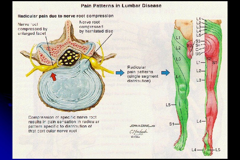
Radiation l l Up back To sacrum To buttocks Down leg

Other Symptoms l l Cough/valsalva exacerbation Distal neuro sx weakness/paresthesia Perianal paresthesia Bowel/bladder sx

Other History l l Prior treatments and response Prior h/o back pain Exercise habits Occupation/recreational activities

Examination l l Walk Standing Sitting Supine

Walking l Gait l l length of stride arm swing trunk motion ? pelvic tilt

Standing

Posture l l l Kyphosis Hyperlordosis Scoliosis
")
Range of Motion l l FF ~90 o (reversal of lumbar lordosis with FF) Ext ~15 -20 o Side bend ~ 30 o Trunk rotation

Palpation l l Spinous processes Dorsal lumbar fascia/soft tissues

Other l Single leg extension l l Gastroc strength l l l Stork Test Toe raises Squat Standing single-leg balance (nl 15 -30 sec)

Sitting l l Distracted SLR DTR - patellar & Achilles Strength - EHL, TA, Peroneals, quads, hip flexors Sensation

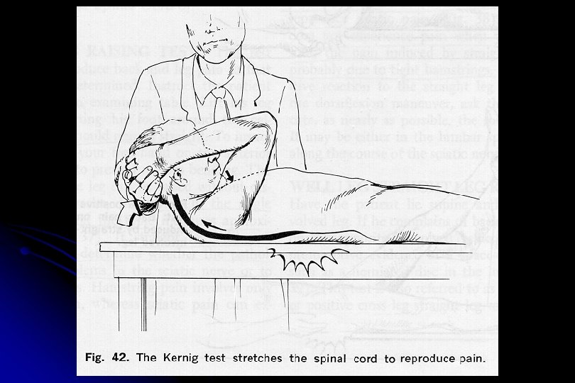
Supine Tests to Stretch the Spinal Cord or Sciatic Nerve l Straight Leg Raise l Cross Leg SLR l Kernig Test
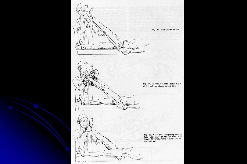
 Leg lengths l l measured ASIS to")
Supine l l Hamstring flexibility (Popliteal angle) Leg lengths l l measured ASIS to Med Mal estimated
 Non-anatomic superficial tenderness l Non-anatomic weakness or sensory loss")
Non-organic Physical Signs (“Waddell’s signs”) Non-anatomic superficial tenderness l Non-anatomic weakness or sensory loss l Simulation tests with axial loading and en bloc rotation producing pain l Distraction test or flip test in which pt has no pain with full extension of knee while seated, but the supine SLR is markedly positive l Over-reaction verbally or exaggerated body language l Waddell, et al. Spine 5(2): 117 -125, 1980.

Neurologic Testing l Primary focus on the L 5 and S 1 never roots, since 98 percent of clinically important disc herniations occur at L 4 L 5 and L 5 -S 1

Sacral Plexus l L 4 l l l Quads/Tibialis Anterior Patellar reflex Sensory Great toe and medial leg

Sacral Plexus l L 5 l l l Strength of Ankle and great toe dorsiflexion Extensor Hallucis Longus Sensory to dorsum of foot

Sacral Plexus l S 1 l l Ankle reflexes and sensation of posterior calf and lateral foot Peroneals/Gastroc Achilles reflex Sensory to lateral and plantar foot

Other l l l Rectal tone Anal wink Cremasteric reflex

Diagnostic Studies l Radiographs l Early if ominous signs l Fever l night l pain l age extremes l h/o Ca l wt loss l Trauma osteoporosis Symptoms present > 1 month

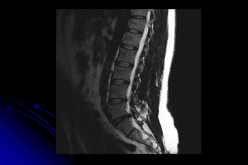
Diagnostic Studies l MRI l l l l More sensitive for infection and cancer > 12 weeks of pain Herniated discs Spinal Stenois order if hx/exam confusing roadmap for surgeon more costly, increased time to scan, problem with claustrophobic patients
 l suspect for stress fx, Ca, inflammation")
Diagnostic Studies l Bone Scan (SPECT) l suspect for stress fx, Ca, inflammation

Diagnostic Studies l EMG/NCV l l r/o peripheral neuropathy localize nerve injury correlate with radiographic changes order after 4 weeks of symptoms

Lab Studies l l CBC, ESR, UA Avoid RF, ANA or others unless indicated

Treatment

Treatment Recommendations Based on the Joint Clinical Practice Guidelines from the American College of Physicians and the American Pain Society l Level of evidenced reviewed and graded l Guidelines published in Annals of Internal Medicine in 2007 l

Recommendations A Panel Strongly recommends l B Panel recommends consideration for eligible patients l C Panel makes no recommendation l D Panel recommends against l I Panel found insufficient evidence l

Acute Mnagement l Medications l Pain control Acetaminophen/NSAID’s l Minimize use of opioids l 2007 joint guidelines from ACP and APS recommend against steroids l l Muscle relaxers l Short term use of benzo or non benzodiazepine muscle relaxers in combination with NSAIDs/acetaminophen

Acute Management l Back Exercises l l l There is no evidence that suggests that back exercises are helpful during acute pain and may actually be counterproductive Upon recovery, back exercises may be useful in preventing recurrence Resume normal activity as quickly as

Subacute Management l l l Continue patient education Mechanics - lifting technique, sport, . . . Avoid l l l prolonged sitting/standing recurrent bending twisting

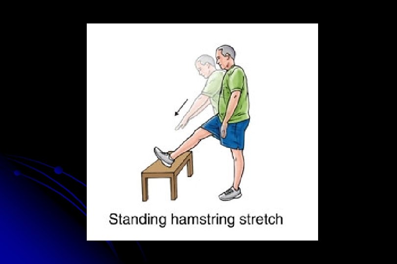
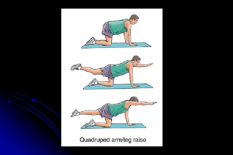
Conditioning l ACTIVITY & CONDITIONING l l l walking Stretching - HS, hip extensors, erector spinae Strengthening - abs, erector spinae

Chronic Low Back Pain > 3 months l Treatment goals: l l Control pain l Maintain function l Prevent disability

Evidenced-Based Reasonable Therapies for Chronic Low Back Pain l l l Acetaminophen NSAIDS TCAs +- Opioids +- Benzodiazepines l l l Physical therapy Exercise therapy Interdisciplinary rehab Spinal Manipulation Yoga Massage
 Ominous signs/sx - fever, weakness, bowel/bladder")
Referral l l Fractures HNP (> 8 weeks) Ominous signs/sx - fever, weakness, bowel/bladder dysfunction Refractory sx > 12 weeks

Referral to… l l PM&R Pain Clinic Neurosurgery Orthopedics

Caveats of Management l l Adequate/complete initial evaluation Follow-up evaluations l l 1 -3 days for acute pain 4 -6 weeks for chronic pain Activity Survey for Red Flags

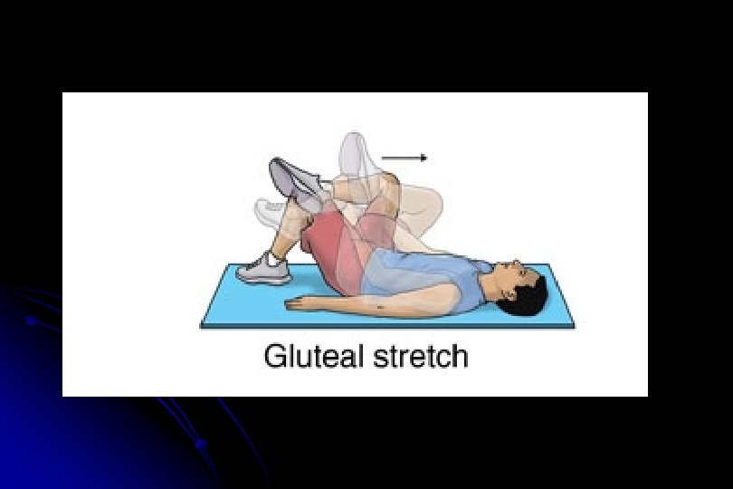
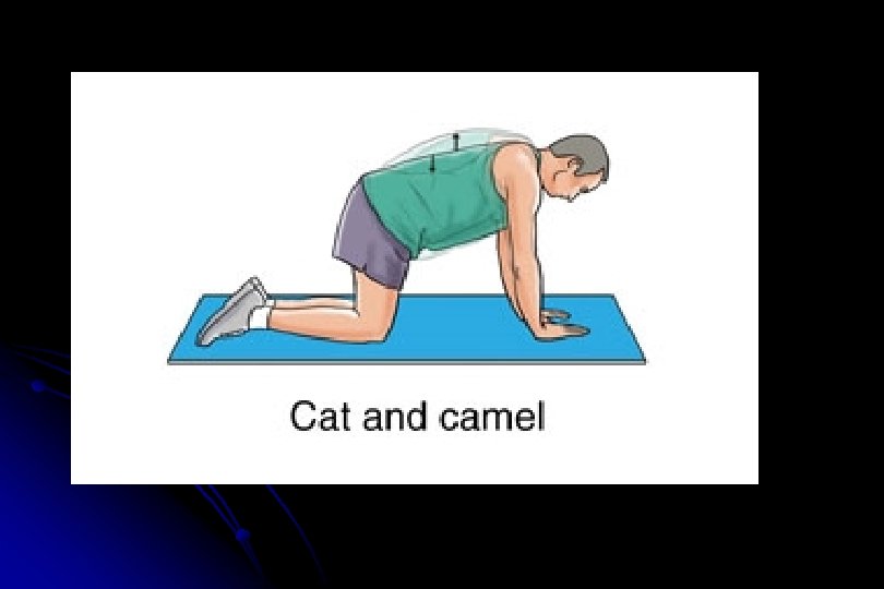
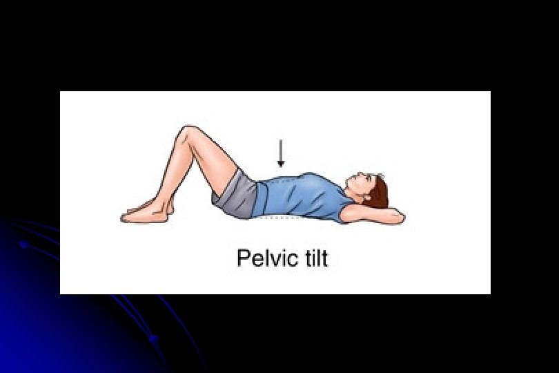
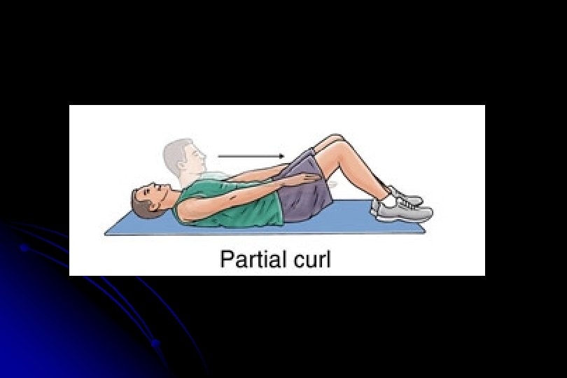
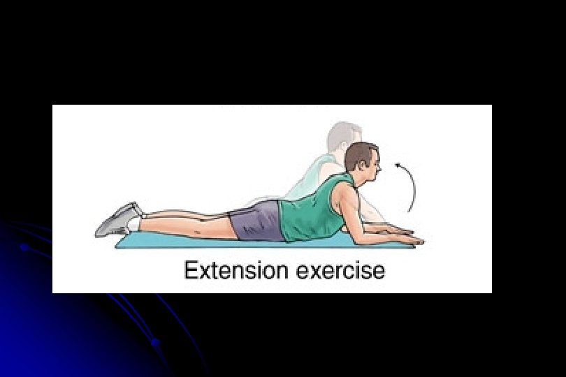
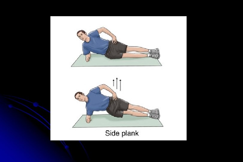
Rehabilitation Exercises for Chronic Back Pain

All of the following are Red Flags EXEPT? 1. 2. 3. 4. > 50 years of age History of Cancer Weight loss Radicular symptoms 10

Indications for an MRI include: 1. 2. 3. 4. Initial trauma evaluation A history of osteoporosis Reassurance >= 12 weeks of pain 10

Effective treatment for acute low back pain includes all except: 1. 2. 3. 4. Acetaminophen NSAIDS TCAs Physical therapy

Treatment goals of Chronic Low Back Pain include: 1. 2. 3. 4. Cure problem Alleviate pain Restore function Prevent disability

What is the lifetime incidence of low back pain 1. 2. 3. 4. >30% >40% >50% >60% 10

In what percentage of patients can the cause of low back pain not be determined? 1. 65% 2. 75% 3. 85% 4. 95% 10

Conclusions l l Describe the clinically relevant anatomy of the lumbar spine Discuss the “red flags” of lower back pain their associated clinical significance Discuss the common causes of low back pain Review and practice physical examination of the lower back and common rehabilitation exercises
5bb09663894624698c389b4a1661b791.ppt