eeedde21e3bae62f58c52a48c12d9d69.ppt
- Количество слайдов: 103


LOGO Chapter 2 Basic Function of the Cell Shenyang Medical college www. themegallery. com

Cells are the structural and functional units of the body. Each type of cell is specially adapted to perform one particular function. Many cells of the body may differ from each other, but all of them have certain basic characteristics that are alike.

This chapter focuses on basic functions of cells: l The transport of molecules across cell membranes; l Cellular signal transduction; l Bioelectrical phenomena of cells; l Mechanisms of muscular excitation and contraction.

Section 1 Cell membrane structure and membrane transport of substances

Molecular structure of the Cell Membrane Compositions of cell membrane protein 55% phospholipids 25% cholesterol 13% other lipids 4% carbohydrates 3% 0% 20% 40% 60% 80% Cell membrane serves as a permeability barrier that allows the cell to maintain the compositions of the cytoplasm. 100%

Fluid mosaic model 1972, Singer and Nicholson Main point: a lipid bilayer as the frame of cell membrane; Interspersed in a lipid bilayer are larger globular protein molecules.

lipid bilayer Lipids are amphipathic No np ola r Polar

Lipids spontaneously form structures A lipid bilayer is a stable, low energy structure Self sealing structure/eliminate free edge What drives this structured association? Exclusion of Lipids from Water… not lipid association
 -to attach only to one surface of")
Membrane proteins a. Extrinsic protein (peripheral protein) -to attach only to one surface of the membrane, and do not penetrate. -associated by weak electrostatic bonds to membrane proteins or lipids, may be dynamic: transient, and regulated. Function: almost entirely as enzyme p. ser. arg --+++
 To penetrate all the way through membrane.")
Membrane proteins b. Intrinsic protein (integral protein) To penetrate all the way through membrane. Function: as enzyme and ionic channel, ionic pump, carrier, controller (G protein).

Membrane carbohydrates l Occur almost in combination with proteins or lipids to form glycoproteins or glycolipids. l They play a major role in immune reactions and often act as receptors for binding hormones.

Transport of substances through the membrane Different substances are transported through the cell membrane by different processes. These processes range from relatively simple process of diffusion to extremely complex mechanisms that require the presence of special molecules within cell membranes.

Simple diffusion

Simple diffusion l Small substances that are soluble in lipids can be transported through a cell membrane from high to low concentrations of them. l such as oxygen, carbon dioxide, alcohol, urea, etc l No energy source is required, so this is referred to as a passive transport mechanism

Simple diffusion is mainly affected by the following factors: l Transmembrane solute concentration gradients, i. e. the difference in solute concentration across membrane. l Membrane permeability to the solute.

Simple diffusion

Protein-mediated Transport l The transport for slight large polar molecules or electrical charged ions is mediated by proteins within the membrane. l Two types of mediated transport can be distinguished as facilitated diffusion and active transport.

l The facilitated diffusion uses a transporter called a channel or a carrier to move solute "downhill" from a higher to a lower concentration across a membrane, which belongs to passive transport. l The active transport uses a transporter that is coupled to an energy source to move solute "uphill' across a membrane against its electrochemical gradient. The active transport is divided into primary active transport and secondary active transport.

Facilitated diffusion Some small water-soluble substances pass through the cell membrane down their concentration or electrical gradients by aid of membrane proteins. Content Title Carrier-mediated diffusion. glucoses, amino acids Channel-mediated diffusion Na+, K+, Ca 2+, etc

Carrier-mediated diffusion. a “ferry” or “shuttle” process

Carrier-mediated diffusion specificity Carrier proteins in the cell membrane selectively bind to a specific conformational substrate. Competitive inhibition Molecules with similar chemical structures compete for carrier site. Substrate Characteristics Saturation there is limitation of facilitated diffusion to Vmax level, relating to quantity of carrier proteins

Channel-mediated diffusion l Ions such as Na+, K+, Cl- and Ca 2+ diffuse across plasma membranes at rates that are much faster than would be predicted from their very low solubility in membrane lipids. l The protein component of the membrane that is responsible for these permeability differences.

characteristics of these channels n have selectivity, less than carriers n opened or closed by gates

Channel-mediated diffusion mechanical – gated channel Types of channels Voltagegated channel chemicalgated channel
 the membrane may affect the")
mechanical – gated channel Voltagegated channel Physically deforming (stretching) the membrane may affect the conformation of some channel proteins.

Voltage-gated channel Changes in the membrane potential can cause movement of the charged regions on a channel protein, altering its shape.

chemical-gated ACh Channels Binding of specific molecules to channel proteins may directly or indirectly produce a change in the shape of the channel protein.

Active transport Substances are transported through a cell membrane against their concentration or electrical gradients mediated by membrane protein carriers. Such carriers must use energy from some other source to do the necessary work According to the source of the energy used to transport, the active transport is divided into primary active transport and secondary active transport.

Primary active transport the energy used to cause the transport is derived directly from the hydrolysis of ATP or some other high-energy phosphate compound. The membrane protein carriers are called pumps. The pumps can hydrolyze ATP, so they are called ATPase , e. g. Always requires the input of energy. For example: Na+/K+ pump or Na+/K+ ATPase. Ca 2+ pump proton pump

Na+/K+ ATPase -hydrolysis of 1 molecule ATP -pumps 3 Na+ ion outward -pumps 2 K+ ion from outside to inside -inhibitor of Na+ /K+ pump is Ouabain.

Importance of the Na+-K+ Pump l Maintain high intracellular K+ concentration gradients across the membrane. l Control the volume of cell. l Provides energy for secondary active transport. l Develop and maintain Na+ and K+ concentration gradients across the membrane which are essential for bioelectricity activity. l Electrogenic action influences membrane potential.

Intracellular vs extracellular ion concentrations Ion Intracellular Extracellular Na+ K+ Mg 2+ Ca 2+ H+ 5 -15 m. M 140 m. M 0. 5 m. M 10 -7. 2 M (p. H 7. 2) 145 m. M 1 -2 m. M 10 -7. 4 M (p. H 7. 4) Cl. Fixed anions 5 -15 m. M high 110 m. M

Secondary active transport l The energy is derived secondarily from energy that has been stored in the form of ionic concentration differences between the two sides of the membrane, created in the first place by primarily active transport l Coupled transport. l Energy needed for “uphill” movement obtained from “downhill” transport of Na+. l Hydrolysis of ATP by Na+/K+ pump required indirectly to maintain [Na+] gradient.

Secondary active transport Na+-linked symporters import amino acids and glucose into many animal cells
 out Na+ glucose counter-transport (antiport) in out in Na+")
Secondary active transport co-transport (symport) out Na+ glucose counter-transport (antiport) in out in Na+ H+ Glucose in the same direction as H+ in the opposite direction to the Na+.

Exocytosis and endocytosis. l Exocytosis. Substances with large molecular size inside the cell are excreted from the cell in the form of membrane-bound vesicles, which is called exocytosis. l Endocytosis. Large membrane-impermeable substances outside the cell can be moved in cells in the form of membrane-bound vesicles, which is called endocytosis. Endocytosis is the reverse process of exocytosis.

Exocytosis and endocytosis Phagocytosis: solid material such as bacteria, dead tissue is engulfed by cells. Pinocytosis: fluid material is engulfed by cells in the body.

Key point Simple diffusion Facilitated diffusion Active transport characteristics of carrier-mediated difusion characteristics of channel-mediated difusion

Section 2 Signal transduction across cell membrane

l The operation of control systems requires that cells be able to communicate with each other, often over long distances. l Much of this intercellular communication is mediated by chemical messengers. l These messengers include chemicals such as hormones, neurotransmitters, cytokines, and so on.

Signal transduction pathways differ between lipid soluble and lipid-insoluble messengers. l Most lipid-soluble messengers are hormones, they diffuse across the plasma membrane and bind to the intracellular receptors. l The very large members of lipid-insoluble combine with plasma membrane receptors, causing the changes in the conformation of receptors and subsequent cascade response.

Lipid-soluble messengers activate receptors that are located inside the cell. The signal transduction of lipid-insoluble messengers is mediated by three different pathways, which are G-protein-linked receptor, ionotropic receptor and enzyme-linked receptor.
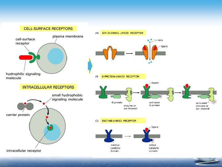

ionotropic receptor Receptors may open or close membrane ion channels, leading to changes in the ion conductances of a cell. This is particularly a feature of neurotransmitter receptors in excitable tissue such as nerve and muscle. l The receptor may act directly on the channel. l Ion-channel responses can be very rapid (millisecondes).

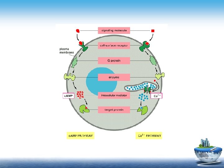
Signal transduction mediated by G -proteinlinked receptor The signal transduction mediated by Gprotein linked receptor is achieved by the cascade activities of the membranous receptors, G protein effector, second messenger and other molecules in the cell membrane and cytoplasm.

Pathways of signal transduction mediated by GProtein-Linked receptor A biological active substance which specifically binds membranous receptor is called ligand. The important pathways are as follows: l The receptor-G protein–c. AMP pathway; l The receptor-G protein–Ca 2+ pathway;

Signal transduction mediated by enzyme-linked receptor The receptors in this category of plasma membrane receptors have intrinsic enzyme activity. There are two most important receptors: tyrosine kinase receptor and guanylyl cyclase receptor.

Section 3 Bioelectrical phenomena of cell

Excitation and Excitable Cell l Excitation is used to describe responses of a cell to stimuli. l Excitability is the ability of excitable cells to response to stimulation or produce action potentials after stimulation. l Excitable cell means the cell which can produce action potentials after stimulation.

l Response means changes in the activity of a living organism induced by stimuli. Types of responses: l Excitation signifies an increase in the activity or occurrence of an activity or an action potential induced by stimuli. l Inhibition means a decrease or cessation in the activity induced by stimuli.

l Stimulation means changes in the external or internal environment, which can elicit response of the body. l A stimulus has three elements (its intensity, its duration--the time period used by the stimulus, and the changing rate of its intensity with respect to its duration).

l If a stimulus is rectangle, a weaker stimulus needs a correspondingly longer time to be effective. l The threshold of a stimulus means the minimal intensity of a stimulus that is needed for eliciting a minimal reaction or an action potential in an excitable tissue with a fixed duration and a fixed changing rate of its intensity with respect to its duration.

• Threshold of a stimulus is commonly used as a measure of a tissue’s excitability. • The higher the threshold of a stimulus, the lower the excitability, and vice versa.

• The stimulus with threshold intensity is called threshold stimulus. • Suprathreshold stimulus means the stimulus whose intensity is bigger than threshold. • Subthreshold stimulus means the stimulus whose intensity is smaller than threshold.

In the modern physiology, the excitation has almost the same meanings as the action potential or the process that action potential produced. “Excitable cells or tissues” for instant: nerve and muscle cells can generate rapidly changing electrochemical impulses at the membranes. It can be used to transmit signals along membrane. Other cells: glandular cells, macrophage, ciliated cell etc. Local changes in membrane potentials play significant roles in controlling many of cell’s functions.

How the membrane potential generate? 1936 Young : Squid Giant Axon : 1 mm 1939 Hodgkin & Huxley Intracellular recording use microelectrode 0+ 0 m. V 0+ RP -90 m. V S 0+ AP
. There is a potential difference across the cell membrane with")
Resting membrane potential (RP). There is a potential difference across the cell membrane with the inside negative with respect to the outside at rest. We say that the resting membrane potential is minus voltage. In neurons, it is about – 70 m. V. In muscle cells, it is about – 90 m. V. 50 (m. V) 0 50 70 0 1 2 3 (ms) 4 5

Several physiological terms l Polarization means the potential inside the cell is negative with respect to the outside at rest. l Hyperpolarization means the potential inside the cell becomes more negative than normal. l Depolarization means the potential inside the cell becomes less negative than normal. We say the membrane potential has reduced. l Reversal polarization means the potential inside the cell is positive with respect to the outside. l Overshoot means the membrane potential above zero. l Repolarization means the membrane potential falls back to the normal resting potential after depolarization.

Origin of the Resting Potential Concentration Difference l The resting potential exists because ions are concentrated on different sides of the membrane. l Na+ and Cl- outside the cell. l K+ and organic anions inside the cell. Na + Na Organic anions (-) K+ Cl- + Na+ K Organic anions (-) + Cl- outside inside Organic anions (-)

l The concentration differences for sodium and potassium are established by the action of the sodium -potassium pump (Na+/K+-ATPase) that pumps more positive charges to the outside than to the inside. Na + Na+ outside K+ K+ inside

Permeability l. The membrane permeabilities to these ions are restricted to different degrees. l Intracellular anions are mostly large impermeable organic anions. l The membrane is far more permeable to potassium than to sodium (50 -100 times) at resting condition.

conductance The permeability of the cell membrane to a given ion is described by conductance(g). Conductance (g) is defined as the inverse of the resistance (g=1/r). permeability to a particular ion ↑, resistance to current carried by that ion↓, while conductance ↑.

Electrochemical Driving Force and Potassium Equilibrium Potential l There are two driving forces for ions diffusion, which were caused by concentration difference and electrical potential difference across the membrane. l The algebraic sum of two forces mentioned above is called electrochemical driving force. Electrochemical driving force Electrostatic force Concentration force Electrochemical summation driving force

l The chemical driving force equals the electrical driving force but their direction is reverse, and the electrochemical driving force is zero. l The membrane potential in this situation is called K+ equilibrium potential (Ek). l The Ek primarily depends on the concentration difference across the membrane.
![The equilibrium potential for K+ (Ek). • Ek depends on the ratio of [K+]](https://present5.com/presentation/eeedde21e3bae62f58c52a48c12d9d69/image-66.jpg "The equilibrium potential for K+ (Ek). • Ek depends on the ratio of [K+]")
The equilibrium potential for K+ (Ek). • Ek depends on the ratio of [K+] on either side of the membrane and can be calculated using the Nernst equation: • Ek=RT / ZF loge [K+]o / [K+]I Where R=ideal gas constant (8. 31 Joules/℃), T=absolute temperature (℃+273 K), F=Faraday’s constant (number of coulombs per mole of charge, 96500 coulombs), Z=ionic valency (+1 for K+) [K+]o = K+ concentration outside the cell [K+]i = K+ concentration inside the cell
![Equation can be simplified as follow: EK = 60 log [K+]o [K+]i (m. V)](https://present5.com/presentation/eeedde21e3bae62f58c52a48c12d9d69/image-67.jpg "Equation can be simplified as follow: EK = 60 log [K+]o [K+]i (m. V)")
Equation can be simplified as follow: EK = 60 log [K+]o [K+]i (m. V) The larger the concentration gradient, the larger the equilibrium potential.

• In fact, the measured value of the resting membrane potential (-60 m. V, never cell) is more positive than the calculated value for Ek (-75 m. V, never cell). Other ions can also cross the membrane, particularly Na+ at rest. The Na+ gradient tends to make the membrane potential more positive than it would otherwise be.

• The concentration gradient for Na+ is in the opposite direction to K+ and the calculated value for ENa is approximately +55 m. V. • because the resting membrane is much more permeable to K+ than Na+, the resting potential is much closer to Ek than ENa.

Mechanism of RP genesis a. At resting state, the cell membrane has permeability mainly to K+ ion. b. Concentration difference of K+ ion exists. c. K+ diffuses outwards Membrane d. A potential gradient (inside -, outside +) builds up. e. The potential gradient opposes further diffusion of the positive K+ Finally, an equilibrium state, the equilibrium potential for K+ (Ek) is achieved. Rp is closer to Ek. inside A- K A- + K - K A + + A- K K A- + AA- K outside + _ _ K+ K+ + + + K+ K+

Factors of affecting the level of resting potential K+ concentration outside the membrane Hyperkalemia [K+]o↑→Rp↓ Hypokalemia [K+]o↓→Rp↑ Membrane permeability to K+ and Na+ more permeable to K+ →the potential↑ more permeable to Na+ →the potential↓ The activity level of Na + / K+ pump The Na+ / K+ ATPase pumps 3 Na+ out of the cell for every 2 K+ transported in. It is an electrogenic pump.
. l When a cell is stimulated, a rapid, reversible, propagated change")
Action potential (AP). l When a cell is stimulated, a rapid, reversible, propagated change in membrane potential occurs, which is called action potential. l AP is a feature of nerves, muscles and glands, which typifies excitable tissues.

Stages of Action potential +50 0 Depolarization phase Repolarization phase -55 -70 Resting state Time
 -70 positive")
Stages of Action potential +50 0 Spike potential -55 Negative after-potential (after-depolarization) -70 positive after-potential (after-hyperpolarization) Time

Properties of the Action Potential Subthreshold stimuli don’t elicit Ap All or none A threshold or suprathreshold stimulus will lead to a full Ap. It is always the same size. Propagation Once an AP has been generated by an external stimulus at one site, it will be automatically conducted over the whole of the excitable membrane.

Generation of an action potential The action potential is initiated by a transient change in membrane ion permeability, which allows sodium and potassium ions to move down their concentration gradients.

How to elicit an action potential l The action potential begins with a partial depolarization (e. g. from firing of another neuron or an adequate stimulus ) l Threshold (intensity): means the minimal intensity of a stimulus that is needed for eliciting an action potential in an excitable tissue.

How to elicit an action potential l A threshold or suprathreshold stimulus applied to a nerve cell elicits the membrane depolarization that reaches a certain level, known as the threshold potential, resulting in open or activation of more sodium channels and a rapid increase in membrane permeability to Na+.

+50 0 Vm -55 -70 Time

Stimulation Positive feedback loop Reach “threshold”? If YES, then. . .

The mechanisms of AP generation l The mechanisms responsible for action potential production depend on two main features: l Transmembrane ionic electrochemical driving forces. l Membrane conductance changes in the course of Ap.

Electrochemical driving forces Resting state Depolarization to +30 mv If Em is -70 mv, ENa is +60 mv, EK is -90 mv Em-ENa=-130 mv Em-EK=+20 mv Em-ENa=-30 Em-EK=+120 Na tend to flow into the cell K tend to flow outside the cell

Membrane conductance changes in the course of Ap

Depolarization l When partial depolarization reaches the threshold potential, voltage-gated sodium ion channels open. l Sodium ions rush in. l The membrane potential changes from -70 m. V to +50 m. V. + - - Na+ Na+ +

Depolarization

The overshoot value l The concentration gradient for Na+ is in the opposite direction to K+ and the calculated value for ENa is approximately +55 m. V. l The overshoot value of Ap is closer to ENa.

Repolarization l Sodium ion channels close and become refractory. l Depolarization triggers opening of voltage-gated potassium ion channels. l K+ ions rush out of the cell, repolarizing and then hyperpolarizing the membrane. Na+ Na + K Na+ + K+ K+ + -

Repolarization

mechanisims of Ap genneration +50 0 Depolarization Repolarization Sodium ions rush in K+ rush out -55 -70 Resting state Time Na-K transporter

Patch Clamp and Membrane Conductance l A technique was developed to allow investigators to monitor the properties of single ion channels, known as patch clamp. l Membrane conductance is the reciprocal of membrane resistance, and consistent with the membrane permeability of the charged ions. l With the patch clamp technique, we can record the electrical flow of single ion channel, calculate the conductance, and understand the permeability change of channels during action potential.

Patch clamp

Patch clamp recording Suction "Giga-seal" 1 µm Cytoplasm Glass microelectrode Ion channels Cell Membrane
 Two gates m m +")
Voltaged-gate Na+ channel: Hodgkin and Huxley model (H-H model) Two gates m m + Three states m h h Close m activation h h h inactivation 时间、电压依赖 Close

Voltage gated potassium channel • Potassium channels don’t inactivate. The fall in g. K following repolarization is simply caused by reduced activation at negative membrane potentials. • Tetraethylammonium (TEA) is a specific blocker of potassium channels. Tetrodotoxin (TTX) is a specific blocker of sodium channels.
. During the")
Excitability changes during AP Absolute refractory period (i. e. spike potential period). During the early part of the action potential there is an absolute refractory period when no stimulus is effective. So spike potentials can not summate. Relative refractory period (i. e. the early part of after-depolarization). Absolute refractory period is followed by the relative refractory period during which an AP can be stimulated by a stronger stimulus than normal.
. For a short time after")
Supernormal period (i. e. the later part of afterdepolarization). For a short time after relative refractory period, an AP can be elicited by a slightly smaller stimulus than normal. Subnormal period (i. e. after-hyperpolarization). The threshold for excitation is slightly higher than normal.

Excitability changes during AP 1. State of sodium channels determines the excitability. ARP: absolute refractory period Na+ channels inactivation, excitability equal to zero. RRP: relative refractory period 0 SB NP SP NP RRP SBNP: subnormal period 1 ARP 2. Distance between the MP and TP determines the excitability. SPNP: supranormal period RP Excitability Inactivated Na+ channels begin to recover TP

Action potential propagation unmyelinated nerve fibers l - is usually explained with the local current theory. The potential gradient is opposite to that for the resting membrane in the excited membrane, current flows from positive to negative, setting up local currents. The transfer of positive charge depolarizes the membrane adjacent to the Ap and, once threshold is reached, an Ap is generated in that region.

myelinated nerve fibers Saltatory Conduction l Myelinated regions of axon are electrically insulated. l Electrical charge moves along the axon rather than across the membrane. l Action potentials occur only at unmyelinated regions: nodes of Ranvier. myelin Node of Ranvier

Action potential propagation

Local response l When the stimulus intensity is subthreshold, a localized subthreshold membrane depolarization appears in the vicinity of the stimulation site, which is called a local response or local potential such as end plate potential.
 It s a graded potential 2) Its propagation is")
Characteristics of local response 1) It s a graded potential 2) Its propagation is electronic conduction 3) It can be summed by two ways A. Spatial summation B. Temporal summation

Key point Excitability Resting potential Action potential Threshold stimulus Threshold potential Absolute refractory period Mechanism of RP genesis Mechanism of AP genesis The difference between Ap and local response
eeedde21e3bae62f58c52a48c12d9d69.ppt