

4. ГЛ.ПС. ГАГ.ppt
- Количество слайдов: 42
 Лекция 4 протеогликаны, полисахариды, липополисахариды; гликолипиды, углевод-углеводное узнавание
") GPI-якорь (Glycosyl Phospho Inositol anchor)
GPI-якорь (Glycosyl Phospho Inositol anchor)
 варианты GPI
 процессинг GPI
 протеогликаны
 Комплекс антитромбина с пентасахаридным фрагментом гепарина протеогликан Glc. NAc-6 Su; Glc. A; Glc. N-Su-3, 6 Su 2; Idu. A-2 Su; Glc. N-S-6 Su
 Примеры полисахаридов млекопитающих. Пространственная структура Hyaluronic acid
 грам-отрицательные и грамположительные бактерии
 строение липополисахарида S-форма липополисахарида SR-форма липополисахарида первое повторяющееся олигосахарид кора липид A O-полисахарид звено O-полисахарида химическое биологическое повторяющееся звено - моносахарид внешний внутренний кор - фосфатная группа - жирная кислота
 LPS, жирные кислоты полисахарид
 LPS взаимодействует с TLR 4
") гликоглицеролипиды и гликосфинголипиды (GSL)
гликоглицеролипиды и гликосфинголипиды (GSL)
 ГЛИКОСФИНГОЛИПИДОВ · LACTO (Lc) Galb 1 -3 Glc. NAcb 1 -3") ОСНОВНЫЕ “СЕРИИ” (КОРЫ) ГЛИКОСФИНГОЛИПИДОВ · LACTO (Lc) Galb 1 -3 Glc. NAcb 1 -3 Galb 1 -4 Glc-Cer · Neo. LACTO (n. Lc) Galb 1 -4 Glc. NAcb 1 -3 Galb 1 -4 Glc-Cer · GLOBO (Gb) Gala 1 -4 Galb 1 -4 Glc-Cer · iso. GLOBO (i. Gb) Gala 1 -3 Galb 1 -4 Glc-Cer · MUCO (Mc) Galb 1 -3 Galb 1 -4 Glc-Cer • GANGLIO (Gg) Gal. NAcb 1 -4 Galb 1 -4 Glc-Cer
ОСНОВНЫЕ “СЕРИИ” (КОРЫ) ГЛИКОСФИНГОЛИПИДОВ · LACTO (Lc) Galb 1 -3 Glc. NAcb 1 -3 Galb 1 -4 Glc-Cer · Neo. LACTO (n. Lc) Galb 1 -4 Glc. NAcb 1 -3 Galb 1 -4 Glc-Cer · GLOBO (Gb) Gala 1 -4 Galb 1 -4 Glc-Cer · iso. GLOBO (i. Gb) Gala 1 -3 Galb 1 -4 Glc-Cer · MUCO (Mc) Galb 1 -3 Galb 1 -4 Glc-Cer • GANGLIO (Gg) Gal. NAcb 1 -4 Galb 1 -4 Glc-Cer
 номенклатуры “ose” номенклатура Symbol
 «продвинутая» OSE номенклатура
") Номенклатура Свеннерхольма (для ганглиозидов)
Номенклатура Свеннерхольма (для ганглиозидов)
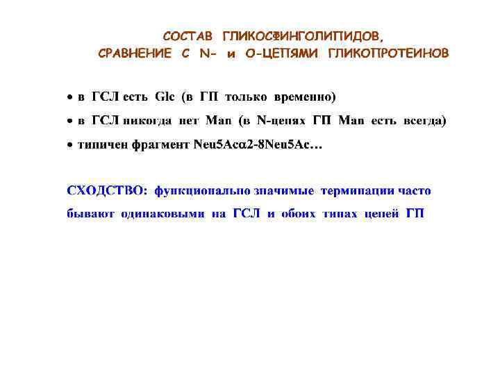
 Сходные терминации
 Lc (лакто-) Gb (глобо-) n. Lc (неолакто-)") модели четырёх коровых фрагментов GSL Gg (ганглио-) Lc (лакто-) Gb (глобо-) n. Lc (неолакто-)
модели четырёх коровых фрагментов GSL Gg (ганглио-) Lc (лакто-) Gb (глобо-) n. Lc (неолакто-)
 A 405 Холестерин как триггер взаимодействия Gb 3 с Gb 3 -связывающими белками
 GSL на поверхности мембраны организованы в кластеры
 рафты, кавеолы, гликосинапсы: ГСЛ хорошо организованы на мембране
 Гликолипиды формируют устойчивые комплексы
 биосинтез ГСЛ, на примере ганглиозидов Cer Lac. Cer Gal Glc. NAc Neu 5 Ac
 как можно модифицировать клетку neoglycolipid modified HOOH HO O O Ac. HN HOOH O H 3 С OH HO O O OН O H N O N H H N O O P O OН O O
 Дальше: гликоландшафт natural glycocalix mucin neoglyco lipid Glyc-PAA pseudo -mucin Hyal. U integral GP glycolipid
 углевод-углеводное взаимодействие
 Le. X = Galb 1 -4(Fuca 1 -3)Glc. NAc") Le. X в эмбриогенезе (мышь) Le. X = Galb 1 -4(Fuca 1 -3)Glc. NAc
Le. X в эмбриогенезе (мышь) Le. X = Galb 1 -4(Fuca 1 -3)Glc. NAc
 гипотеза об узнавании Le. X специфическими белками второй клетки
 углевод-углеводное взаимодействие: GM 3/Gg 3 и др. Слабые, но многочисленные…
 кластеры ГСЛ взаимодействуют и передают сигнал
 и транс- (b) (а) (b)") углевод-углеводное взаимодействие: цис- (а) и транс- (b) (а) (b)
углевод-углеводное взаимодействие: цис- (а) и транс- (b) (а) (b)
 углевод-углеводное взаимодействие strong repulsive
 углевод-углеводное взаимодействие протеогликанов в губках
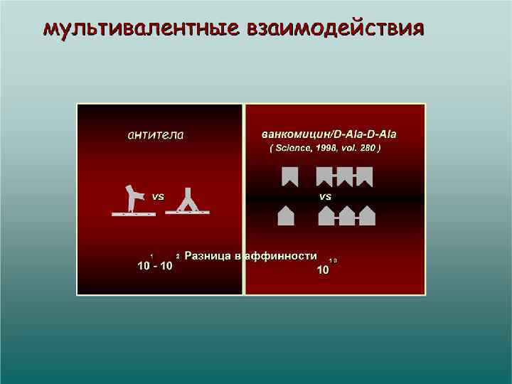
 • мультивалентные • часто динамические,") углевод-углеводные и углевод-белковые взаимодействия • cлабые (на моновалентном уровне) • мультивалентные • часто динамические, первичные в ступенчатом процессе
углевод-углеводные и углевод-белковые взаимодействия • cлабые (на моновалентном уровне) • мультивалентные • часто динамические, первичные в ступенчатом процессе
 коллектины как пример мультивалентного белка
 the end
") углевод-углеводное взаимодействие с участием полисахаридов )
углевод-углеводное взаимодействие с участием полисахаридов )
 углевод-углеводное взаимодействие: простые методы исследования
 клеткам иммунной системы белок CD 1 сульфатид") презентация гликолипидов (на DC, MФ) клеткам иммунной системы белок CD 1 сульфатид
презентация гликолипидов (на DC, MФ) клеткам иммунной системы белок CD 1 сульфатид
 contacts") GSLs in glycosynapse, involved in cell-cell adhesion. When glycosynapse of one cell (“a”) contacts glycosynapse of another cell (“b”), cell adhesion occurs by two mechanisms: (1) GSL-to-GSL interaction, which induces activation of signal transducer (STD), leading to change of cellular phenotype. In this process, proteolipid protein (PLP) [18, 35] may stabilize conformation of GSL. (2) GSL binds to GSL-binding protein, which induces activation of STD, leading to change of cellular phenotype. Each process is further explained below. 1. GSL-to-GSL interaction, either homotypic or heterotypic. A typical example of homotypic interaction is cell adhesion based on Lex-to-Lex interaction, observed in autoaggregation of embryonic stem cells D 3 M, or embryonal carcinoma cells F 9, in the presence of Ca 2+. Occurrence of such cell adhesion was further confirmed using D 3 M or F 9 cells whose E-cadherin gene was knocked out. These cells still displayed strong Lex-dependent autoaggregation and adhesion to Lex GSL-coated plates. which were eliminated by si. RNA of fucosyltransferase-9, involved in Lex synthesis. Lex-to-Lex interaction has been extensively studied and confirmed by various biophysical procedures, including atomic force microscopy, aggregation of gold glyconanoparticles with Lex, and adhesion energy change based on contact angle (Δ; θc) of two Lex vesicles. Heterotypic carbohydrate-to-carbohydrate interactions were found between GM 3 and Gg 3, mediating adhesion of melanoma cells to lymphoma cells; and between GM 3 and Lac. Cer, mediating binding of melanoma cells to microvascular endothelial cells, i. e. , cancer metastatic process. Gold lactosyl nanoparticles were found to inhibit melanoma cell metastasis in vivo. Interaction of Gal. Cer with sulfatide (3 -O-sulfated Gal. Cer), previously observed on various biophysical bases, was found to mediate adhesion of interfacing membranes of oligodendrocytes. Adhesion based on both homotypic and heterotypic carbohydrate interaction induces activation of signal transducers at cytoplasmic site, to alter cellular phenotype. Hakomori Page 13 FEBS Lett. Author manuscript; available in PMC 2011 May 3. NIH-PA Author Manuscript 2. Structure and function of carbohydrate-binding proteins expressed at cell surface membrane involved in cell-cell adhesion, or interaction of cell with its microenvironment, are well established by many studies. Three major classes of carbohydrate-binding proteins are: (i) Selectins (E-, P-, and L- types) having different structures and functions, and recognizing different glycosyl epitopes containing Lac. NAc backbone with fucosyl, sialosyl, or sulfate residue. Selectins play a major role in inflammatory processes and cancer progression, particularly metastasis (for review see. (ii) Sialic acid-binding lectins, abbreviated as “siglecs”, expressed at lymphocytes and myelocytes. Their function is to maintain the internal microenvironment (for review see. (iii) Galectins, comprising a huge number of variants, that recognize galactose or galactosamine. Their functions are varied, and many are still unclear (for review see
GSLs in glycosynapse, involved in cell-cell adhesion. When glycosynapse of one cell (“a”) contacts glycosynapse of another cell (“b”), cell adhesion occurs by two mechanisms: (1) GSL-to-GSL interaction, which induces activation of signal transducer (STD), leading to change of cellular phenotype. In this process, proteolipid protein (PLP) [18, 35] may stabilize conformation of GSL. (2) GSL binds to GSL-binding protein, which induces activation of STD, leading to change of cellular phenotype. Each process is further explained below. 1. GSL-to-GSL interaction, either homotypic or heterotypic. A typical example of homotypic interaction is cell adhesion based on Lex-to-Lex interaction, observed in autoaggregation of embryonic stem cells D 3 M, or embryonal carcinoma cells F 9, in the presence of Ca 2+. Occurrence of such cell adhesion was further confirmed using D 3 M or F 9 cells whose E-cadherin gene was knocked out. These cells still displayed strong Lex-dependent autoaggregation and adhesion to Lex GSL-coated plates. which were eliminated by si. RNA of fucosyltransferase-9, involved in Lex synthesis. Lex-to-Lex interaction has been extensively studied and confirmed by various biophysical procedures, including atomic force microscopy, aggregation of gold glyconanoparticles with Lex, and adhesion energy change based on contact angle (Δ; θc) of two Lex vesicles. Heterotypic carbohydrate-to-carbohydrate interactions were found between GM 3 and Gg 3, mediating adhesion of melanoma cells to lymphoma cells; and between GM 3 and Lac. Cer, mediating binding of melanoma cells to microvascular endothelial cells, i. e. , cancer metastatic process. Gold lactosyl nanoparticles were found to inhibit melanoma cell metastasis in vivo. Interaction of Gal. Cer with sulfatide (3 -O-sulfated Gal. Cer), previously observed on various biophysical bases, was found to mediate adhesion of interfacing membranes of oligodendrocytes. Adhesion based on both homotypic and heterotypic carbohydrate interaction induces activation of signal transducers at cytoplasmic site, to alter cellular phenotype. Hakomori Page 13 FEBS Lett. Author manuscript; available in PMC 2011 May 3. NIH-PA Author Manuscript 2. Structure and function of carbohydrate-binding proteins expressed at cell surface membrane involved in cell-cell adhesion, or interaction of cell with its microenvironment, are well established by many studies. Three major classes of carbohydrate-binding proteins are: (i) Selectins (E-, P-, and L- types) having different structures and functions, and recognizing different glycosyl epitopes containing Lac. NAc backbone with fucosyl, sialosyl, or sulfate residue. Selectins play a major role in inflammatory processes and cancer progression, particularly metastasis (for review see. (ii) Sialic acid-binding lectins, abbreviated as “siglecs”, expressed at lymphocytes and myelocytes. Their function is to maintain the internal microenvironment (for review see. (iii) Galectins, comprising a huge number of variants, that recognize galactose or galactosamine. Their functions are varied, and many are still unclear (for review see