Lecture 7: Membrane Transport Essential Cell Biology Fourth

Lecture 7: Membrane Transport Essential Cell Biology Fourth Edition Chapter 12
Membrane Transport SIMPLE DIFFUSSION – Small hydrophobic molecules Can pass right through the bilayer (eg. OXYGEN, CO2) 2. PASSIVE (FACILITATED) TRANSPORT – Substances Carried via a protein, molecule by Molecule. The TRANSPORT PROTEIN must have a binding site for the specific molecule transported. 3. ION FLOW THROUGH CHANNELS – ION CHANNELS are Membrane proteins that can open and close. When Open they act like a tunnel that can let through A stream (thousands) of ions. 4. ACTIVE TRANSPORT – Membrane proteins called PUMPS move molecules against a concentration Gradient; a source of chemical energy is required (ATP).
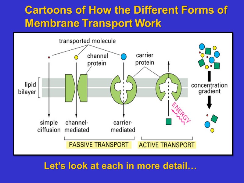
Cartoons of How the Different Forms of Membrane Transport Work Let’s look at each in more detail…
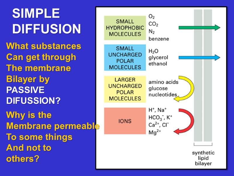
What substances Can get through The membrane Bilayer by PASSIVE DIFUSSION? Why is the Membrane permeable To some things And not to others? SIMPLE DIFFUSION
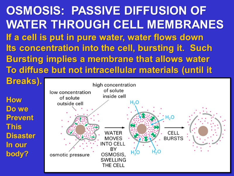
OSMOSIS: PASSIVE DIFFUSION OF WATER THROUGH CELL MEMBRANES If a cell is put in pure water, water flows down Its concentration into the cell, bursting it. Such Bursting implies a membrane that allows water To diffuse but not intracellular materials (until it Breaks). How Do we Prevent This Disaster In our body?
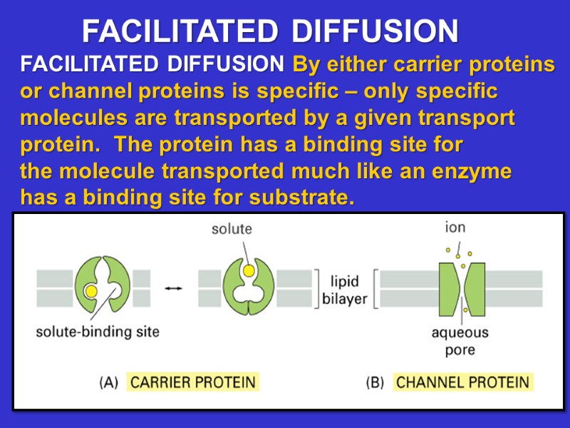
FACILITATED DIFFUSION By either carrier proteins or channel proteins is specific – only specific molecules are transported by a given transport protein. The protein has a binding site for the molecule transported much like an enzyme has a binding site for substrate. FACILITATED DIFFUSION
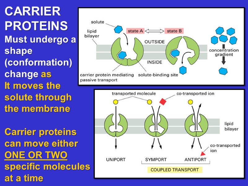
CARRIER PROTEINS Must undergo a shape (conformation) change as It moves the solute through the membrane Carrier proteins can move either ONE OR TWO specific molecules at a time
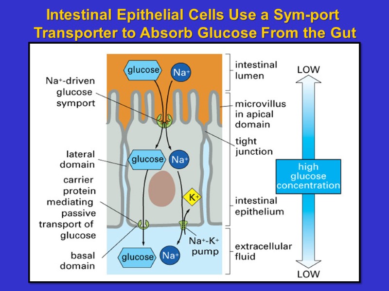
Intestinal Epithelial Cells Use a Sym-port Transporter to Absorb Glucose From the Gut
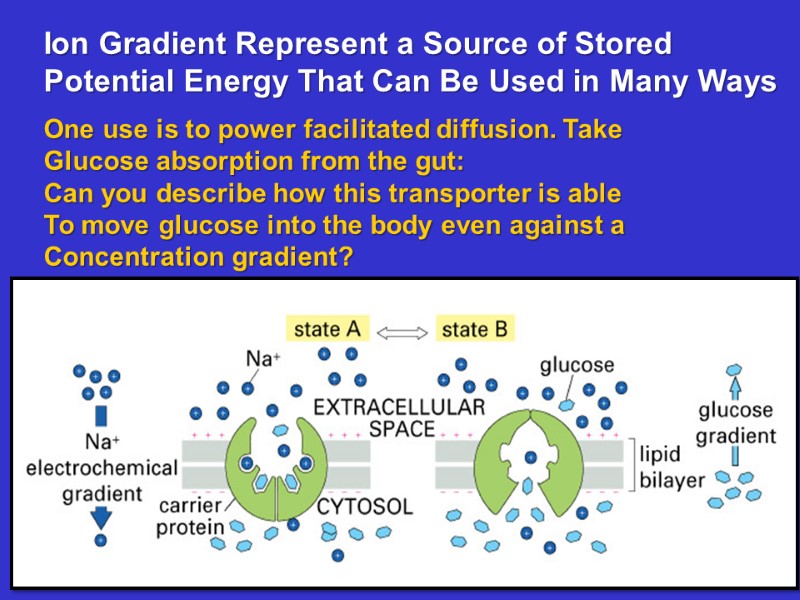
Ion Gradient Represent a Source of Stored Potential Energy That Can Be Used in Many Ways One use is to power facilitated diffusion. Take Glucose absorption from the gut: Can you describe how this transporter is able To move glucose into the body even against a Concentration gradient?
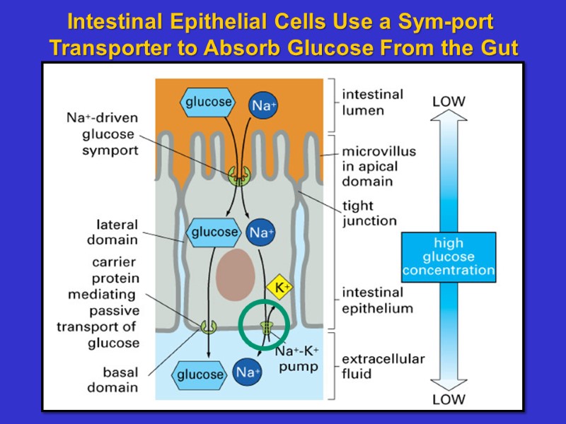
Intestinal Epithelial Cells Use a Sym-port Transporter to Absorb Glucose From the Gut
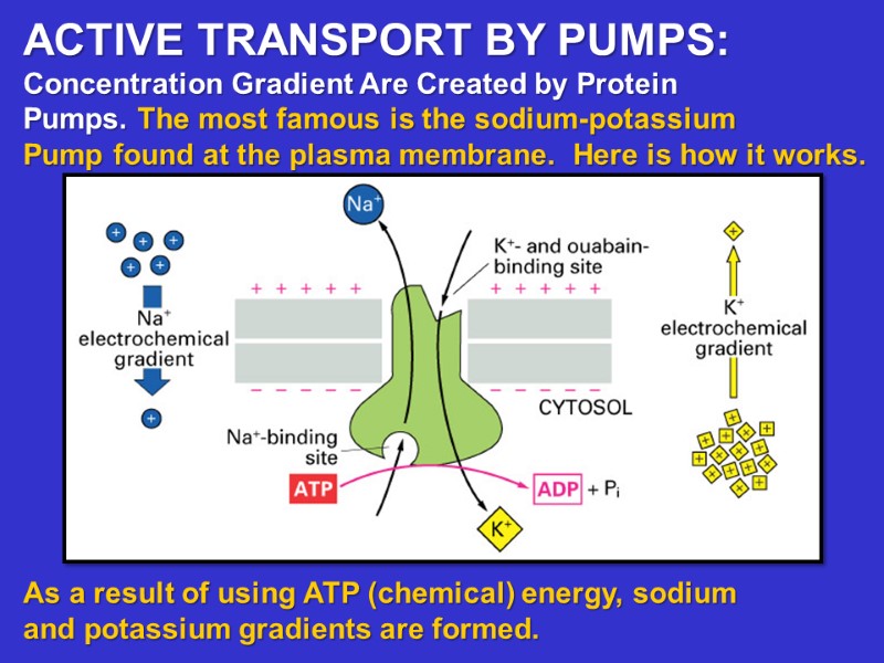
ACTIVE TRANSPORT BY PUMPS: Concentration Gradient Are Created by Protein Pumps. The most famous is the sodium-potassium Pump found at the plasma membrane. Here is how it works. As a result of using ATP (chemical) energy, sodium and potassium gradients are formed.
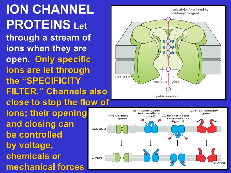
ION CHANNEL PROTEINS Let through a stream of ions when they are open. Only specific ions are let through the “SPECIFICITY FILTER.” Channels also close to stop the flow of ions; their opening and closing can be controlled by voltage, chemicals or mechanical forces
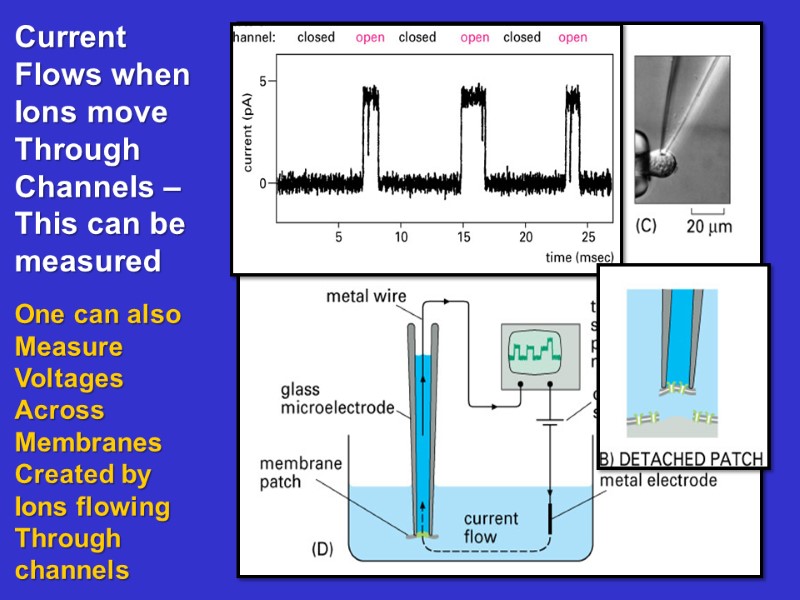
Current Flows when Ions move Through Channels – This can be measured One can also Measure Voltages Across Membranes Created by Ions flowing Through channels
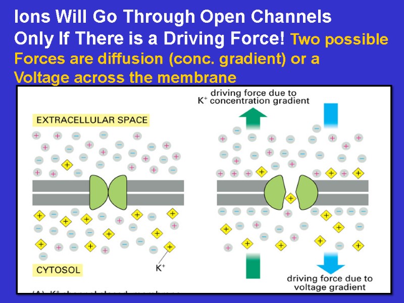
Ions Will Go Through Open Channels Only If There is a Driving Force! Two possible Forces are diffusion (conc. gradient) or a Voltage across the membrane
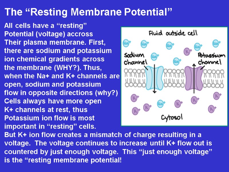
The “Resting Membrane Potential” All cells have a “resting” Potential (voltage) accross Their plasma membrane. First, there are sodium and potassium ion chemical gradients across the membrane (WHY?). Thus, when the Na+ and K+ channels are open, sodium and potassium flow in opposite directions (why?) Cells always have more open K+ channels at rest, thus Potassium ion flow is most important in “resting” cells. But K+ ion flow creates a mismatch of charge resulting in a voltage. The voltage continues to increase until K+ flow out is countered by just enough voltage. This “just enough voltage” is the “resting membrane potential!
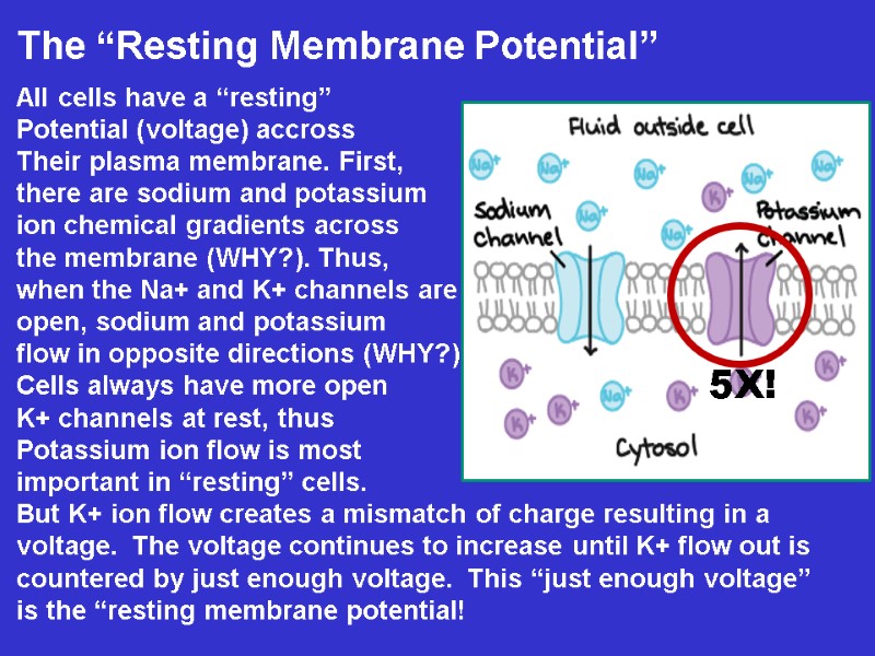
The “Resting Membrane Potential” All cells have a “resting” Potential (voltage) accross Their plasma membrane. First, there are sodium and potassium ion chemical gradients across the membrane (WHY?). Thus, when the Na+ and K+ channels are open, sodium and potassium flow in opposite directions (WHY?) Cells always have more open K+ channels at rest, thus Potassium ion flow is most important in “resting” cells. But K+ ion flow creates a mismatch of charge resulting in a voltage. The voltage continues to increase until K+ flow out is countered by just enough voltage. This “just enough voltage” is the “resting membrane potential!
Here is the same explanation in slightly Different words: Most “resting” cells have many more potassium Channels open than sodium channels. Potassium ions flow down their gradient until An electrochemical EQUILIBRIUM is reached. At equilibrium, flow of potassium out and flow In become equal so they are balanced – A STEADY STATE. But this balance will not occur until potassium Ion flow has moved a little bit of charge across The membrane to set up a voltage difference Capable of providing a force that balances Diffusion. This voltage across the membrane Can be measured! (How do you think this is done?)
THE ACTION POTENTIAL IN MUSCLE AND NERVE If a new set of channels open this will change the “resting” membrane potential! This is exactly what happens during an ACTION POTENTIAL in a nerve axon. A new set of sodium channels open (“activation”) and after a few milliseconds they close (“inactivation”). Sodium will Flow into the cell and this will change the membrane potential temporarily. Why? This temporary change reduces the membrane potential from -80 mV past 0 to +40 mV. This temporary “depolarization” is called the ACTION POTENTIAL. The Action Potential is temporary because as soon as the Na+ channels close, K+ channels open: what happens then?
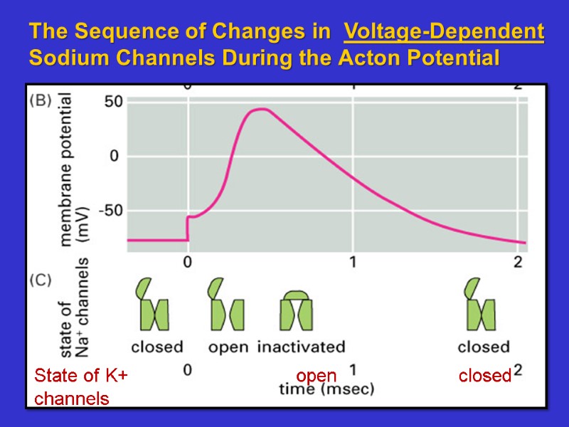
The Sequence of Changes in Voltage-Dependent Sodium Channels During the Acton Potential State of K+ open closed channels
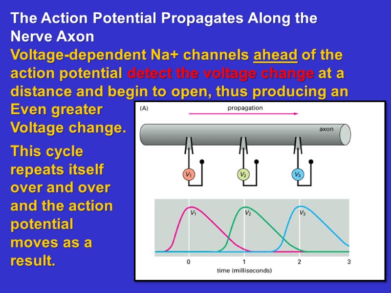
The Action Potential Propagates Along the Nerve Axon Voltage-dependent Na+ channels ahead of the action potential detect the voltage change at a distance and begin to open, thus producing an Even greater Voltage change. This cycle repeats itself over and over and the action potential moves as a result.
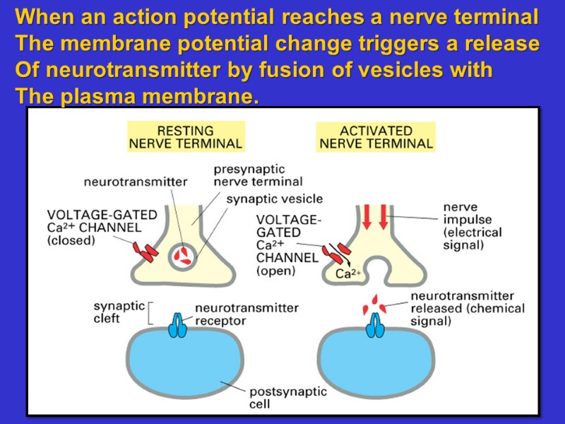
When an action potential reaches a nerve terminal The membrane potential change triggers a release Of neurotransmitter by fusion of vesicles with The plasma membrane.
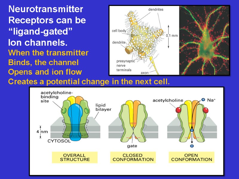
Neurotransmitter Receptors can be “ligand-gated” Ion channels. When the transmitter Binds, the channel Opens and ion flow Creates a potential change in the next cell.
15098-2017_lecture_7_bio_353_membrane_transport_bb.ppt
- Количество слайдов: 22

