Lecture 5. Thyroid and Parathyroid disorders.


lecture_5_thyroid_gland_diseases.ppt
- Размер: 1.7 Mегабайта
- Количество слайдов: 37
Описание презентации Lecture 5. Thyroid and Parathyroid disorders. по слайдам
 Lecture 5. Thyroid and Parathyroid disorders. Graves’ disease, Hypothyroidism, Hashimoto’s thyroiditis, Hypoparathyroidism : aetiology and pathogenesis, signs and symptoms, diagnosis and treatment. Associate professor Roman Ilnytskyi. STUDY GUIDE OF THE LECTURES COURSE Seventh semester, fourth academic year
 Key points of the lecture • Graves’ disease: definition, history, causes, symptoms and signs, taking diagnosis and treatment. • Hypothyroidism: definition, causes, symptoms and signs, taking diagnosis and treatment. • Hashimoto’s thyroiditis: definition, epidemiology, symptoms and signs, treatment. • Hypoparathyroidism: definition, causes, symptoms and signs, taking diagnosis, treatment, first aid.
 The main thyroid and parathyroid disorders include: • Graves’ disease • Hypothyroidism • Hashimoto’s thyroiditis • Hypoparathyroidism
 Graves’ disease • Graves’ disease is an autoimmune disease of a thyroid. • It most commonly affects the thyroid, frequently causing it to enlarge to twice its size or more (goitre), become overactive, with related hyperthyroid symptoms such as increased heartbeat, muscle weakness, disturbed sleep, and irritability. • It can also affect the eyes, causing bulging eyes (proptosis). It affects other systems of the body, including the skin, heart, circulation and nervous system.
 History • Graves’ disease owes its name to the Irish doctor Robert James Graves, who described a case of goiter with exophthalmos in 1835. • However, the German Karl Adolph von Basedow independently reported the same constellation of symptoms in 1840. • As a result, on the European Continent, the terms Basedow’s syndrome , or Basedow’s disease are more common than Graves’ disease.
 Epidemiology and causes • Graves ‘ disease affects up to 2% of the female population, sometimes appears after childbirth, and has a female: male incidence of 5: 1 to 10: 1. • It has a strong hereditary component; when one identical twin has Graves’ disease, the other twin will have it 25% of the time. • Smoking and exposure to second-hand smoke is associated with the eye manifestations but not the thyroid manifestations.
 Pathophysiology • There appears to be a genetic predisposition for Graves’ disease. • Graves’ disease is an autoimmune disorder, in which the body produces antibodies to the receptor for thyroid-stimulating hormone (TSH). • These antibodies cause hyperthyroidism because they bind to the TSH receptor and chronically stimulate it. The TSH receptor is expressed on the follicular cells of the thyroid gland, and the result of chronic stimulation is an abnormally high production of T 3 and T 4.
 Exophthalmos • It`s protrusion of the eyeball from the orbit, caused by disease, especially hyperthyroidism, or injury. • The infiltrative exophthalmos that is frequently encountered has been explained by postulating that the thyroid gland the extraocular muscles share a common antigen which is recognized by the antibodies.
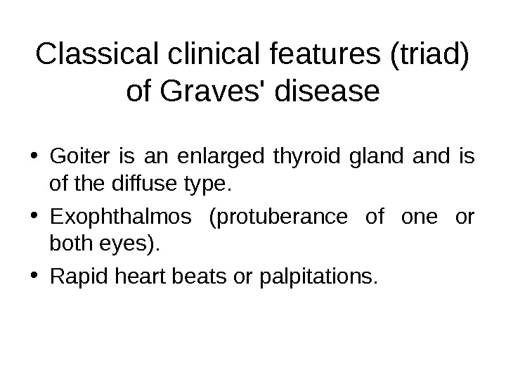
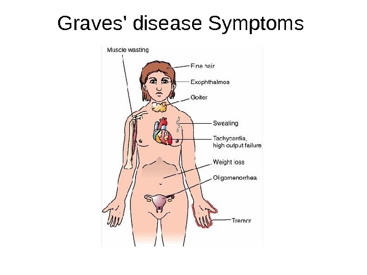
 Classical clinical features (triad) of Graves’ disease • Goiter is an enlarged thyroid gland is of the diffuse type. • Exophthalmos (protuberance of one or both eyes). • Rapid heart beats or palpitations.
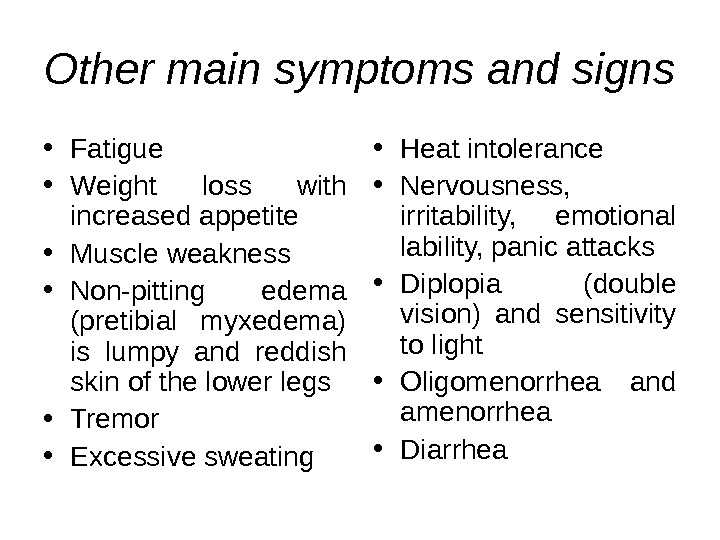
 Other main symptoms and signs • Fatigue • Weight loss with increased appetite • Muscle weakness • Non-pitting edema (pretibial myxedema) is lumpy and reddish skin of the lower legs • Tremor • Excessive sweating • Heat intolerance • Nervousness, irritability, emotional lability, panic attacks • Diplopia (double vision) and sensitivity to light • Oligomenorrhea and amenorrhea • Diarrhea
 Graves’ disease Symptoms
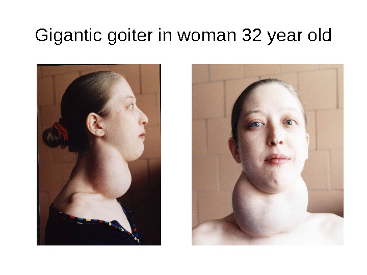
 Gigantic goiter in woman 32 year old
 Graves’ ophthalmopathy • Thyroid-associated ophthalmopathy is one of the most typical symptoms of Graves’ disease. • The ocular manifestations that are relatively specific to Graves’ disease include soft tissue inflammation, proptosis (protrusion of one or both globes of the eyes), corneal exposure, and optic nerve compression. • These more general symptoms include lid retraction, lid lag, and a delay in the downward excursion of the upper eyelid, during downward gaze.
 Diagnosis • Another sign of Graves’ disease is hyperthyroidism – overproduction of the thyroid hormones T 3 and T 4. • Other useful laboratory measurements in Graves’ disease include thyroid-stimulating hormone (TSH, usually low in Graves’ disease due to negative feedback from the elevated T 3 and T 4), and protein-bound iodine (elevated). • Thyroid-stimulating antibodies may also be detected serologically.
 Treatment • Treatment of Graves’ disease includes antithyroid drugs which reduce the production of thyroid hormone, radioiodine (radioactive iodine I-131), and thyroidectomy (surgical excision of the gland). • As operating on a frankly hyperthyroid patient is dangerous, prior to thyroidectomy preoperative treatment with antithyroid drugs is given to render the patient «euthyroid» ( i. e. normothyroid). • Treatment with antithyroid medications must be given for six months to two years, in order to be effective.
 Antithyroid drugs • The main antithyroid drugs are carbimazole (in the UK), methimazole (in the US), and propylthiouracil/PTU. These drugs block the binding of iodine and coupling of iodotyrosines. • The most dangerous side-effect is agranulocytosis. • The most common side effects are rash and peripheral neuritis.
 Radioiodine • Radioiodine (radioactive iodine-131) was developed in the early 1940 s at the Mallinckrodt General Clinical Research Center. • Indications for radioiodine are: failed medical therapy or surgery and where medical or surgical therapy are contraindicated. • Contraindications to RAI are pregnancy (absolute), ophthalmopathy (relative; it can aggravate thyroid eye disease), solitary nodules.
 Surgery • This modality is suitable for young patients and pregnant patients. • Indications are: a large goitre (especially when compressing the trachea), suspicious nodules or suspected cancer (to pathologically examine thyroid) and patients with ophthalmopathy. • Both bilateral subtotal thyroidectomy and the Hartley-Dunhill procedure (hemithyroidectomy on one side and partial lobectomy on other side) are possible.
 Hypothyroidism • Hypothyroidism is the disease state in humans and in vertebrates caused by insufficient production of thyroid hormones by the thyroid gland. Cretinism is a form of hypothyroidism found in infants. • About three percent of the general population is hypothyroidic. Factors such as iodine deficiency or exposure to iodine-131 (I-131) can increase that risk.
 Causes • Iodine deficiency is the most common cause of hypothyroidism worldwide. • In iodine-replete individuals hypothyroidism is generally caused by Hashimoto’s thyroiditis, or otherwise as a result of either an absent thyroid gland or a deficiency in stimulating hormones from the hypothalamus or pituitary. • Hypothyroidism can result from postpartum thyroiditis, a condition that affects about 5% of all women within a year of giving birth.
 Early symptoms and signs in adults • Poor muscle tone (muscle hypotonia) • Fatigue • Cold intolerance, increased sensitivity to cold • Depression • Muscle cramps and joint pain • Carpal Tunnel Syndrome • Goitre • Thin, brittle fingernails • Thin, brittle hair • Paleness • Osteoporosis • Decreased sweating • Dry, itchy skin • Weight gain and water retention • Bradycardia (low heart rate – fewer than sixty beats per minute) • Constipation
 Late symptoms • Slow speech and a hoarse, breaking voice – deepening of the voice can also be noticed • Dry puffy skin, especially on the face • Abnormal menstrual cycles • Low basal body temperature • Impaired memory • Impaired cognitive function (brain fog) and inattentiveness • Hair loss
 Diagnostic testing • To diagnose primary hypothyroidism, many doctors simply measure the amount of thyroid-stimulating hormone (TSH) being produced by the pituitary gland. High levels of TSH indicate that the thyroid is not producing sufficient levels of thyroid hormone (mainly as T 4 and smaller amounts of T 3). • However, measuring just TSH fails to diagnose secondary and tertiary hypothyroidism, thus leading to the following suggested blood testing if the TSH is normal and hypothyroidism is still suspected: Free triiodothyronine (f. T 3), Free levothyroxine (f. T 4), Total T 3, Total T 4.
 Hashimoto’s thyroiditis • Hashimoto’s thyroiditis is an autoimmune disease in which the thyroid gland is gradually destroyed by a variety of cell and antibody mediated immune processes. It was the first disease to be recognised as an autoimmune disease. • It was first described by Dr. Hakaru Hashimoto in Japan in 1912. Japanese physician Hakaru Hashimoto (1881− 1934) was teacher of the medical school at Kyushu University.
 Epidemiology • This disorder is believed to be the most common cause of primary hypothyroidism in North America. An average of 1 to 1. 5 in a 1000 people have this disease. It occurs far more often in women than in men (between 10: 1 and 20: 1), and is most prevalent between 45 and 65 years of age. • In European countries, an atrophic form of autoimmune thyroiditis (Ord’s thyroiditis) is more common than Hashimoto’s thyroiditis.
 Causes • The family history of thyroid disorders is common, with the HLA-DR 5 gene most strongly implicated conferring a relative risk of 3 in the UK. • The underlying specifics of the immune system destruction of thyroid cells is not clearly understood. Various autoantibodies may be present against thyroid peroxidase, thyroglobulin and TSH receptors, although a small percentage of patients may have none of these antibodies present.
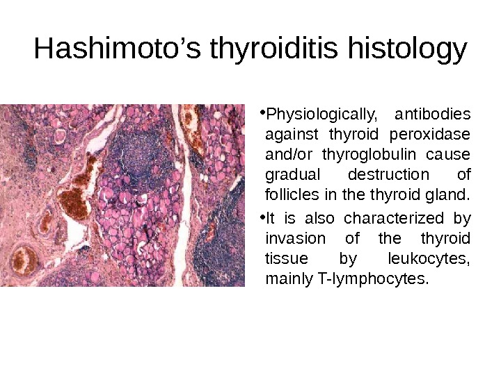
 Hashimoto’s thyroiditis histology • Physiologically, antibodies against thyroid peroxidase and/or thyroglobulin cause gradual destruction of follicles in the thyroid gland. • It is also characterized by invasion of the thyroid tissue by leukocytes, mainly T-lymphocytes.
 Symptoms and signs • Symptoms of Hashimoto’s thyroiditis include weight gain, depression, mania, sensitivity to heat and cold, fatigue, panic attacks, bradycardia, tachycardia, high cholesterol, reactive hypoglycemia, constipation, migraines, muscle weakness, cramps, memory loss, infertility and hair loss. • Testing for thyroid-stimulating hormone (TSH) and anti-thyroid antibodies can resolve any diagnostic difficulty.
 Treatment • Hypothyroidism caused by Hashimoto’s Thyroiditis is treated with thyroid hormone replacement agents such as levothyroxine or desiccated thyroid extract. • A tablet taken once a day generally keeps the thyroid hormone levels normal. In most cases, the treatment needs to be taken for the rest of the patient’s life.
 Hypoparathyroidism • In medicine (endocrinology), hypoparathyroidism is decreased function of the parathyroid glands, leading to decreased levels of parathyroid hormone (PTH) and hypocalcemia. • The consequence, hypocalcemia, is a serious medical condition.
 Causes of the hypoparathyroidism • Removal of or trauma to the parathyroid glands in thyroid surgery (thyroidectomy) or other neck surgeries is a recognized cause. • Autoimmune invasion and destruction is the most common non-surgical cause. It can occur as part of autoimmune polyendocrine syndromes. • Magnesium deficiency.
 Signs and symptoms • Tingling lips, fingers, and toes • Muscle cramps • Pain in the face, legs, and feet • Abdominal pain • Dry hair • Brittle nails • Dry, scaly skin • Weakened tooth enamel (in children) • Muscle spasms called tetany (can lead to spasms of the larynx, causing breathing difficulties) • Convulsions (seizures) • Tetanic contractions
 Additional symptoms that may be associated with this disease include • Painful menstruation • Hand or foot spasms • Decreased consciousness • Delayed or absent tooth formation
 Diagnosis • Diagnosis is by measurement of calcium, serum albumin (for correction) and PTH in blood. • PTH degrades rapidly at ambient temperatures and the blood sample therefore has to be transported to the laboratory on ice. • Other tests include ECG for abnormal heart rhythms, and measurement of blood magnesium levels.
 Treatment • Severe hypocalcemia, a potentially life-threatening condition, is treated as soon as possible with intravenous calcium (e. g. as calcium gluconate). Generally, a central venous catheter is recommended, as the calcium can irritate peripheral veins and cause phlebitis. • Long-term treatment of hypoparathyroidism is with calcium and Vitamin D 3 supplementation (D 1 is ineffective in the absence of renal conversion).
 First aid • In the event of a life-threatening attack of low calcium levels or tetany (prolonged muscle contractions), calcium is administered by intravenous infusion. • Precautions are taken to prevent seizures or larynx spasms. • The heart is monitored for abnormal rhythms until the person is stable. • When the life-threatening attack has been controlled, treatment continues with medicine taken by mouth as often as four times a day.
 Thanks for attention!

