
Классификация опухолей ЦНС ВОЗ 2016 г. Выполнила: студентка


 Astrocytomas Pilocytic astrocytoma Pleomorphic xanthoastrocytoma Subependimal giant cell astrocytoma Diffuse")
 Astrocytomas Pilocytic astrocytoma Pleomorphic xanthoastrocytoma Subependimal giant cell astrocytoma Diffuse")


")


 Вторичная (secondary)")


 Craig")


 meningiomas are characterised histologically by 2007 год • 4 to")


klassifikaciya_opuholey_cns_voz_2016.pptx
- Размер: 8.6 Мб
- Автор: Наталья Николаева
- Количество слайдов: 85
Описание презентации Классификация опухолей ЦНС ВОЗ 2016 г. Выполнила: студентка по слайдам
Классификация опухолей ЦНС ВОЗ 2016 г. Выполнила: студентка 6 курса ЛФ ПМГМУ им И. М. Сеченова Ахмадуллина Диляра
Классификации ВОЗ опухолей ЦНС 1 издание, 1979 2 издание, 1993 3 издание, 2000 4 издание, 2007 , , Диагнозы классификация определение степени злокачественности основаны только на морфологии
После 2007 года Накопление большого объема данных о молекулярных основах патогенеза Клиническое приложение этих данных отстает
« I believe that we have reached the tipping point, the point at which we know enough about the significance of molecular events that we can begin to use them for classification and for transitioning away from the old systems» David. N. Louis,
ISN-Haarlem Guidelines 2014 “ A critical question with major practical consequences has therefore arisen: how should clinically relevant molecular information be incorporated into nervous system tumor classification? ”
ISN-Haarlem Guidelines 2014 – “integrated diagnosis” Гистологичес кая картина Молекулярно- генетический профиль. WHO Grade
• В 2016 году выходит пересмотр 4 издания классификации опухолей ЦНС ВОЗ • Над ним работают 122 автора из 19 стран • Коллектив авторов возглавляет рабочая группа из 35 неврологов, нейроонкологов, клиницистов и ученых из 10 стран
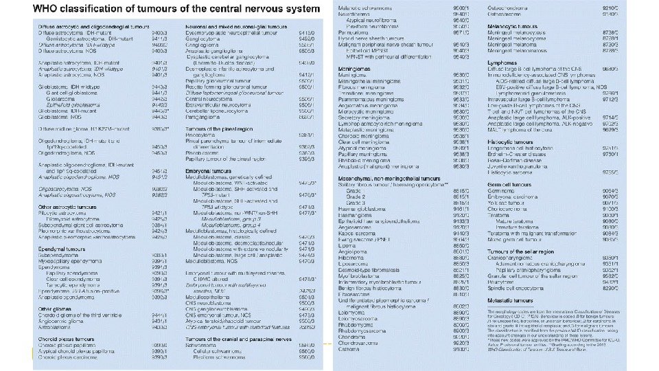
Структура и номенклатура
Diffuse astrocytic and oligodendroglial tumours Diffuse astrocytoma, IDH-mutant Gemistocytic astrocytoma, IDH-mutant Diffuse astrocytoma, IDH-wildtype Diffuse astrocytoma, NOS Anaplastic astrocytoma, IDH-mutant Anaplastic astrocytoma, IDH-wildtype Anaplastic astrocytoma, NOS Glioblastoma, IDH-wildtype Giant cell glioblastoma Gliosarcoma Epithelioid glioblastoma Glioblastoma, IDH-mutant Glioblastoma, NOS Diffuse midline glioma, H 3 K 27 M-mutant Oligodendroglioma, IDH-mutant and 1 p/ 19 q-codeleted Oligodendroglioma, NOS Anaplastic oligodendroglioma, IDH-mutant and Ip/19 q-codeleted Anaplastic oligodendroglioma, NOS Oligoastrocytoma, NOS Anaplastic oligoastrocytoma, NOS
Как формулируется диагноз? Diffuse astrocytoma, IDH-mutant WHO Grade II
Варианты обозначения молекулярного профиля опухоли • Diffuse astrocytoma, IDH-mutant Mutant • Diffuse astrocytoma, IDH-wildtype Wildtype • Diffuse astrocytoma, NOS = not otherwise specified • Ependimoma, RELA fusion-positive. Positive • Medulloblastoma, WNT-activated Altered • Embryonal tumour with multilayered rosettes, C 19 MC-a. Itered Activated
WHO Grade III WHO Grade IVDiffuse astrocytic and oligodendroglial tumours Diffuse astrocytoma, IDH-mutant II Anaplastic astrocytoma, IDH-mutant III Glioblastoma, IDH-wildtype IV Glioblastoma, IDH-mutant IV Diffuse midline glioma , H 3 K 27 M-mutant IV Oligodendroglioma, IDH-mutant and 1 p/19 q codeleted II Anaplastic oligodendroglioma, IDH-mutant and 1 p/19 q codeleted III Other astrocytic tumours Pilocytic astrocytoma I Subependymal giant cell astrocytoma I Pleomorphic xanthoastrocytoma II Anaplastic pleomorphic xanthoastrocytoma III Ependymal tumours Subependymoma I Myxopapillary ependymoma I Ependymoma II Ependymoma, RELA fusion-positive II or III Anaplastic ependymoma III Other gliomas Angiocentric glioma I Chordoid glioma of third ventricle II Степень злокачественности
Что нового?
Сформулирована концепция структуры диагноза опухолей ЦНС в молекулярную эру • Диагноз базируется на гистологической картине и молекулярно-генетическом анализе • При этом генетическая характеристика всегда будет «побеждать» гистологическую картину • В диагнозе указывается сначала фенотип опухоли, затем через запятую генетические свойства
Почему нельзя основываться только на молекулярном анализе? Сначала необходимо сузить круг поиска, чтобы понять, какие генетические маркеры мы будем искать Градация опухолей ВОЗ по степени злокачественности основана на гистологии Существуют отдельные виды опухолей, для которых на данный момент не определены молекулярные маркеры
Реорганизация отдельных разделов Добавление вновь выделенных форм и вариантов, паттернов Исключение старых форм, вариантов
Глиомы GLIAL NEOPLASMS (2007) Astrocytomas Pilocytic astrocytoma Pleomorphic xanthoastrocytoma Subependimal giant cell astrocytoma Diffuse astrocytoma Anaplastic astrocytoma Glioblastoma astrocytoma Oligodenrogliomas Oligodendroglioma Anaplastic oligodendroglioma Oligoasrocytomas
Глиомы GLIAL NEOPLASMS (2007) Astrocytomas Pilocytic astrocytoma Pleomorphic xanthoastrocytoma Subependimal giant cell astrocytoma Diffuse astrocytoma Anaplastic astrocytoma Glioblastoma astrocytoma Oligodenrogliomas Oligodendroglioma Anaplastic oligodendroglioma Oligoasrocytomas GLIAL NEOPLASMS (2016) Diffuse astrocytic and oligodendroglial tumours Diffuse astrocytoma Anaplastic astrocytoma Glioblastoma astrocytoma Oligodendroglioma Anaplastic oligodendroglioma Oligoasrocytoma Anaplastic oligoasrocytoma Diffuse midline glioma, H 3 K 27 M-mutant Other astrocytomas Pilocytic astrocytoma Pleomorphic xanthoastrocytoma Subependimal giant cell astrocytoma
Диффузные глиомы. Зависимость прогноза от молекулярного типа опухоли IDH-mutant 1 p/19 q codeletion TERT Олигодендр оглиомы, WHO II/III Прогноз хороший 1 p/19 q intact TP 53 ATRX Дифф. астроцитома , WHO II-IV Прогноз средний IDH-wildtyp e EGFR PTEN CKDN 2 A Дифф. астроцитома , WHO II-IV Прогноз плохой
Clinical Outcomes According to Histologic Class and Grade According to Molecular Subtype Comprehensive, Integrative Genomic Analysis of Diffuse Lower-Grade Gliomas. The Cancer Genome Atlas Research Network. N Engl J Med 2015; 372: 2481 —
Диффузные глиомы. Зависимость прогноза от молекулярных свойств опухоли Прогноз у пациентов с «IDH-mutant» типом лучше, чем у пациентов с «IDH-wildtype» типом, независимо от степени злокачественности (WHO Grade) Christian Hartmann et al. Patients with IDH 1 wild type anaplastic astrocytomas exhibit worse prognosis than IDH 1 -mutated glioblastomas, and IDH 1 mutation status accounts for the unfavorable prognostic effect of higher age: implications for classification of gliomas. Acta Neuropathol (2010) 120: 707–
Molecular taxonomy of IDH -mutant glioma progression Hiroaki. Wakimoto et al. Targetable Signaling Pathway Mutations Are Associated with Malignant Phenotype in IDH -Mutant Gliomas. Clin Cancer Res. June 1 2014(20)(11)2898 —
A novel view of the genetic landscape of glioblastomas Parsons DW et al. Anintegratedgenomicanalysisofhumanglioblastoma multiforme. Science. 2008 Sep 26; 321(5897): 1807 -12.
Гены IDH 1 – 2 q 34 IDH 2 – 15 q 26.
Изомеры белка IDH (изоцитратдегидрогеназа)
Соотношение различных мутаций в генах IDH 1 и IDH 2 • 90% — мутантный белок R 132 H • 10% — другие мутации 95% — IDH 1 5% — IDH
Мутантный белок R 132 H Lenny Dang et al. Cancer-associated IDH 1 mutations produce 2 -hydroxyglutarate. Nature, 2010; 465(7300): 966. Strong reaction of IDH 1 R 132 H antibody clone H 09 in tumor center of anaplastic oligoastrocytoma.
Нормальные ферменты IDH vs Мутантные формы IDH Andrew R. Mullen and Ralph J. De. Berardinis. Genetically-defined metabolic reprogramming in cancer. Cell, 2012; 23(11): p 552–
Роль R 2 -HG в развитии опухолевого процесса Michael Heuser et al. Enigmas of IDH mutations in hematology/oncology. Experimental Hematology, 43(8): p 685– 697 Figueroa ME et al. Leukemic IDH 1 and IDH 2 mutations result in a hypermethylation phenotype, disrupt TET 2 function, and impair hematopoietic differentiation. Cancer Cell, 2010; 18(6): 553 —
Diffuse astrocytic and oligodendroglial tumours Diffuse astrocytoma, IDH-mutant Gemistocytic astrocytoma, IDH-mutant Diffuse astrocytoma, IDH-wildtype Diffuse astrocytoma, NOS Anaplastic astrocytoma, IDH-mutant Anaplastic astrocytoma, IDH-wildtype Anaplastic astrocytoma, NOS Glioblastoma, IDH-wildtype Giant cell glioblastoma Gliosarcoma Epithelioid glioblastoma Glioblastoma, IDH-mutant Glioblastoma, NOS Diffuse midline glioma, H 3 K 27 M-mutant Oligodendroglioma, IDH-mutant and 1 p/ 19 q-codeleted Oligodendroglioma, NOS Anaplastic oligodendroglioma, IDH-mutant and Ip/19 q-codeleted Anaplastic oligodendroglioma, NOS Oligoastrocytoma, NOS Anaplastic oligoastrocytoma, NOS
Diffuse astrocytoma WHO Grade II/ Anaplastic astrocytoma WHO Grade III IDH-mutant – положительн ые результаты Иммуногистохимия на мутантный протеин R 132 H IDH 1 Секвенирование 132 кодона IDH 1 Секвенирование 172 кодона IDH 2 IDH-wildtype – отрицательны е результаты Иммуногистохимия на мутантный протеин R 132 H IDH 1 Секвенирование 132 кодона IDH 1 Секвенирование 172 кодона IDH 2 NOS Если проведение молекулярно-генет ического анализа невозможно Если молекулярно-генет ический анализ был выполнен не в полном обеме
IDH-mutant Секвенирование IDH 1 и IDH 2 Мутация найдена IDH-mutant Мутация не найдена IDH-wildtype
Diffuse astrocytoma, IDH-mutant WHO Grade II 2007 год • Gemistocyti c astrocytom a • Protoplasm atic astrocytom a • Fibrillary astrosytom a 2016 год • Gemistocyti c astrocytom a, IDH-mutant
Gemistocytic astrocytoma, IDH-mutant WHO Grade II Гемистоциты характеризуются насыщенной эозинофильной цитоплазмой и эксцентрично расположенным ядром Количество гемистоцитов(тучных астроцитов) должно быть более 20— 35% отклеточного состава опухоли
Diffuse astrocytic and oligodendroglial tumours Diffuse astrocytoma, IDH-mutant Gemistocytic astrocytoma, IDH-mutant Diffuse astrocytoma, IDH-wildtype Diffuse astrocytoma, NOS Anaplastic astrocytoma, IDH-mutant Anaplastic astrocytoma, IDH-wildtype Anaplastic astrocytoma, NOS Glioblastoma, IDH-wildtype Giant cell glioblastoma Gliosarcoma Epithelioid glioblastoma Glioblastoma, IDH-mutant Glioblastoma, NOS Diffuse midline glioma, H 3 K 27 M-mutant Oligodendroglioma, IDH-mutant and 1 p/ 19 q-codeleted Oligodendroglioma, NOS Anaplastic oligodendroglioma, IDH-mutant and Ip/19 q-codeleted Anaplastic oligodendroglioma, NOS Oligoastrocytoma, NOS Anaplastic oligoastrocytoma, NOS 90% 10%
Сравнительная характеристика глиобластом IDH-wildtype glioblastoma 90% IDH-mutant glioblastoma 10% Синонимы Первичная (primary) Вторичная (secondary) Медиана возраста при постановке диагноза 62 года 44 года Средняя продолжительность жизни — операция + лучевая терапия + химиотерапия 9, 9 месяцев 15 месяцев 24 месяца 31 месяц Локализация Супратенториально Лобные доли Некрозы Обширные Ограниченные TERT promoter mutations 72% 26% TP 53 mutations 27% 81% ATRX mutations В порядке исключения 71% EGFR amplification 35% В порядке исключения PTEN mutations 24% В порядке исключения
Glioblastoma WHO Grade IV IDH-wildty pe Если пациент старше 55 лет, для постановки диагноза достаточно отрицательного результата иммуногистохимического анализа на R 132 H IDH 1 Если пациент младше 55 лет, для постановки диагноза потребуются отрицательные результаты иммуногистохимии, а также секвенирования IDH 1 и IDH 2 IDH-mutant – положительн ый результат любого из исследований Иммуногистохимия на мутантный протеин R 132 H IDH 1 Секвенирование 132 кодона IDH 1 Секвенирование 172 кодона IDH 2 NOS Если проведение молекулярно-генетического анализа невозможно Если молекулярно-генетический анализ был выполнен не в полном объеме
Glioblastoma, IDH-wildtype WHO Grade IV 2007 год • Giant cell glioblasto ma • Gliosarco ma 2016 год • Giant cell glioblasto ma • Gliosarco ma • Epithelioi d glioblasto ma
Epithelioid glioblastoma В основном, возникает у детей и молодых людей. В типичных случаях расположена поверхностно в полушариях или в диэнцефальной области Представлена большими эпителиодными клетками с обильной эозинофильной цитоплазмой и заметными ядрышками Возможно присутствие рабдоидных клеток (указаны стрелками) Мутация BRAF V 600 E Отсутствуют типичные свойства глиобластом, IDH-wildtype (EGFR and LOH 10) GFAP S-100 OLIG 2 BRAF V 600 E mutant protein
Glioblastoma/Astrocytoma patterns На 2007 год • Small cell glioblastoma/astroc ytoma • Granular cell glioblastoma/astroc ytoma На 2016 год • Small cell glioblastoma/astroc ytoma • Granular cell glioblastoma/astroc ytoma • Glioblastoma with primitive neuronal component
Glioblastoma with primitive neuronal component Обычно содержит клетки астроцитарного ряда и состоит из хорошо отделенных узлов, в которых обнаруживают примитивные клетки на ранней стадии нейрональной дифференцировки (розетки Гомера-Райта, synap+, GFAP-) Возможна амплификация генов MYC и MYCN Опухоли с таким паттерном имеют тенденцию к распространению по ЦСЖ В 25% случаев возникает вторично из менее злокачественных предшественников. OLIG 2 — Амплификаци я MYCNSynaptophysin + IDH 1 R 132 H mutant protein розетки Гомера-Райт а
Diffuse astrocytic and oligodendroglial tumours Diffuse astrocytoma, IDH-mutant Gemistocytic astrocytoma, IDH-mutant Diffuse astrocytoma, IDH-wildtype Diffuse astrocytoma, NOS Anaplastic astrocytoma, IDH-mutant Anaplastic astrocytoma, IDH-wildtype Anaplastic astrocytoma, NOS Glioblastoma, IDH-wildtype Giant cell glioblastoma Gliosarcoma Epithelioid glioblastoma Glioblastoma, IDH-mutant Glioblastoma, NOS Diffuse midline glioma, H 3 K 27 M-mutant Oligodendroglioma, IDH-mutant and 1 p/19 q-codeleted Oligodendroglioma, NOS Anaplastic oligodendroglioma, IDH-mutant and Ip/19 q-codeleted Anaplastic oligodendroglioma, NOS Oligoastrocytoma, NOS Anaplastic oligoastrocytoma, NOS
Oligodendroglioma WHO Grade II/ Anaplastic oligodendroglioma WHO Grade III IDH-mutant and 1 p/19 q-codeleted Иммуногистохимия на мутантный протеин R 132 H IDH 1 Секвенирование 132 кодона IDH 1 Секвенирование 172 кодона IDH 2 Коделеция 1 p/19 q NOS Проведение молекулярно-генетического анализа невозможно Отрицательные результаты молекулярно-генетического анализа исследования
1 p 19 q co-deletion in gliomas occurs through an unbalanced whole-arm translocation between chromosomes 1 and 19 [t(1; 19) (q 10; p 10)]. It is hypothesised that translocation creates two derivative chromosomes and is followed by loss of the derivative chromosome containing 1 p and 19 q [+] Pinkham MB, et al. , FISHing Tips: What Every Clinician Should Know About 1 p 19 q Analysis in Gliomas Using Fluorescence in situ Hybridisation, Clinical Oncology (2015), http: //dx. doi. org/10. 1016/j. clon. 2015. 04.
1 p 19 q codeletion in oligodendrogliomas via fluorescence in situ hybridization (FISH) Craig Horbinski et al. Gone FISHing: Clinical Lessons Learned in Brain Tumor Molecular Diagnostics over the Last Decade. Brain Pathology, 2011; 21(1): 57 -73.
Oligodendroglioma WHO Grade II/ Anaplastic oligodendroglioma WHO Grade III IDH-mutant and 1 p/19 q-codeleted Иммуногистохимия на мутантный протеин R 132 H IDH 1 Секвенирование 132 кодона IDH 1 Секвенирование 172 кодона IDH 2 Коделеция 1 p/19 q NOS Проведение молекулярно-генетического анализа невозможно Отрицательные результаты молекулярно-генетического анализа исследования
Diffuse astrocytic and oligodendroglial tumours Diffuse astrocytoma, IDH-mutant Gemistocytic astrocytoma, IDH-mutant Diffuse astrocytoma, IDH-wildtype Diffuse astrocytoma, NOS Anaplastic astrocytoma, IDH-mutant Anaplastic astrocytoma, IDH-wildtype Anaplastic astrocytoma, NOS Glioblastoma, IDH-wildtype Giant cell glioblastoma Gliosarcoma Epithelioid glioblastoma Glioblastoma, IDH-mutant Glioblastoma, NOS Diffuse midline glioma, H 3 K 27 M-mutant Oligodendroglioma, IDH-mutant and 1 p/ 19 q-codeleted Oligodendroglioma, NOS Anaplastic oligodendroglioma, IDH-mutant and Ip/19 q-codeleted Anaplastic oligodendroglioma, NOS Oligoastrocytoma, NOS Anaplastic oligoastrocytoma, NOS
Oligoastrocytoma, NOS WHO Grade II/ Anaplastic oligoastrocytoma, NOS WHO Grade III Из 43 случаев олигоастроцитом после молекулярно-генетического анализа диагноз подтвердился только у одного пациента
Diffuse astrocytic and oligodendroglial tumours Diffuse astrocytoma, IDH-mutant Gemistocytic astrocytoma, IDH-mutant Diffuse astrocytoma, IDH-wildtype Diffuse astrocytoma, NOS Anaplastic astrocytoma, IDH-mutant Anaplastic astrocytoma, IDH-wildtype Anaplastic astrocytoma, NOS Glioblastoma, IDH-wildtype Giant cell glioblastoma Gliosarcoma Epithelioid glioblastoma Glioblastoma, IDH-mutant Glioblastoma, NOS Diffuse midline glioma, H 3 K 27 M-mutant Oligodendroglioma, IDH-mutant and 1 p/ 19 q-codeleted Oligodendroglioma, NOS Anaplastic oligodendroglioma, IDH-mutant and Ip/19 q-codeleted Anaplastic oligodendroglioma, NOS Oligoastrocytoma, NOS Anaplastic oligoastrocytoma, NOS
Diffuse midline glioma, H 3 K 27 M-mutant WHO Grade IV Встречается, в основном, среди детей Мутация гистона H 3 гена H 3 F 3 A или реже гена HIST 1 H 3 B Диффузный рост Локализация: таламус, ствол (особенно мост), спинной мозг. K 27 M TP 53 + ATRX —
Глиоматоз головного мозга Исключен из классификации ВОЗ 2016 года как отдельный вид опухоли На настоящий момент считается скорее характером роста опухоли, который может наблюдаться у различных глиом, включая IDH-mutant астроцитомы и олигодендроглиомы и IDH-wildtype глиобластомы
Diffuse astrocytic and oligodendroglial tumours Алгоритм постановки диагноза.
Other astrocytic tumours Pilocytic astrocytoma Pilomyxoid astrocytoma Subependymal giant cell astrocytoma Pleomorphic xanthoastrocytoma Anaplastic pleomorphic xanthoastrocytoma
Anaplastic pleomorphic xanthoastrocytoma WHO Grade III Предыдущее название: плейоморфная ксантоастроцитома с анапластическим компонентом Гистологические критерии: 1. Пять или более митозов в 10 полях зрения 2. Участки некроза M. R. Patibandla et al. Pleomorphic xanthoastrocytoma with anaplastic features: A rare case report and review of literature with reference to current management. Asian J Neurosurg, 2016; 11(3): 319. (a) × 100, стрелка указывает на гигантскую плейоморфную клетку (b) × 400, стрелка указывает на митоз (c) × 400 (d) CD 68 — (e) GFAP (глиофибриллярный кислый белок) +
Ependymal tumours Subependymoma Myxopapillary ependymoma Ependymoma Papillary ependymoma Clear cell ependymoma Tanycytic ependymoma Cellular ependymoma Ependymoma, RELA fusion—positive Anaplastic ependymoma
Ependymoma, RELA fusion—positive WHO Grade II or III • Большая часть супратенториальных опухолей детей • Экспрессия L 1 CAM обсуждается как потенциальный маркер для данной формы • Members of the nuclear factor-κB (NF-κB) family of transcriptional regulators are central mediators of the cellular inflammatory response. RELA is the principal effector of canonical NF-κB signalling. Matthew Parker et al. C 11 orf 95–RELA fusions drive oncogenic NF-k. B signalling in ependymoma. Nature,
Neuronal and mixed neuronal-glial tumours Dysembryoplastic neuroepithelial tumour Gangliocytoma Ganglioma Anaplastic ganglioma Dysplastic cerebellar gangliocytoma (Lhermitte-Duclos disease) Desmoplastic infantile astrocytoma and ganglioma Papillary glioneuronal tumour Rosette-forming glioneuronal tumour Diffuse leptomeningeal glioneuronal tumour Central neurocytoma Extraventricular neurocytoma Cerebellar liponeurocytoma Paraganglioma
Diffuse leptomeningeal glioneuronal tumour Чаще встречается у детей или подростков Гистологическая картина сходна с олигодендроглиомами: мономорфные светлые клетки (d) OLIG 2 и S-100 позитивны, часть экспрессирует синаптофизин Обычно с BRAF fusions, делециями 1 p (+/- 19 q) Мутации IDH отсутствуют OLIG 2 1 p deletion BRAF g – 1 p – красный сигнал, 1 q – зеленый h – BRAF – красный, KIAA 1549 — зеленыйсинаптофизин
Multinodular and vacuolated pattern Множественные узлы опухоли с заметной вакуолизацией Дифференцировка клеток по типу глии и/или нейрональной ткани Jason T. Huse et al. Multinodular and Vacuolating Neuronal Tumors of the Cerebrum: 10 Cases of a Distinctive Seizure-Associated Lesion. Brain Pathology, 2013; 23: 515–
Embryonal tumours. Medulloblastomas
Embryonal tumours. Medulloblastomas Taylor MD, Northcott PA, Korshunov A, et al. Molecular subgroups of medulloblastoma: the current consensus. Acta Neuropathol 2012; 123: 465 -472.
Embryonal tumours. Medulloblastomas, genetically defined • Medulloblastoma, WNT-activated • Medulloblastoma, SHH-activated and TP 53 -mutant • Medulloblastoma, SHH-activated and TP 53 -wi. Idtype • Medulloblastoma, non-WNT/non-SHH • Medulloblastoma, group 3 • Medulloblastoma, group 4 Medulloblastomas, histologically defined • Medulloblastoma, classic • Medulloblastoma, desmoplastic/nodular • Medulloblastoma with extensive nodularity • Medulloblastoma, large cell / anaplastic • Medulloblastoma, NOS
Корреляция между генетическим профилем, морфологической картиной и прогнозом для пациента.
Other Embryonal tumours Embryonal tumour with multilayered rosettes, C 19 MC-a. Itered Embryonal tumour with multilayered rosettes, NOS Medulloepithelioma CNS neuroblastoma CNS ganglioneuroblastoma CNS embryonal tumour, NOS Atypical teratoid/rhabdoid tumour CNS embryonal tumour with rhabdoid features
Embryonal tumour with multilayered rosettes R Neelima et al. Embryonal tumor with multilayered rosettes: Two case reports with a review of the literature. Neurogy India, 2012; 60(1): 96 —
Embryonal tumour with multilayered rosettes C 19 MC-altered Выявлена амплификация участка C 19 MC NOS Проведение молекуло-генетическо го анализа невозможно Не выявлено амплификации участка C 19 M
Other Embryonal tumours Embryonal tumour with multilayered rosettes, C 19 MC-a. Itered Embryonal tumour with multilayered rosettes, NOS Medulloepithelioma CNS neuroblastoma CNS ganglioneuroblastoma CNS embryonal tumour, NOS Atypical teratoid/rhabdoid tumour CNS embryonal tumour with rhabdoid features
Atypical teratoid/rhabdoid tumour CNS embryonal tumour with rhabdoid features AK Taneja et al. Meningeal presentation of an atypical teratoid/rhabdoid tumor. Neurol India, 2010 Jul-Aug; 58(4): 681 —
Atypical teratoid/rhabdoid tumour CNS embryonal tumour with rhabdoid features Atypical teratoid/rhabdoid tumour Повреждения INI 1 Повреждения BRG 1 CNS embryonal tumour with rhabdoid features Не выявлены данные аномалии
Tumours of the cranial and paraspinal nerves Schwannoma Cellular schwannoma Plexiform schwannoma Melanotic schwannoma Neurofibroma Atypical neurofibroma Plexitorm neurofibroma Perineurioma Hybrid nerve sheath tumours Malignant peripheral nerve sheath tumour Epithelioid MPNST with perineurial differentiation
Melanotic schwannoma Встречается редко, в литературе описано около 20 случаев. Чаще всего поражает корешки спинномозговых нервов, но может наблюдаться и в других локализациях. В некоторых случаях ассоциирована с комплексом Карни и геном PRKAR 1 A Michael Khoo et al. Melanotic schwannoma: an 11 -year case series. Skeletal Radiol, 2016; 45: 29–
Tumours of the cranial and paraspinal nerves Schwannoma Cellular schwannoma Plexiform schwannoma Melanotic schwannoma Neurofibroma Atypical neurofibroma Plexitorm neurofibroma Perineurioma Hybrid nerve sheath tumours Malignant peripheral nerve sheath tumour Epithelioid MPNST with perineurial differentiation
Hybrid nerve sheath tumours Представляет собой не один вид, а скорее целую группу опухолей, так как возможны различные комбинации. На рисунке представлена опухоль с нейрофиброматозным и периневральным компонентами. A — Immunohistochemical staining for S-100 protein B — Immunohistochemical staining for EMA Denisa Kacerovska et al. Hybrid Peripheral Nerve Sheath Tumors, Including a Malignant Variant in Type 1 Neurofibromatosis. Am J Dermatopathol 2013; 35: 641–
Tumours of the cranial and paraspinal nerves Schwannoma Cellular schwannoma Plexiform schwannoma Melanotic schwannoma Neurofibroma Atypical neurofibroma Plexitorm neurofibroma Perineurioma Hybrid nerve sheath tumours Malignant peripheral nerve sheath tumour Epithelioid MPNST with perineurial differentiation
Malignant peripheral nerve sheath tumour variants Epithelioid MPNST with perineurial differentiation Vickie Y. Jo et al. Epithelioid Malignant Peripheral Nerve Sheath Tumor Clinicopathologic Analysis of 63 Cases. Am J Surg Pathol, 2015; 39 (5): 673 —
Meningiomas Meningioma Meningothelial meningioma Fibrous meningioma Transitional meningioma Psammomatous meningioma Angiomatous meningioma Microcystic meningioma Secretory meningioma Lymphoplasmacyte-rich meningioma Metaplastic meningioma Chordoid meningioma Clear cell meningioma Atypical menimgioma Papillary meningiama Rhabdoid meningioma Anaplastic (malignant) meningioma
Atypical (WHO grade II) meningiomas are characterised histologically by 2007 год • 4 to 19 mitoses per ten high-power fields • 3 or more of the following 5 histologic features: • necrosis • sheet-like growth • small cell change • increased cellularity • prominent nucleoli 2016 год • 4 to 19 mitoses per ten high-power fields • 3 or more of the following 5 histologic features: • necrosis • sheet-like growth • small cell change • increased cellularity • prominent nucleoli • direct invasion into brain parenchyma
Mesenchymal, non-meningothelial tumours Solitary fibrous turnour / haemangiopericytoma Grade 1 Grade 2 Grade 3 Haemangioblastoma Haemangiorna Epithelioid haernangioendothelioma Angiosarcoma Kaposi sarcoma Ewing sarcoma / PNET Lipoma Angiolipoma Hibernoma Liposarcoma Desmoid-type fibromatosis Myofibroblastoma Inflammatory myofibroblastic turrour Benign fibrous histiocytoma Fibrosarcoma Undifferentiated pleomorphic sarcoma/malignant fibrous histocytoma Leiomyosarcoma Rhabdomyoma Rhabdornyosarcoma Chondroma Crondrosarcoma Osteochondroma Osteosarcoma
Солитарная фиброзная опухоль твердой мозговой оболочки/ Гемангиоперицитома 2007 год • Солитарная фиброзная опухоль WHO Grade I • Гемангиоперицит ома WHO Grade II • Анапластическая гемангиоперицит ома WHO Grade III 2016 год • Солитарная фиброзная опухоль / Гемангиоперици тома • WHO Grade III
Солитарная фиброзная опухоль твердой мозговой оболочки/ Гемангиоперицитома Leonille Schweizer et al. Meningeal hemangiopericytoma and solitary fibrous tumors carry the NAB 2 -STAT 6 fusion and can be diagnosed by nuclear expression of STAT 6 protein. Acta Neuropathol, 2013; 125: 651–
Солитарная фиброзная опухоль твердой мозговой оболочки/ Гемангиоперицитома NAB 2 STAT 6 HPC SFT Leonille Schweizer et al. Meningeal hemangiopericytoma and solitary fibrous tumors carry the NAB 2 -STAT 6 fusion and can be diagnosed by nuclear expression of STAT 6 protein. Acta Neuropathol, 2013; 125: 651–
Ключевые моменты 1. Мы перешли в эру постановки диагноза с помощью молекулярной генетики 2. Обязательно определение молекулярного типа глиом (IDH-mutant или IDH-wildtype, а также наличие 1 p/19 q коделеции при гистологической картине олигодендроглиомы) 3. Молекулярный профиль опухоли помогает нам лучше определять прогноз, выбирать тактику лечения и разрабатывать таргетную терапию
Основной источник по изменениям в классификации: Остальные источники указаны в нижней части слайда