
Kharkov National Medical University Head of Microbiology,


introduction_in_medical_microbiology-2012.ppt
- Размер: 7 Mегабайта
- Количество слайдов: 35
Описание презентации Kharkov National Medical University Head of Microbiology, по слайдам
Kharkov National Medical University Head of Microbiology, Virology and Immunology Department Minukhin Valeriy Vladimirivich
• Tkachenko Victoria • 1, 5, 11, 14, 19, 21, 30 • Kovalenko Natalia • 2, 12, 25, 29 • Siritsa Anna • 6, 8, 15, 16, 20, 22, 28 • Kon Katerina • 3, 7, 10, 18, 24, 26, 27 • Mozgovaya Yulia • 4, 9, 13, 17,
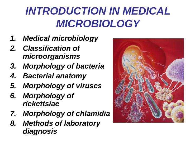
INTRODUCTION IN MEDICAL MICROBIOLOGY 1. Medical microbiology 2. Classification of microorganisms 3. Morphology of bacteria 4. Bacterial anatomy 5. Morphology of viruses 6. Morphology of rickettsiae 7. Morphology of chlamidia 8. Methods of laboratory diagnosis
• Medical microbiology is the study of causative agents of infectious diseases of humans and their reactions to such infections. In other words it deals with etiology, pathogenesis, laboratory diagnosis, specific treatment and control of infection (immunization).
Modern medical microbiology • Bacteriology – the science of bacteria, the causative agents of a member of infectious diseases. • Virology – the science of viruses, non-cellular living systems, capable of causing infectious diseases in man. • Immunology – the science which concerned with mechanisms of body protection against pathogenic microorganisms and foreign cells and substances. • Mycology – the study of fungi pathogenic for man. • Protozoology – which deals with pathogenic unicellular animal organisms.
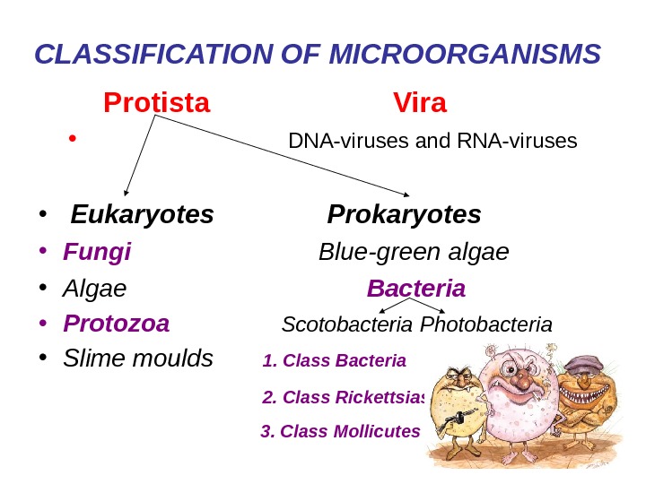
CLASSIFICATION OF MICROORGANISMS Protista Vira • DNA-viruses and RNA-viruses • Eukaryotes Prokaryotes • Fungi Blue-green algae • Algae Bacteria • Protozoa Scotobacteria Photobacteria • Slime moulds 1. Class Bacteria 2. Class Rickettsias 3. Class Mollicutes
Microbiological nomenclature • In microbiology the binominal system of nomenclature is accepted where each species has a generic and a specific name. The generic name is written with a capital letter, and the specific name – with a small letter. For example: the anthrax bacillus – Bacillus anthracis ; the tetanus bacillus – Clostridium tetani.
The size of bacteria • The size of bacteria is measured in micrometer ( m) or micron ( ) (1 micron or micrometer is one thousandth of a millimeter) and varies from 0. 1 to 16 -18 . Most pathogenic bacteria measure from 0. 1 to 10 . • The other units of measurement of microorganisms are millimicron (m ) or nanometer (nm) (one millionth of a millimeter) and 1 Angstrom (Å) (one tenth of nanometer).
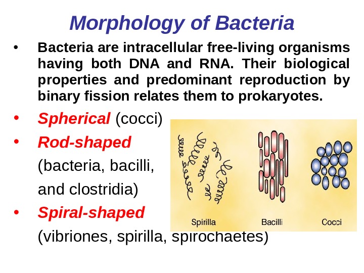
Morphology of Bacteria • Bacteria are intracellular free-living organisms having both DNA and RNA. Their biological properties and predominant reproduction by binary fission relates them to prokaryotes. • Spherical (cocci) • Rod-shaped (bacteria, bacilli, and clostridia) • Spiral-shaped (vibriones, spirilla, spirochaetes)
Spherical (cocci) bacteria 1. Micrococci 2. Diplococci 3. Streptococci 4. Staphylococci 5. Tetracocci 6. Sarcine
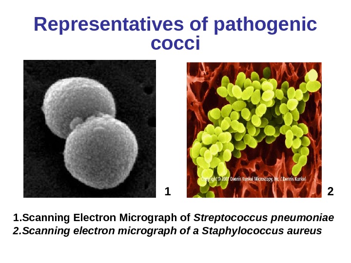
Representatives of pathogenic cocci 1. Scanning Electron Micrograph of Streptococcus pneumoniae 2. Scanning electron micrograph of a Staphylococcus aureus
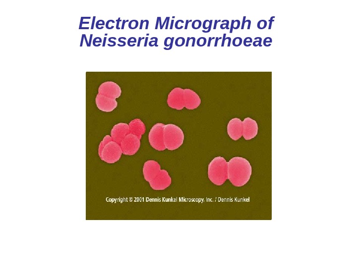
Electron Micrograph of Neisseria gonorrhoeae
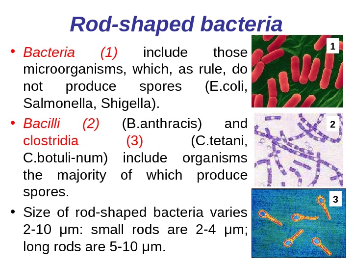
Rod-shaped bacteria • Bacteria (1) include those microorganisms, which, as rule, do not produce spores (E. coli, Salmonella, Shigella). • Bacilli (2) (B. anthracis) and clostridia (3) (C. tetani, C. botuli-num) include organisms the majority of which produce spores. • Size of rod-shaped bacteria varies 2 -10 μ m: small rods are 2 -4 μ m; long rods are 5 -10 μ m.
ARRANGEMENT OF ROD-SHAPED BACTERI
Rod-shaped bacteria 1. Single Rod 2. Streptobacillus
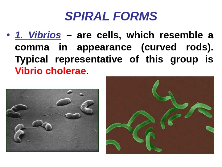
SPIRAL FORMS • 1. Vibrios – are cells, which resemble a comma in appearance (curved rods). Typical representative of this group is Vibrio cholerae.
2. Spirilla – are coiled forms of bacteria. Pathogenic species: Spirillum minus (1) – which is responsible for a disease in humans transmitted through the bite of rats – rat-bite fever – sodoku; Helicobacter pylori (2) – causative agent of ulcer disease of stomach.
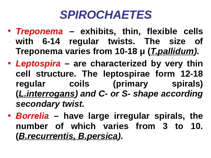
SPIROCHAETES • Treponema – exhibits, thin, flexible cells with 6 -14 regular twists. The size of Treponema varies from 10 -18 μ ( T. pallidum ). • Leptospira – are characterized by very thin cell structure. The leptospirae form 12 -18 regular coils (primary spirals) ( L. interrogans ) and C- or S- shape according secondary twist. • Borrelia – have large irregular spirals, the number of which varies from 3 to 10. ( B. recurrentis, B. persica ).
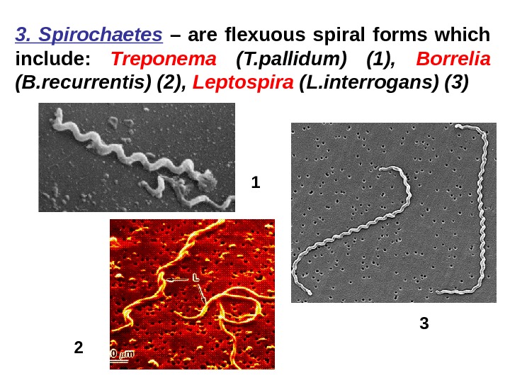
3. Spirochaetes – are flexuous spiral forms which include: Treponema (T. pallidum) (1), Borrelia (B. recurrentis) (2), Leptospira (L. interrogans) (3)
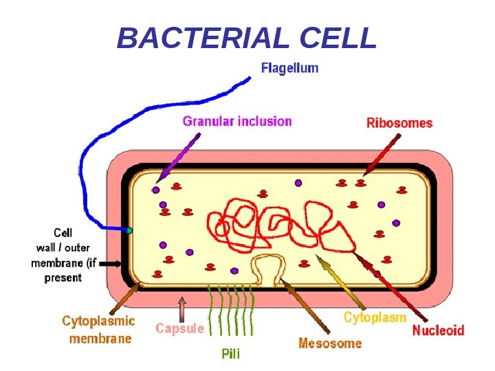
BACTERIAL CELL
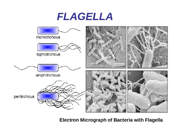
FLAGELLA Electron Micrograph of Bacteria with Flagella
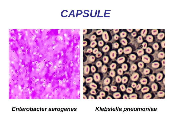
CAPSULE Klebsiella pneumoniae. Enterobacter aerogenes
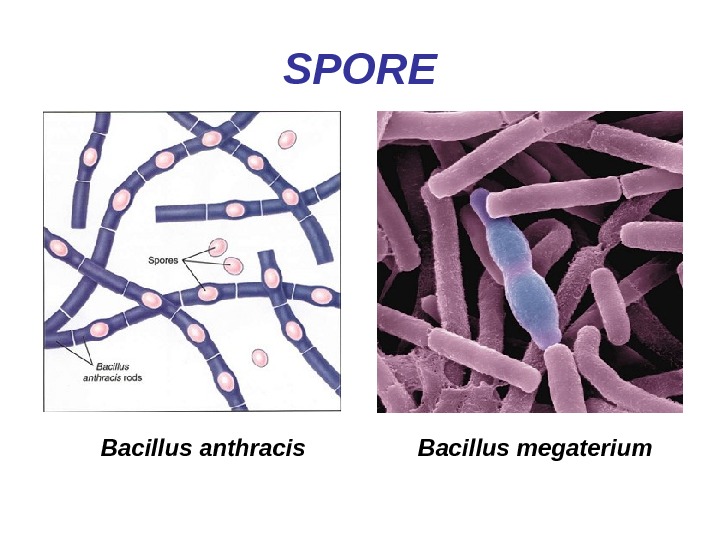
SPORE Bacillus megaterium Bacillus anthracis
Cell wall • In addition to conferring rigidity upon bacteria, the cell wall protects against osmotic damage • Chemically, the rigid part of the cell wall is peptidoglycan • First described by Gram in 1884. It is’ used to study morphologic appearance of bacteria. Gram’s stain differentiates all bacteria into two distinct groups: • a. Gram-positive organisms • b. Gram-negative organisms
fuchsine Gram Staining Technique
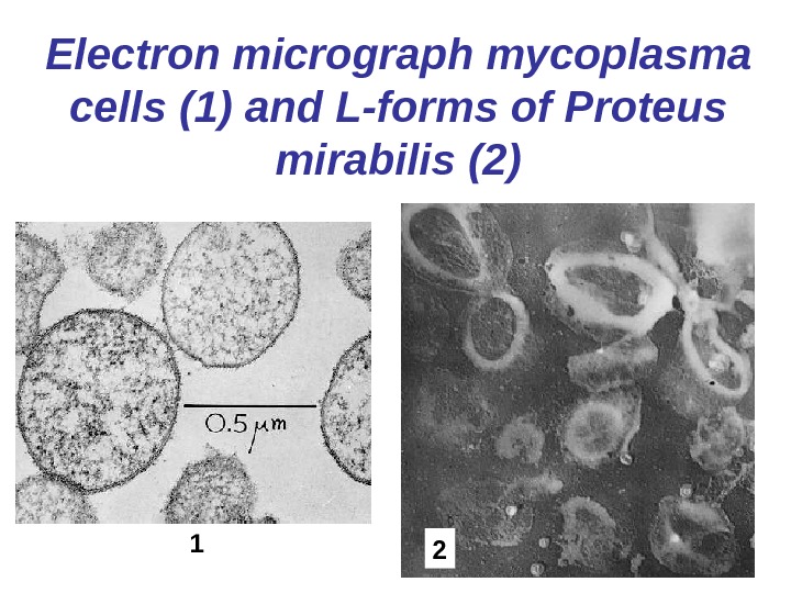
Bacteria with deficient cell walls • Mycoplasma : a genus of naturally occurring bacteria which lack cell walls • L-forms : cell-wall-deficient forms of bacteria, usually produced in the body of patients treated with penicillin • Spheroplasts : derived from Gram-negative bacteria; produced artificially by lysozyme or by growth with penicillin or any other agent capable of breaking down the peptidoglycan layer • Protoplasts : derived from Gram-positive bacteria and totally lacking cell walls; produced artificially by lysozyme and hypertonic medium
Electron micrograph mycoplasma cells (1) and L-forms of Proteus mirabilis (2)
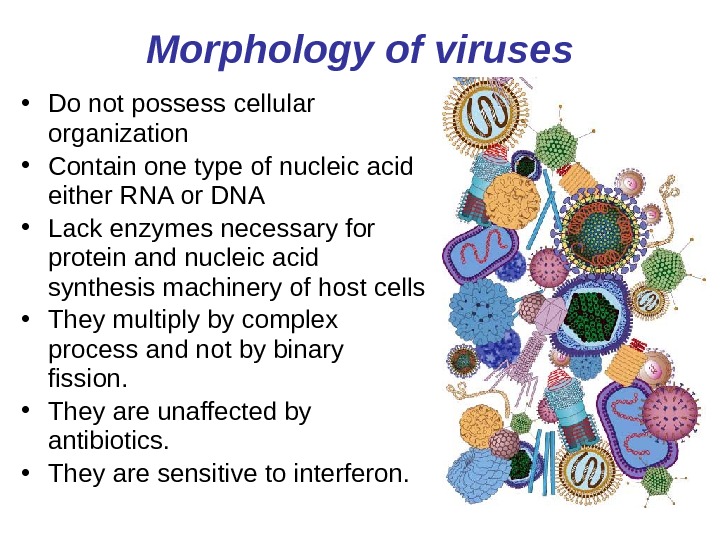
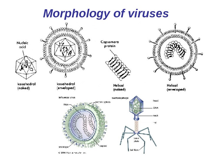
Morphology of viruses • Do not possess cellular organization • Contain one type of nucleic acid either RNA or DNA • Lack enzymes necessary for protein and nucleic acid synthesis machinery of host cells • They multiply by complex process and not by binary fission. • They are unaffected by antibiotics. • They are sensitive to interferon.
Morphology of viruses
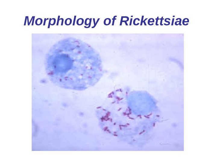
Morphology of Rickettsiae. • They are minute organisms having properties in between bacteria and viruses. • It contains both DNA and RNA. • Contains enzymes for metabolic functions. • Multiplies by binary fission. • It is coccobacilli 300 x 600 nm in size, non-motile, non-capsulated and is Gram-negative. • Sensitive to many antibiotics. • Can multiply only inside living cells.
Morphology of Rickettsiae
Morphology of chlamydia. • Chlamydiae are Gram-negative. They lack some important mechanisms for the production of metabolic energy, so they are intracellular parasites. There are 2 morphological forms of chlamydia: • Elementary bodies • Initial bodies
Methods of laboratory diagnosis 1. Bacterioscopical 2. Bacteriological 3. Detection sensitivity of bacteria to antibiotics 4. Serological 5. Biological 6. DNA-technology test (PCR)