Презентация lecture 1.pptx
- Количество слайдов: 23


Introduction lecture. The Subject and Problems of Microbiology. A brief history of microbiology. Classification of microorganisms. Functional anatomy of prokaryotic cells. G. A. Abdulina G. A.

Woese сlassification • Woese attempted to establish In 1990, the name "domain" was proposed for the highest rank. [ 1. Domain Bacteria (prokaryotes) → 2. Domain Archaea (prokaryotes) → 3. Domain Eukarya • Kingdom Protoctista or Protista • Kingdom Plantae • Kingdom Fungi • Kingdom Animalia Kingdom Bacteria Kingdom Archaea

The taxonomic classification In Berge’s Manual bacteria are divided into four divisions: • 1. Gracilicutes • 2. Firmicutes • 3. Tenericutes • 4. Mendosicutes • Each division is divided into classis; classis into orders; orders into families; families into genera; genera into species.

BACTERIAL SIZE • Individual bacterial cells are invisible to the naked eye. • In general, the sizes of bacteria isolated from humans fall within a limited range. Spherical bacteria (cocci) are between 0. 5– 1. 5 µm in diameter. Rod shaped have diameters of 0. 2– 1 µm by 0. 5– 5 µm in length. • One micron (µm) is a thousand fold smaller than a millimetre (1 mm= 1000 µm) or a metre ( 1 m = 1000000 µm).
 • Rod-shaped (bacteria, bacilli and")
Shapes of bacteria Three main shapes: • Spherical (cocci) • Rod-shaped (bacteria, bacilli and clostridia) • Spiral-shaped (vibrios and spirilla).

Figure 1. Structure of typical bacterial cell
, filamentous surface appendages about 12 to 30")
Flagella are long (3 to 12 µm), filamentous surface appendages about 12 to 30 nm in diameter. A flagellum consists of three parts: (1) the long filament, ; (2) the hook structure; and (3) the basal body. Chemically, They are constructed of proteins, called flagellins and numerous of proteins. Function: 1. locomotion ; 2. immunogenic – H (Flagella) antigens Detection: direct method – Loffler staining indirect method –hanging drop and crash drop
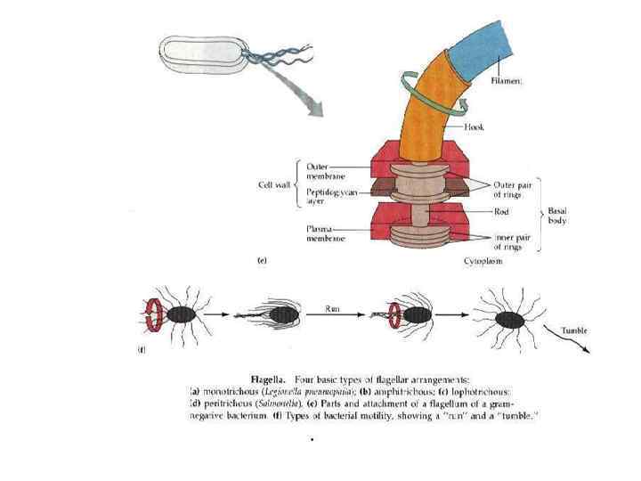

Types of the flagella Monotrichous -a single flagellum at one pole; Peritrichous -distributed over the entire cell; Lophotrichous-a tuft of flagella coming from one pole; Amphitrichous -flagella at both poles of the cell;

Pili The terms pili and fimbriae are hairlike appendages on the surface of many Gram-negative bacteria and consists of proteins -pilins. Pili can come in two types: 1. short, abundant common pili or fimbriae with as many as 200 per cell 2. a small number (one to six) of very long pili known as sex pili. Function of the common pili : to adhere to various epithelial surfaces, to red blood cells (colonization factors) The sex pili attach male to female bacteria during conjugation.

Glycocalyx , Capsule , Slime layer The bacterial glycocalyx is a viscous , gelatinous polymer that is external to the cell wall and composed of polysaccharide, polypeptide, or both. . Glycocalyx , unorganized and only loosely attached to the cell wall is described as a slime layer. The glycocalyx , organized and is firmly attached to the cell wall, is described as a capsule.

Function: A glycocalyx helps cells attach to their target is called an extracellular polymeric substance (EPS) Capsules protect pathogenic bacteria from phagocytosis. Detection: Burry Ginse, India Ink, Capsule swelling test

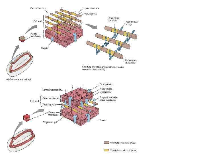
Cell wall The cell wall of the bacterial cell is a complex, semirigid structure The chemical composition of the cell wall is used to differentiate major types of bacteria. Gram-Positive Cell Walls Many layers of peptidoglycan, forming a thick, rigid structure. In addition, the cell walls of gram-positive bacteria contain teichoic acid. Gram-Negative Cell Walls One or a very few layers of peptidoglycan and an outer membrane. The outer membrane of the gram-negative cell consists of lipopolysaccharides (LPS), lipoproteins, and phospholipid

The function of the cell wall is to • prevent bacterial cells from rupturing • help maintain the shape of a bacterium • serve as a point of anchorage for flagella. • gram-negative bacteria release lipid A (LPS), which functions as an endotoxin Clinically, the cell wall is important because it contributes to the ability of some species to cause disease and is the site of action of some antibiotics. Detection: the Gram Stain
 CPM of prokaryotes consists primarily of phospholipids proteins. One exception")
The Cytoplasmic Membrane (CPM) CPM of prokaryotes consists primarily of phospholipids proteins. One exception is the wall-less prokaryote Mycoplasma, which contains membrane sterols. Functions • to serve as a selective barrier through which materials enter and exit the cell • to break nutrients • to produce energy.

Nucleoid The nucleoid of a bacterial cell (usually contains a single long, continuous. and frequently circularly arranged thread of double-stranded DNA called the bacterial chromosome. Nucleoid do not include histones , nuclear membrane and nucleoli. Function: • to keep cell's genetic information; • to control cell activity Detection: • The nuclear substance can be identified by Robinow's and Feulgen's microchemical test

Ribosomes A ribosome is comprised of protein and ribosomal RNA. There are thousands of ribosomes in the cell. Prokaryotic ribosomes are relatively small (70 S) and less dense than eukaryotic ribosomes (80 S) Functions : synthesize polypeptide Ribosomes are targets for antibiotics to inhibit the bacterium’s protein synthesis. Detection: Ribosomes are identified by their sedimentation rate
 An inclusion is a storage area that serves as a reserve for")
Inclusions (granules) An inclusion is a storage area that serves as a reserve for lipids, nitrogen, phosphate, starch, and sulfur within the cytoplasm. The other type of storage granule is that found in Corynebacterium spp. , known as Volutin containing metaphosphate (or metachromatic) granules which stain with methylene blue or Neisser stain.
 Spores are small spherical or oval bodies formed within the cell. The")
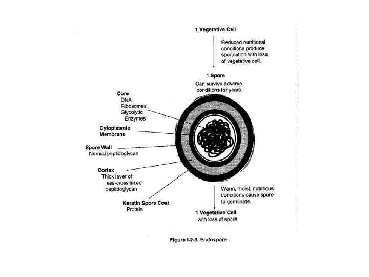
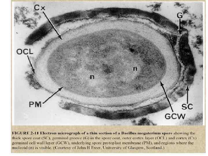
ENDOSPORES (spores) Spores are small spherical or oval bodies formed within the cell. The spore protects bacteria and withstand boiling, chemical treatments and radiation Some microorganisms, principally rodshaped (bacilli and clostridia), are capable of sporulation. These include the causative agents of anthrax, tetanus, gas gangrene, botulism Special Auesky staining procedures are used to Identify spore
;")
Spores are located • in the centre of the cell (causative agent of anthrax); • terminally, at the ends of the rod (causative agent of tetanus); • subterminaly, towards the ends (Causative agents of botulism, gas gangrene, etc. ). Sporulation is completed within 18 to 20 hours Usually germination takes place more quickly than sporulation (within 4 to 5 hours).
Презентация lecture 1.pptx