

27b45a640c4728526629a3de05c0c9de.ppt
- Количество слайдов: 22
 Informática en Anatomía Patológica Sergi Serrano Universitat Autònoma de Barcelona Hospital del Mar-IMIM Barcelona, 11 Junio
![[1586] A 5 -Year Evaluation of Barcoding in Surgical Pathology at a Teritary Care](https://present5.com/presentation/27b45a640c4728526629a3de05c0c9de/image-2.jpg "[1586] A 5 -Year Evaluation of Barcoding in Surgical Pathology at a Teritary Care") [1586] A 5 -Year Evaluation of Barcoding in Surgical Pathology at a Teritary Care Hospital. Arivarasan Karunamurthy, Anil Parwani, Luke T Wiehagen, Samuel A Yousem, Anthony L Piccoli, Liron Pantanowitz. University of Pittsburgh Medical Center, Pittsburgh, PA
[1586] A 5 -Year Evaluation of Barcoding in Surgical Pathology at a Teritary Care Hospital. Arivarasan Karunamurthy, Anil Parwani, Luke T Wiehagen, Samuel A Yousem, Anthony L Piccoli, Liron Pantanowitz. University of Pittsburgh Medical Center, Pittsburgh, PA
 Background: Our aim was to study the impact of technologyrelated errors following barcoding in surgical pathology at our institution. Design: We analyzed quality assurance data of reported adverse patient events over a 5 -year period ( pre-barcode: Jan 2008 to Jan 2009, barcode implementation period: Feb 2009 to Dec 2009, and post-barcode: Jan 2010 to Dec 2012).
 Pre-barcode period (Jan 2008 -Jan") Reported errors Error rate (events per 10, 000 specimens) Pre-barcode period (Jan 2008 -Jan 2009) Implementation Post-barcode phase(Feb 2009 period(Jan 2010 -Dec 2009) -Dec 2012) 7. 7 6. 4 6. 0 34 (44%) 43 (76%) 135 (75%) Hardware errors 4 (5%) 3 (5%) 8 (4%) Downtime errors Other (unspecified) errors Total errors 0 (0%) 3 (2%) 39 (51%) 11 (19%) 35 (19%) 77 57 181 Human related errors
Reported errors Error rate (events per 10, 000 specimens) Pre-barcode period (Jan 2008 -Jan 2009) Implementation Post-barcode phase(Feb 2009 period(Jan 2010 -Dec 2009) -Dec 2012) 7. 7 6. 4 6. 0 34 (44%) 43 (76%) 135 (75%) Hardware errors 4 (5%) 3 (5%) 8 (4%) Downtime errors Other (unspecified) errors Total errors 0 (0%) 3 (2%) 39 (51%) 11 (19%) 35 (19%) 77 57 181 Human related errors
 Conclusions: Implementation of barcoding helped reduce, but did not eliminate, misidentification-related errors in our AP laboratory. Such errors may persist or even increase due to better reporting of adverse events (e. g. electronic alerts and heightened user awareness), non-compliance of users (during grossing and block/slide retrieval), and as a result of technology failures (e. g. misplaced barcoded labels, printer errors and system outage).
![[1595] Auto. Stitcher. TM: An Automated Program for Accurate Reconstruction of Digitized Whole Histological](https://present5.com/presentation/27b45a640c4728526629a3de05c0c9de/image-6.jpg "[1595] Auto. Stitcher. TM: An Automated Program for Accurate Reconstruction of Digitized Whole Histological") [1595] Auto. Stitcher. TM: An Automated Program for Accurate Reconstruction of Digitized Whole Histological Sections From Tissue Fragments
Gregory Penzias, Andrew Janowczyk, Asha Singanamalli, Mirabela Rusu, Natalie Shih, Michael Feldman, Satish Viswanath, Anant Madabhushi. Case Western Reserve University, Cleveland, OH; University of Pennsylvania, Philadelphia, PA
[1595] Auto. Stitcher. TM: An Automated Program for Accurate Reconstruction of Digitized Whole Histological Sections From Tissue Fragments
Gregory Penzias, Andrew Janowczyk, Asha Singanamalli, Mirabela Rusu, Natalie Shih, Michael Feldman, Satish Viswanath, Anant Madabhushi. Case Western Reserve University, Cleveland, OH; University of Pennsylvania, Philadelphia, PA
 Background: We recently presented a memory efficient, flexible tool called Histo. Sticher. TM for manual reassembly of image fragments. However, this technique still requires laboriously manually identifying matching locations ("fiducials") prior to reconstruction. We present preliminary results of an automated method of reconstructing PWMHSs (pseudo whole-mount histological sections) from tissue fragment images, thus removing a major impediment to pathology-radiology correlation research.
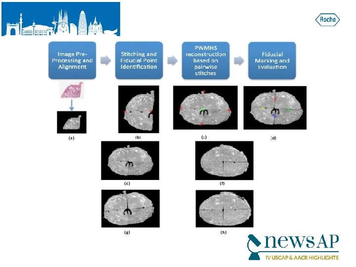
 Results: Auto. Stitcher results were compared to manual PWMHS reconstructions obtained via Histo. Stitcher (Figure 1). Quantitative evaluation required computing the mean distance between fiducials identified at the midpoints of each fragment's edge (and thus independent of fiducials used for PWMHS reconstruction by either Auto. Stitcher or Histo. Stitcher). Auto. Stitcher demonstrated a mean error of 7. 10 pixels compared to an error of 8. 13 pixels for Histo. Stitcher in a cohort of 4 ex vivo prostate sections.
 Conclusions: We have presented preliminary results of a completely automated algorithm that successfully reconstructs PWMHS images from digitized tissue fragments, with relatively low error compared to manual reconstruction.
![[1602] Diagnostic Time in Digital Comparative Study on 400 Cases. Pathology: A Aleksandar Vodovnik.](https://present5.com/presentation/27b45a640c4728526629a3de05c0c9de/image-11.jpg "[1602] Diagnostic Time in Digital Comparative Study on 400 Cases. Pathology: A Aleksandar Vodovnik.") [1602] Diagnostic Time in Digital Comparative Study on 400 Cases. Pathology: A Aleksandar Vodovnik. Førde Central Hospital, Førde, Norway
[1602] Diagnostic Time in Digital Comparative Study on 400 Cases. Pathology: A Aleksandar Vodovnik. Førde Central Hospital, Førde, Norway
 Background: Numerous validation studies in digital pathology confirmed its value as a diagnostic tool. However, a longer time to diagnosis than traditional microscopy has been currently seen as a significant barrier to the routine use of digital pathology.
 in histology, nongynaecological") Design: One senior staff pathologist reported 400 consecutive cases (1396 slides) in histology, nongynaecological and fine needle aspiration cytology, by means of digital pathology and traditional microscopy (20 sessions, 20 cases/session), over four weeks time. Primary diagnosis was digital, followed by traditional microscopy, six months later.
Design: One senior staff pathologist reported 400 consecutive cases (1396 slides) in histology, nongynaecological and fine needle aspiration cytology, by means of digital pathology and traditional microscopy (20 sessions, 20 cases/session), over four weeks time. Primary diagnosis was digital, followed by traditional microscopy, six months later.
 and 1956 (97. 8/session)") Results: Digital and microscopic diagnostic time was 1841 (92. 05/session) and 1956 (97. 8/session) minutes, respectively. Digital diagnostic time was shorter than microscopic in 13 sessions. Four sessions with shorter microscopic diagnostic time included more cases requiring an extensive use of magnifications over X 20. The diagnostic time was similar in three session.
Results: Digital and microscopic diagnostic time was 1841 (92. 05/session) and 1956 (97. 8/session) minutes, respectively. Digital diagnostic time was shorter than microscopic in 13 sessions. Four sessions with shorter microscopic diagnostic time included more cases requiring an extensive use of magnifications over X 20. The diagnostic time was similar in three session.
 Conclusions: Diagnostic time in digital pathology can be shorter comparing with traditional microscopy, in the routine diagnostic setting, when adequate and stable network speeds and fully integrated LIMS are achieved. This also relates to better ergonomics, larger viewing field and absence of physical slide handling, with effects on both diagnostic and non-diagnostic time. Possible reasons for differences with previous studies may be their setting, image size, network speed and participant's level of confidence and experience in digital reporting.
![[1577] FISH Digital Image Capture and Automated Segmentation Improves Laboratory Workflow Efficiency. Parker W](https://present5.com/presentation/27b45a640c4728526629a3de05c0c9de/image-16.jpg "[1577] FISH Digital Image Capture and Automated Segmentation Improves Laboratory Workflow Efficiency. Parker W") [1577] FISH Digital Image Capture and Automated Segmentation Improves Laboratory Workflow Efficiency. Parker W Clement, Kristina Moore, Amy Sandoval, Nermin Uvejzovic, Leslie Rowe, Rodney R Miles, Mohamed E Salama. University of Utah and ARUP Laboratories, Salt Lake City, UT
[1577] FISH Digital Image Capture and Automated Segmentation Improves Laboratory Workflow Efficiency. Parker W Clement, Kristina Moore, Amy Sandoval, Nermin Uvejzovic, Leslie Rowe, Rodney R Miles, Mohamed E Salama. University of Utah and ARUP Laboratories, Salt Lake City, UT
 Background: Limitations to FISH interpretation include identification of individual cells showing fluorescent signal without cellular overlap. These limitations are exacerbated by manual counting and interpretation, leaving manual FISH analysis subjective and time consuming. We examined if incorporation of automated segmentation protocols on digitally captured images improves cell detection and laboratory workflow efficiency.

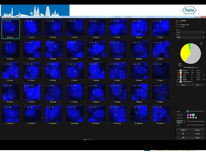
 were analyzed (5 Her 2/neu, 3 FKHR, and") Design: FISH cases (hematologic and non-hematologic) were analyzed (5 Her 2/neu, 3 FKHR, and 8 MYC) using automated FISH analysis systems (Gen. ASIs, Applied Spectral Imaging, Carlsbad, CA). H&E slides were reviewed and marked by a pathologist. Following image capture by Gen. ASIs, manual and automated segmentation protocols were performed on a minimum of 60 cells (hematopoietic cases) and 120 cells (non-hematopoietic cases) from four to five digital image frames. Both methods were compared for efficiency and accuracy
Design: FISH cases (hematologic and non-hematologic) were analyzed (5 Her 2/neu, 3 FKHR, and 8 MYC) using automated FISH analysis systems (Gen. ASIs, Applied Spectral Imaging, Carlsbad, CA). H&E slides were reviewed and marked by a pathologist. Following image capture by Gen. ASIs, manual and automated segmentation protocols were performed on a minimum of 60 cells (hematopoietic cases) and 120 cells (non-hematopoietic cases) from four to five digital image frames. Both methods were compared for efficiency and accuracy
 Results: Automated segmentation by Gen. ASIs resulted in an overall significant technician time reduction (range 13 -19 min, mean 10. 7 min) in comparison to manual segmentation, (range 19 -85 min, mean 37. 2 min) (p= <0. 0001). Automated segmentation maintained significant time reduction over manual segmentation in each test: Her 2/neu (p=<0. 0001), FKHR (p=0. 0004), and MYC (p=0. 06). There was no significant difference in the classification time between methods (p=0. 46). There was high concordance on both signal scoring pattern (P=<0. 0001, r 2=0. 98) as well as 100% concordance on case classification/result interpretation in both methods with gold standard microscopic evaluation
 Conclusions: Digital capture and analysis FISH systems perform automated cell detection much more rapidly than manual segmentation yet maintain high levels of accuracy, thus improving laboratory workflow. Adoption of digital imaging in the clinical laboratory also provides advantages including long term archival and quality assurance measures.