2cd1bcb7068beb38008d27e11d9c3a28.ppt
- Количество слайдов: 63
 Incontri Pitagorici di Cardiologia 2010 Prevenzione cardiovascolare : alla ricerca della placca vulnerabile o del paziente vulnerabile? Giancarlo Casolo g. casolo@usl 12. toscana. it Crotone, 2 Ottobre 2010
 Definizione del problema • Paziente Vulnerabile: Colui il quale presenta una spiccata propensione a sviluppare una sindrome coronarica acuta • Placca Vulnerabile: Lesione della parete del vaso che mostra una spiccata propensione a determinare una sindrome coronarica acuta
 placca Plausibilità della teoria Evidenza scientifica Ricadute assistenziali
 The vulnerable plaque hypothesis • Most myocardial infarctions originate from lesions that are not severely narrowed • There are specific characteristics of a plaque prone to fissuration /disruption / erosion and leading to ACS • The identification of these lesions prior to ACS may promote their treatment by PCI thus preventing the complications
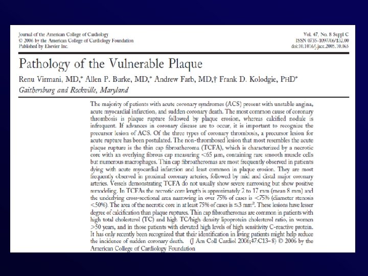
 Schematic figure illustrating the most common type of vulnerable plaque characterized by thin fibrous cap, extensive macrophage infiltration, paucity of smooth muscle cells, and large lipid core, without significant luminal narrowing Naghavi, M. et al. Circulation 2003; 108: 1664 -1672 Naghavi et Al. Circulation 2003 Copyright © 2003 American Heart Association
 – – – – Necrotic lipid") Vulnerable Plaque • Thin Cap Fibro. Atheroma (TCFA) – – – – Necrotic lipid core Thin fibrous cap Rare smooth muscle cells Numerous macrophages Positive remodelling of the vessel Increased Vasa Vasorum neovascularization Spotty calcification Intraplaque Hemorrage (ongoing healing process)
Vulnerable Plaque • Thin Cap Fibro. Atheroma (TCFA) – – – – Necrotic lipid core Thin fibrous cap Rare smooth muscle cells Numerous macrophages Positive remodelling of the vessel Increased Vasa Vasorum neovascularization Spotty calcification Intraplaque Hemorrage (ongoing healing process)
 cap lipid-rich core thrombus cap
 Matter et Al. Eur Heart J 2009
 Tools for Imaging the Vulnerable Plaque Matter et Al. Eur Heart J 2009
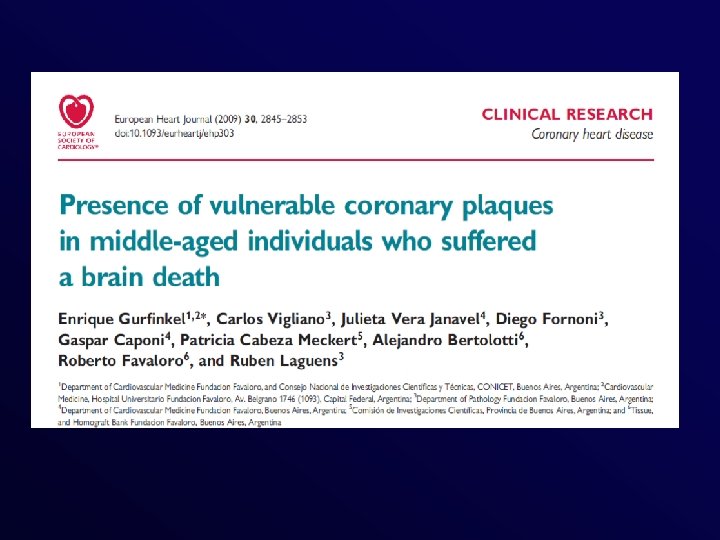
 57% Gurfinkel et Al. Eur Heart J 2009 1. 1 lesioni/pz 32% Calibro
 Applicabilità clinica? …. . Unfortunately, the search for the so-called vulnerable plaque is hampered by the lack of both natural history studies and proven local or regional therapies for these otherwise asymptomatic plaques. Ambrose GA, Srikanth S. Curr Opin Cardiol, 2009
 Applicabilità clinica? The Vulnerable Plaque “Hypothesis” Promise, but Little Progress A Pub. Med search using the terms “vulnerable plaque” or “high-risk” plaque yields 2, 000 references to journal articles published over the past 20 years. During this 20 -year period, many diagnostic techniques designed to “detect” vulnerable plaques have come and gone. These include thermography, spectroscopy, palpography, virtual histology, optical coherence tomography, and many more. A large number of startup companies with “breakthrough” approaches have come and gone, nearly all leaving investors with empty pockets, but no progress. What Nissen SE. Jacc Imag 2009 has gone wrong?
 Il concetto di placca vulnerabile EVIDENZE • La placca che causa occlusione acuta non è severamente stenotica e dunque non è identificabile prima con l’angiografia o con test per valutare la riserva coronarica Si • Studi autoptici hanno evidenziato che la causa dell’IMA spesso è una placca “a rischio Si/Ni • Se trovo la placca a rischio la tratto e prevengo l’evento No
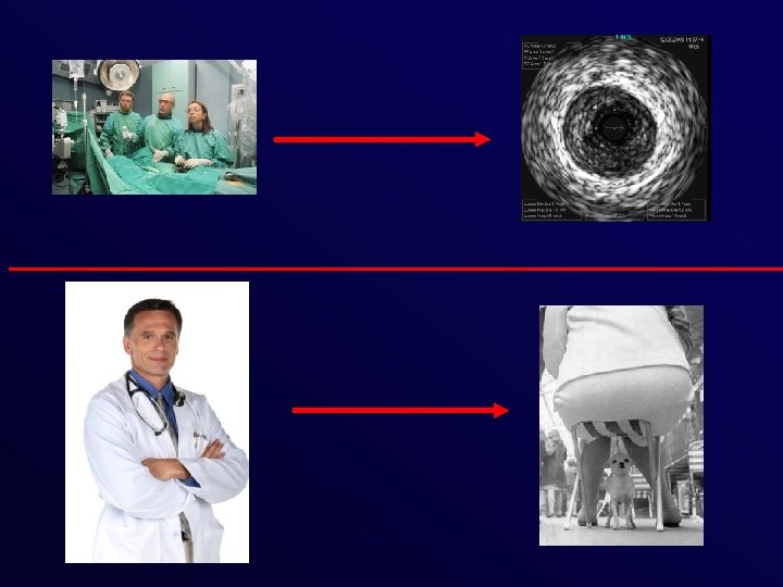
 Il portatore “sano” della placca vulnerabile
 La fine della teoria della placca vulnerabile?
 The risk of a vulnerable patient is affected by vulnerable plaque and/or vulnerable blood and/or vulnerable myocardium Naghavi, M. et al. Circulation 2003; 108: 1664 -1672 Naghavi et Al. Circulation 2003 Copyright © 2003 American Heart Association
 Il paziente vulnerabile • Inizialmente designato come termine per definire il paziente con eventi alla distruzione della placca (non tutti manifestano eventi) • Attribuibile a fattori ematologici, instabilità del miocardio, etc. . ) che possono spiegare il manifestarsi dell’evento
 colui") Il paziente vulnerabile • Attualmente si definisce come paziente vulnerabile (paziente vulnerabile cardiovascolare) colui che è prono a sviluppare eventi • Per costui sarebbe utile un tool adatto a verificare il rischio di SCA e morte Naghavi et Al. Circulation 2003
Il paziente vulnerabile • Attualmente si definisce come paziente vulnerabile (paziente vulnerabile cardiovascolare) colui che è prono a sviluppare eventi • Per costui sarebbe utile un tool adatto a verificare il rischio di SCA e morte Naghavi et Al. Circulation 2003
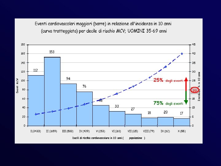
 Sopravvivenza nelle prime 24 ore dall’esordio dei sintomi di IMA 100 14% 39% non ancora ricoverati e vivi 42% 80 Pazienti (%) 68% 35% ricoverati e vivi 60 21% 40 20 4% 14% deceduti dopo il ricovero 21% deceduti prima del ricovero 32% 7% 21% 26% deceduti prima del ricovero deceduti in ospedale 26% 30% 0 0 1 4 8 12 16 20 Prime 24 ore dall’insorgenza dell’IMA Dati del Registro MONICA di Augsburg (mod. ) 24 giorno 28 Eur Heart J 1998; 19: 1140 -64
 Carte di rischio
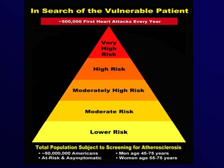
 New CHD Risk Equivalents • CAD • >20% 10 -year risk of CHD (Framingham projections) • Diabetes • Other forms of clinical atherosclerotic disease: – Peripheral arterial disease – Abdominal aortic aneurysm – Carotid artery disease Expert Panel on Detection, Evaluation, and Treatment of High Blood Cholesterol in Adults. JAMA 2001; 285: 2486 -2497.
 Who Has More Cardiovascular Risk Factors? Sir Winston Churchill, 91 Jim Fixx, 53

 Aterosclerosi e Malattia coronarica P. Libby. Circulation. 2001; 104: 365 -372
 Plaque Growth/ Stenotic Area La storia tradizionale Domain of ACS Domain of Angina Asymptomatic Time/Years
 Plaque Growth/ Stenotic Area La storia riveduta Domain of ACS Domain of Angina Asymptomatic Time/Years
 La guarigione delle complicazioni della placca contribuisce alla evoluzione dell’aterosclerosi Burke et Al. Circulation 2001
 Datazione Carbonio 14 Goncalves et Al. Circ Res 2010
 Coronary heart disease epidemics: not all the same Mirzaei M et Al. Heart 2009
 Coronary heart disease epidemics: not all the same Mirzaei M et Al. Heart 2009
 Mirzaei M et Al. Heart 2009
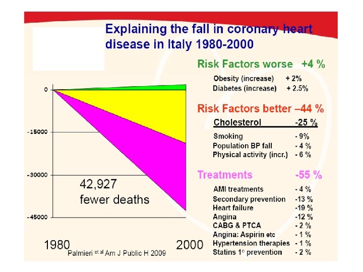
 Contributo delle singole azioni alla riduzione della mortalità per CAD in Svezia 1986 -2002 Bjork L et Al. Eur Heart J 2009
 Percentage of Decrease in Deaths From Coronary Disease Due to Treatments for Established Disease and Changes in Risk Factors Hlatky et Al. JACC 2009
 Placca vulnerabile o paziente vulnerabile?
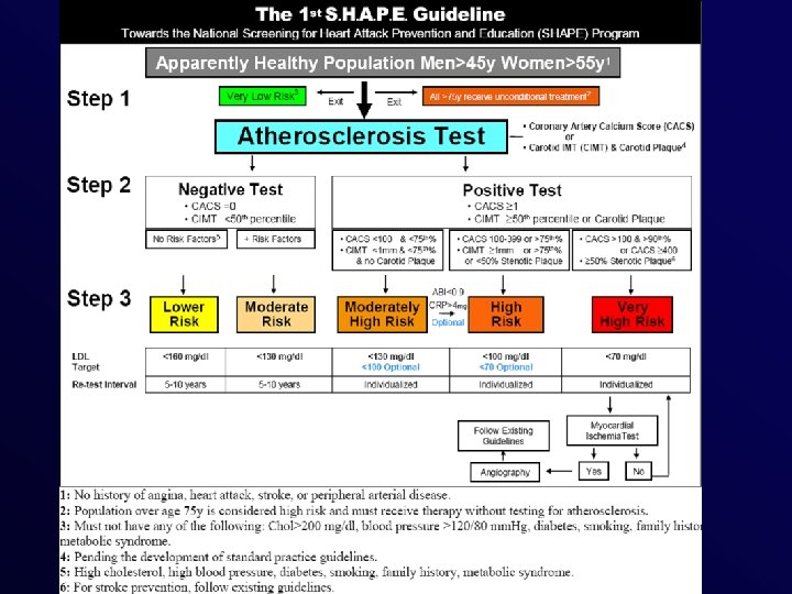
 Circulation 2010; 121: 1447 -1454
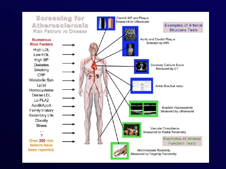
 Peso prognostico aggiuntivo dei segni di danno aterosclerotico subclinico Sehestedt T et Al. European Heart Journal Advance Access published December 23, 2009
 Future approach: the basis • Vulnerable patients do have vulnerable plaques • One may therefore assess the evidence for subclinical plaque disruption or… • Look for molecules, cells or extracellular matrix protein exposed by plaque disruption and…. • The degree of atherosclerotic burden
 Stenosis detection LAD D 2 Diag 2 LAD Diag 2 Distal LAD 1 stenosis Courtesy of MAR Imaging Center, Bnai Zion Hospital, Haifa, Israel LAD Distal LAD 1 stenosis confirmed by cathlab
 Alternative/additive imaging approach • Atherosclerotic burden – Number of plaques – Total plaque volume • Molecular Imaging of Cell Apoptosis • Molecular Imaging of MMP • Nanoparticles (i. e. HDL)
 Multicontrast In Vivo CMR Chu et Al. J. Am. Coll. Cardiol. Img. 2009; 2; 883 -896
 Plaque imaging Lipidic Core Soft Fibrous Remodelling of the vessel 1 1 Mixed 2 Calcific 3 3
 MDCT in ACS Pundzuite et Al. Eur Heart J 2009
 Plaque composition and ACS Motoyama et Al. JACC 2007
 • FU 27+10") Design of the study • 1160 Pts (835 m, 325 f) • FU 27+10 Mo • MSCT 64 – Vessel remodelling (VR) – Plaque consistency (LAP) : <(NCP 30 HU) and intermediate attenuation plaques (30 HU
Design of the study • 1160 Pts (835 m, 325 f) • FU 27+10 Mo • MSCT 64 – Vessel remodelling (VR) – Plaque consistency (LAP) : <(NCP 30 HU) and intermediate attenuation plaques (30 HU
 22. 2% (10) • Pts with") Results Subsequent ACS • Pts with LAP+VR (45) 22. 2% (10) • Pts with LAP or VR (27) 3. 7% (1) • Pts wo LAP or VR (820) 0. 5% (4)
Results Subsequent ACS • Pts with LAP+VR (45) 22. 2% (10) • Pts with LAP or VR (27) 3. 7% (1) • Pts wo LAP or VR (820) 0. 5% (4)
 Results Motoyama et Al. JACC 2009
 Placca vulnerabile o paziente vulnerabile? Placca vulnerabile e paziente vulnerabile?
 Clinicamente • Il paziente che ha già avuto eventi, ha fatto la coronarografia e mostra placche vulnerabili (IVUs) • Il paziente vulnerabile ha caratteristiche in parte identificabili che lo predispongono ad eventi cardiovascolari (es. pz ad alto rischio CV)
 Paziente vs placca vulnerabile Paziente vulnerabile Lesione identificabile Unica o al massimo 2 -3 Elementi distintivi Trattabile Approccio “locale” Imaging complesso Lesione non identificabile Molteplici lesioni Nessuno specifico elemento Approccio multifattoriale Approccio “generale” Imaging facile (!? )
 • Molti eventi sono preceduti da placche vulnerabili • Il “timing” preciso") Conclusioni (1) • Molti eventi sono preceduti da placche vulnerabili • Il “timing” preciso tra queste e gli eventi è sconosciuto • Nessuna evidenza che il loro trattamento procura beneficio • Nessuno conosce la durata della vulnerabilità di una lesione • La diagnosi è invasiva (per ora…)
Conclusioni (1) • Molti eventi sono preceduti da placche vulnerabili • Il “timing” preciso tra queste e gli eventi è sconosciuto • Nessuna evidenza che il loro trattamento procura beneficio • Nessuno conosce la durata della vulnerabilità di una lesione • La diagnosi è invasiva (per ora…)
 • Molti eventi si verificano in pazienti a basso rischio • Molti") Conclusioni (2) • Molti eventi si verificano in pazienti a basso rischio • Molti di questi hanno una ATS sublinica • La presenza di questa riclassifica il rischio • La identificazione più realistica deve essere clinica e quindi strumentale (non invasiva? ) • Il trattamento medico è efficace nel ridurre gli eventi
Conclusioni (2) • Molti eventi si verificano in pazienti a basso rischio • Molti di questi hanno una ATS sublinica • La presenza di questa riclassifica il rischio • La identificazione più realistica deve essere clinica e quindi strumentale (non invasiva? ) • Il trattamento medico è efficace nel ridurre gli eventi