Immunology of transplantation. Reproductive immunology. Transplantation immunology is

15046-immunology_of_transplantation.ppt
- Количество слайдов: 36
 Immunology of transplantation. Reproductive immunology.
 Transplantation immunology is getting increasingly important, from a clinical point of view, now involving cellular grafts (for example, blood transfusion and bone marrow grafts), as well as organ transplants (kidney, heart, liver, pancreas, lungs, cornea, skin, etc.). individual cellular antigens determining tissular compatibility or incompatibility ("histocompatibility") play an important role in immunobiology, as they participate in immune recognition and antigen presentation phenomena.
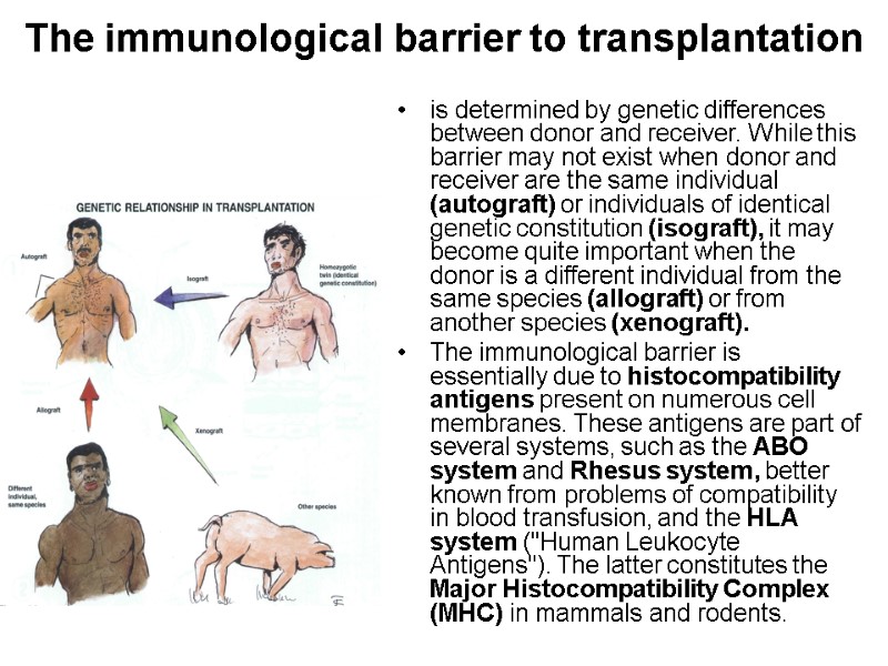
 The immunological barrier to transplantation is determined by genetic differences between donor and receiver. While this barrier may not exist when donor and receiver are the same individual (autograft) or individuals of identical genetic constitution (isograft), it may become quite important when the donor is a different individual from the same species (allograft) or from another species (xenograft). The immunological barrier is essentially due to histocompatibility antigens present on numerous cell membranes. These antigens are part of several systems, such as the ABO system and Rhesus system, better known from problems of compatibility in blood transfusion, and the HLA system ("Human Leukocyte Antigens"). The latter constitutes the Major Histocompatibility Complex (MHC) in mammals and rodents.
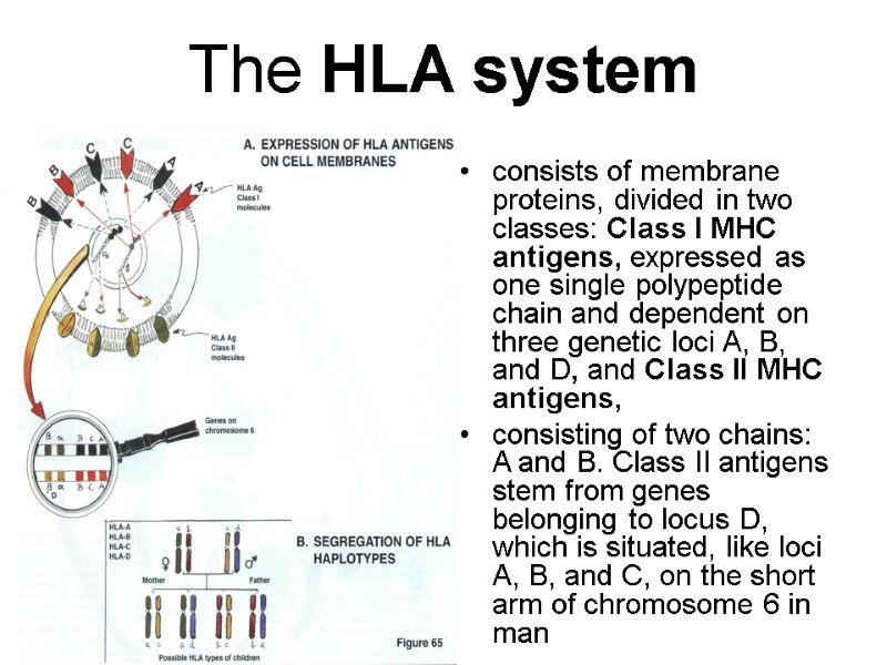
 The HLA system consists of membrane proteins, divided in two classes: Class I MHC antigens, expressed as one single polypeptide chain and dependent on three genetic loci A, B, and D, and Class II MHC antigens, consisting of two chains: A and B. Class II antigens stem from genes belonging to locus D, which is situated, like loci A, B, and C, on the short arm of chromosome 6 in man
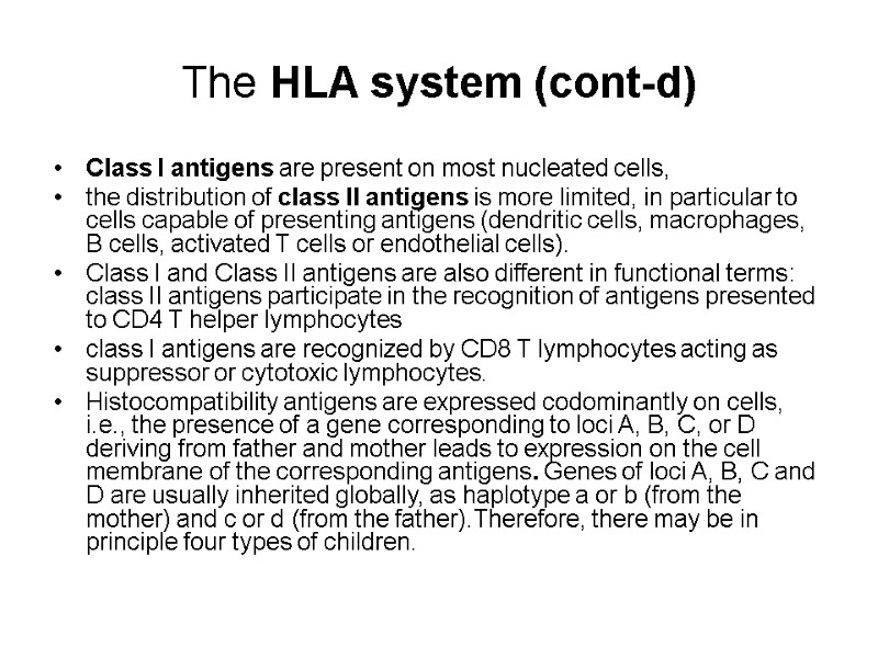
 The HLA system (cont-d) Class I antigens are present on most nucleated cells, the distribution of class II antigens is more limited, in particular to cells capable of presenting antigens (dendritic cells, macrophages, B cells, activated T cells or endothelial cells). Class I and Class II antigens are also different in functional terms: class II antigens participate in the recognition of antigens presented to CD4 T helper lymphocytes class I antigens are recognized by CD8 T lymphocytes acting as suppressor or cytotoxic lymphocytes. Histocompatibility antigens are expressed codominantly on cells, i.e., the presence of a gene corresponding to loci A, B, C, or D deriving from father and mother leads to expression on the cell membrane of the corresponding antigens. Genes of loci A, B, C and D are usually inherited globally, as haplotype a or b (from the mother) and c or d (from the father).Therefore, there may be in principle four types of children.
 Clinical considerations even sister may be with an identical HLA formula. As a result, we are always interested in looking for an HLA identical donor in the patient's own family, in case of transplantation or especially where repeated blood transfusions are involved. In contrast, as there are about 80 known allelic genes for loci A, B and C and 35 for locus D, the chances of finding an HLA identical non-related donor are relatively slim.
 transplantation between a donor and receiver which are not identical for their histocompatibility antigens, leads obligatorily to immunological rejection, if the immune system of the receiver is intact. T lymphocytes are mainly responsible for this phenomenon
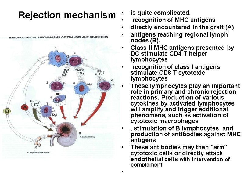
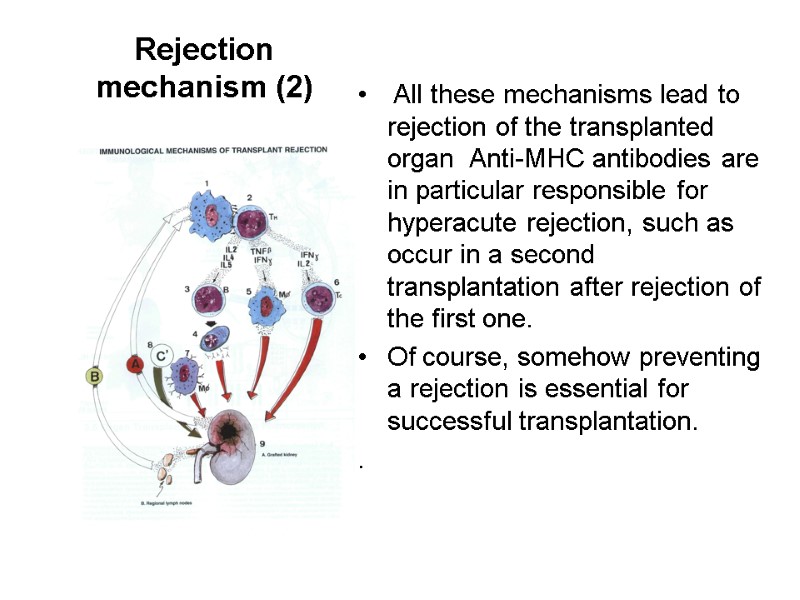
 Rejection mechanism is quite complicated. recognition of MHC antigens directly encountered in the graft (A) antigens reaching regional lymph nodes (B). Class II MHC antigens presented by DC stimulate CD4 T helper lymphocytes recognition of class I antigens stimulate CD8 T cytotoxic lymphocytes These lymphocytes play an important role in primary and chronic rejection reactions. Production of various cytokines by activated lymphocytes will amplify and trigger additional phenomena, such as activation of cytotoxic macrophages , stimulation of B lymphocytes and production of antibodies against MHC antigens These antibodies may then "arm" cytotoxic cells or directly attack endothelial cells with intervention of complement
 Rejection mechanism (2) All these mechanisms lead to rejection of the transplanted organ Anti-MHC antibodies are in particular responsible for hyperacute rejection, such as occur in a second transplantation after rejection of the first one. Of course, somehow preventing a rejection is essential for successful transplantation. .
 Possible ways of prophylaxis A first step reducing as much as possible the MHC differences between donor and receiver, which is possible in elective transplantation (e. g., kidney) by setting up organ banks and receiver networks for potential organ donors. using drugs suppressing cellular activation, in a non-specific manner to minimize rejection, (corticosteroids), cytokine production (cyclosporin) or cellular proliferation (azathioprine). specific interventions are the subject of intense studies, with the purpose of avoiding the frequent side effects of immunosuppressive drugs. Induction of immunological tolerance by administration of antigens from the graft during embriyonal development of the receiver, although possible in immunological experiments is obviously impractical in clinical situations. the administration of histocompatibility antigens, or their corresponding antibodies in adults, under certain conditions, permits the attenuation of rejection (facilitation phenomenon). Attempts to introduce human MHC antigens into animal species, such as pigs, by transgenic technology (introdution of genes from one individual or one species to another), are currently under way. This would enable us to some day perform xenografts.
 Graft-Versus-Host Reaction (GVH) In this type of reaction, it could be said that it is the graft which rejects the host. Immunologically competent T lymphocytes contained in the graft (mainly in bone marrow grafts) attack cells of the receiver, and in acute forms may provoke immunologically severe and even fatal lesions, involving liver, skin, and intestine. Grafts containing aggressive lymphocytes are true Trojan horses introduced into the receiver's circulation, particularly when the receiver suffers from immune deficiency.
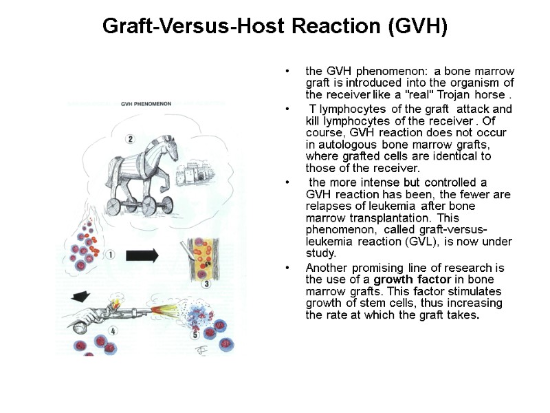
 Graft-Versus-Host Reaction (GVH) the GVH phenomenon: a bone marrow graft is introduced into the organism of the receiver like a "real" Trojan horse . T lymphocytes of the graft attack and kill lymphocytes of the receiver . Of course, GVH reaction does not occur in autologous bone marrow grafts, where grafted cells are identical to those of the receiver. the more intense but controlled a GVH reaction has been, the fewer are relapses of leukemia after bone marrow transplantation. This phenomenon, called graft-versus-leukemia reaction (GVL), is now under study. Another promising line of research is the use of a growth factor in bone marrow grafts. This factor stimulates growth of stem cells, thus increasing the rate at which the graft takes.
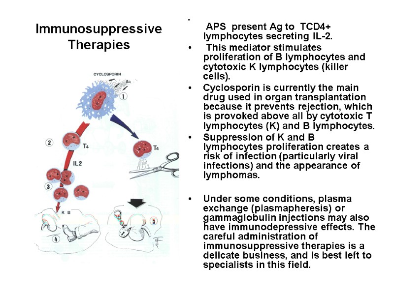
 Immunosuppressive Therapies The main indications for immunosuppressive therapy are organ transplantation and autoimmune diseases. The first immunosuppressive drugs were, on the one hand, antimitotic, i.e., acting on dividing cells, such as azathioprine, 6-mercaptopurine or methotrexate, and, on the other hand, corticosteroids, which suppress production of several lymphokines required for an immune response. The discovery of cyclosporin has represented a major advance and has been followed by new derivatives with similar action, and also by other biological strategies based on antilymphocyte antibodies, immunotoxins or suppressor cytokines (e.g., IL-10). Cyclosporin is an immunosuppressive extract of a mould (Tolypocladium). It has little general toxicity and is not cytostatic. It inhibits mainly the release of interleukin 2 and other lymphokines by T lymphocytes. As a result, there is a considerable reduction in proliferation of T4 lymphocytes and B lymphocytes. It may be recalled that interleukin 2 is synthesized by helper lymphocytes of the ThO and Th1 classes, when they are stimulated by a APC.
 Immunosuppressive Therapies APS present Ag to TCD4+ lymphocytes secreting IL-2. This mediator stimulates proliferation of B lymphocytes and cytotoxic K lymphocytes (killer cells). Cyclosporin is currently the main drug used in organ transplantation because it prevents rejection, which is provoked above all by cytotoxic T lymphocytes (K) and B lymphocytes. Suppression of K and B lymphocytes proliferation creates a risk of infection (particularly viral infections) and the appearance of lymphomas. Under some conditions, plasma exchange (plasmapheresis) or gammaglobulin injections may also have immunodepressive effects. The careful administration of immunosuppressive therapies is a delicate business, and is best left to specialists in this field.
 Immune Response to Pregnancy (Alloimmunity) Function: to alert the mother to react to the baby as a baby, not as an infection. Consequence: blocking antibody production (crossmatch positive by flow cytometry).
 There are five categories of immune problems that can cause pregnancy loss, IVF failures and infertility. Category 1 is the least severe, while Category 5 is the most severe. Without treatment, a woman with Category 1 problems can experience recurrent pregnancy loss, which may activate other categories of immune problems from Category 2, 3, 4 or 5.
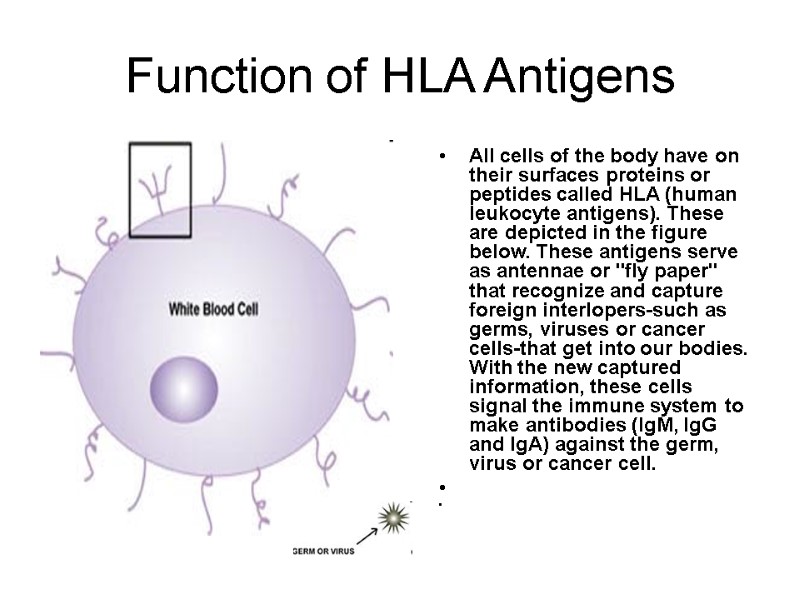
 Function of HLA Antigens All cells of the body have on their surfaces proteins or peptides called HLA (human leukocyte antigens). These are depicted in the figure below. These antigens serve as antennae or "fly paper" that recognize and capture foreign interlopers-such as germs, viruses or cancer cells-that get into our bodies. With the new captured information, these cells signal the immune system to make antibodies (IgM, IgG and IgA) against the germ, virus or cancer cell.
 A pregnancy must also be recognized as a foreign being (father puts HLA antigens on the placenta that are different from those of the mother). When this applies, the mother makes an antibody called a blocking antibody that attaches to the placenta and makes it look to her like a "wolf in sheep's clothing." The antibody she makes in this circumstance does not kill; it protects the baby and makes the placental cells grow faster. When the father's HLA antigens placed on the placenta are too similar to the mother's HLA antigens, she does not make the antibody. In this circumstance the baby is not protected, the placental cells are not stimulated to grow and the baby dies. She interprets the pregnancy as "altered self" (i.e., a cancer cell). Therefore, when the cells of the baby die, she activates other immune problems from Category 2, 3, 4 or 5 where the natural killer cells that she was born with are now misinterpreting the baby as a cancer. This occurs in couples sharing DQ alpha HLA antigens.
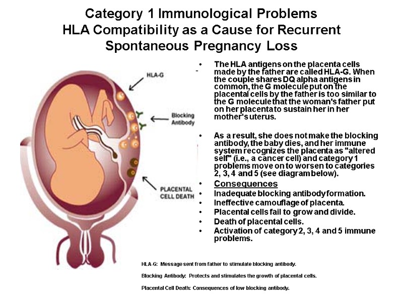
 Category 1 Immunological Problems HLA Compatibility as a Cause for Recurrent Spontaneous Pregnancy Loss The HLA antigens on the placenta cells made by the father are called HLA-G. When the couple shares DQ alpha antigens in common, the G molecule put on the placental cells by the father is too similar to the G molecule that the woman's father put on her placenta to sustain her in her mother's uterus. As a result, she does not make the blocking antibody, the baby dies, and her immune system recognizes the placenta as "altered self" (i.e., a cancer cell) and category 1 problems move on to worsen to categories 2, 3, 4 and 5 (see diagram below). Consequences Inadequate blocking antibody formation. Ineffective camouflage of placenta. Placental cells fail to grow and divide. Death of placental cells. Activation of category 2, 3, 4 and 5 immune problems. HLA-G: Message sent from father to stimulate blocking antibody. Blocking Antibody: Protects and stimulates the growth of placental cells. Placental Cell Death: Consequences of low blocking antibody.
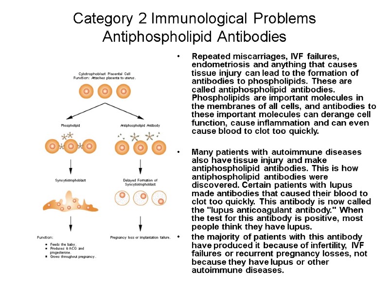
 Category 2 Immunological Problems Antiphospholipid Antibodies Repeated miscarriages, IVF failures, endometriosis and anything that causes tissue injury can lead to the formation of antibodies to phospholipids. These are called antiphospholipid antibodies. Phospholipids are important molecules in the membranes of all cells, and antibodies to these important molecules can derange cell function, cause inflammation and can even cause blood to clot too quickly. Many patients with autoimmune diseases also have tissue injury and make antiphospholipid antibodies. This is how antiphospholipid antibodies were discovered. Certain patients with lupus made antibodies that caused their blood to clot too quickly. This antibody is now called the "lupus anticoagulant antibody." When the test for this antibody is positive, most people think they have lupus. the majority of patients with this antibody have produced it because of infertility, IVF failures or recurrent pregnancy losses, not because they have lupus or other autoimmune diseases.
 The incidence of this problem increases in women by 15% with each pregnancy that is lost. It is a significant consequence of infertility, implantation failures and recurrent pregnancy losses. There are six different phospholipid molecules that have very important functions in cell membranes and intracellular organelles. The phospholipid molecules are Cardiolipin Ethanolamine Glycerol Inositol Phosphatidic Acid Serine Cell death or cell injury can lead to the production of antibodies to all or any one of these molecules. These antibodies disrupt cell functions and increase the clotting speed of blood. This can cause chaos early in pregnancy. Serine and Ethanolamine are phospholipids that serve as clue molecules in allowing the placenta to be securely attached to the uterus during implantation. They also allow the cytotrophoblast to change into a new cell, the syncytiotrophoblast, which begins to feed the baby by transporting nutrition from the mother's blood into the baby. Antibodies to these phospholipids prevent secure attachment or often totally prevent attachment. In addition, antibodies to these phospholipids prevent the cytophoblast from forming into the syncytiotrophoblast, which is needed to feed the baby. there is now a reason thatthis problem is 97% in causing pregnancies to fail early.
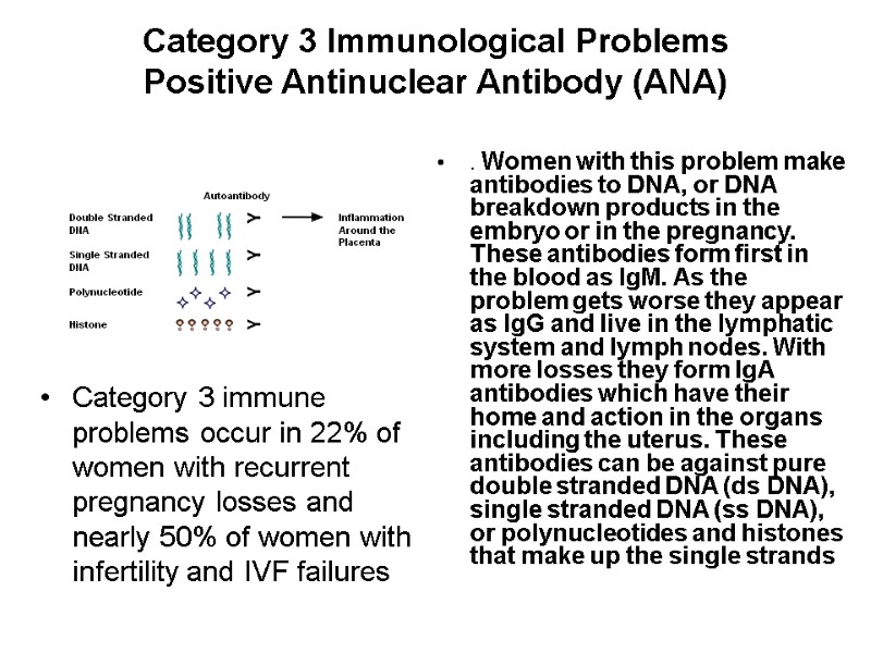
 Category 3 Immunological Problems Positive Antinuclear Antibody (ANA) Category 3 immune problems occur in 22% of women with recurrent pregnancy losses and nearly 50% of women with infertility and IVF failures . Women with this problem make antibodies to DNA, or DNA breakdown products in the embryo or in the pregnancy. These antibodies form first in the blood as IgM. As the problem gets worse they appear as IgG and live in the lymphatic system and lymph nodes. With more losses they form IgA antibodies which have their home and action in the organs including the uterus. These antibodies can be against pure double stranded DNA (ds DNA), single stranded DNA (ss DNA), or polynucleotides and histones that make up the single strands
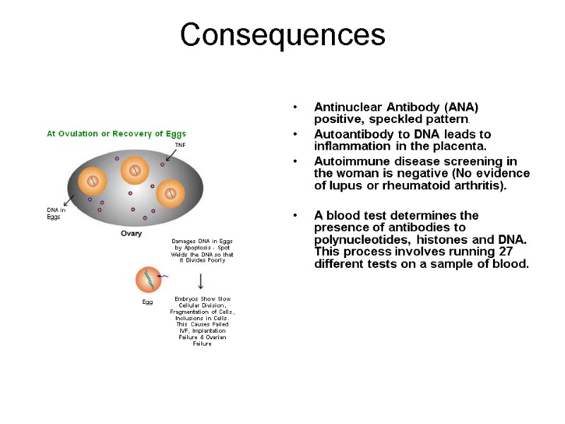
 Consequences Antinuclear Antibody (ANA) positive, speckled pattern. Autoantibody to DNA leads to inflammation in the placenta. Autoimmune disease screening in the woman is negative (No evidence of lupus or rheumatoid arthritis). A blood test determines the presence of antibodies to polynucleotides, histones and DNA. This process involves running 27 different tests on a sample of blood.
 Positive Antinuclear Antibody (ANA) Diagnosis The presence of antibodies is also tested for by doing the ANA test. This is a less sensitive test. The test is reported as a titer and a pattern. Any titer above 1:40 is significant. The titers can get into the thousands such as 1:2,500. The pattern is reported as homogeneous, nucleolar or speckled: Homogeneous: the antibody is to the ss DNA or ds DNA. Nucleolar: the antibody is directed to the polynucleotides. Speckled: the antibody is directed against the histones. Some women demonstrate a mixed pattern of speckled/homogeneous.
 Positive Antinuclear Antibody (ANA) Diagnosis (2) These same antibodies appear positive in women with lupus, rheumatoid arthritis, Crohn's disease and other autoimmune diseases. They are usually in high titers. Pregnancy losses, infertility and IVF failures cause the titers to be much lower. In women with no autoimmune diseases but a positive antibody, the antibody causes inflammation around the embryo at the time of implantation or in the placenta after implantation
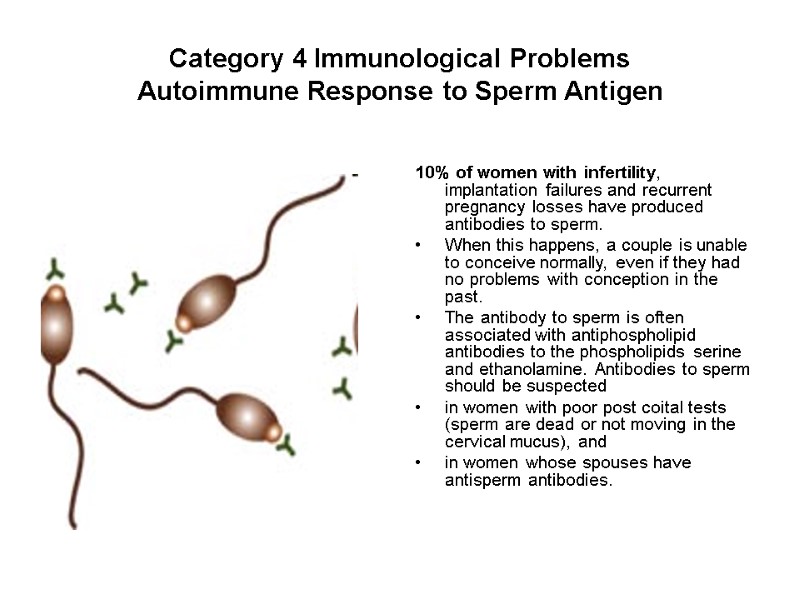
 Category 4 Immunological Problems Autoimmune Response to Sperm Antigen 10% of women with infertility, implantation failures and recurrent pregnancy losses have produced antibodies to sperm. When this happens, a couple is unable to conceive normally, even if they had no problems with conception in the past. The antibody to sperm is often associated with antiphospholipid antibodies to the phospholipids serine and ethanolamine. Antibodies to sperm should be suspected in women with poor post coital tests (sperm are dead or not moving in the cervical mucus), and in women whose spouses have antisperm antibodies.
 Autoimmune Response to Sperm Antigen (2) Being exposed to antibody coated sperm dispensed by the male seems to encourage women to make antisperm antibodies on their own. When antisperm antibodies develop, they will inactivate or attack sperm from the husband and any donor (i.e., they are not partner specific). Testing for antisperm antibodies in women is done from a blood sample. T here are more than five different methods to determine if antisperm antibodies are present. The most sensitive and reliable methods are immunobead binding antisperm antibody assay, and flow cytometry detection of antisperm antibodies. The presence of antisperm antibodies in women strongly predicts that she will also have category 5 immune problems.
 Consequences Sperm antibody test positive. Sperm antibody positive by flow cytometer. Couple is unable to conceive normally. Multiple failed pregnancy through IVF, IUI, GIFT or ZIFT.
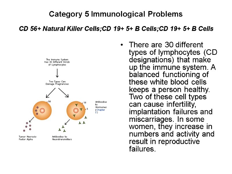
 Category 5 Immunological Problems CD 56+ Natural Killer Cells;CD 19+ 5+ B Cells;CD 19+ 5+ B Cells There are 30 different types of lymphocytes (CD designations) that make up the immune system. A balanced functioning of these white blood cells keeps a person healthy. Two of these cell types can cause infertility, implantation failures and miscarriages. In some women, they increase in numbers and activity and result in reproductive failures.
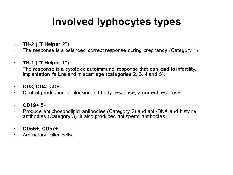
 Involved lyphocytes types TH-2 ("T Helper 2") The response is a balanced correct response during pregnancy (Category 1). TH-1 ("T Helper 1") The response is a cytotoxic autoimmune response that can lead to infertility, implantation failure and miscarriage (categories 2, 3, 4 and 5). CD3, CD4, CD8 Control production of blocking antibody response; a correct response. CD19+ 5+ Produce antiphospholipid antibodies (Category 2) and anti-DNA and histone antibodies (Category 3). It also produces antisperm antibodies. CD56+, CD57+ Are natural killer cells.
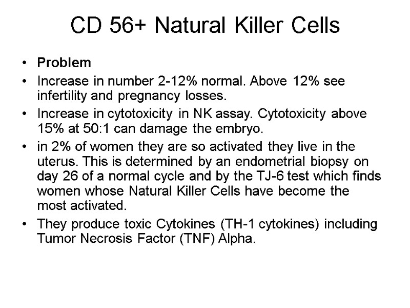
 CD 56+ Natural Killer Cells Problem Increase in number 2-12% normal. Above 12% see infertility and pregnancy losses. Increase in cytotoxicity in NK assay. Cytotoxicity above 15% at 50:1 can damage the embryo. in 2% of women they are so activated they live in the uterus. This is determined by an endometrial biopsy on day 26 of a normal cycle and by the TJ-6 test which finds women whose Natural Killer Cells have become the most activated. They produce toxic Cytokines (TH-1 cytokines) including Tumor Necrosis Factor (TNF) Alpha.
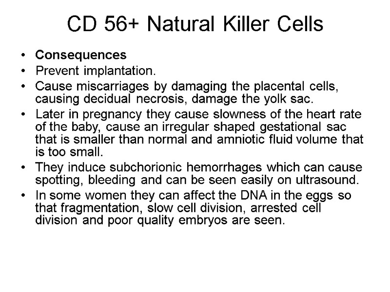
 CD 56+ Natural Killer Cells Consequences Prevent implantation. Cause miscarriages by damaging the placental cells, causing decidual necrosis, damage the yolk sac. Later in pregnancy they cause slowness of the heart rate of the baby, cause an irregular shaped gestational sac that is smaller than normal and amniotic fluid volume that is too small. They induce subchorionic hemorrhages which can cause spotting, bleeding and can be seen easily on ultrasound. In some women they can affect the DNA in the eggs so that fragmentation, slow cell division, arrested cell division and poor quality embryos are seen.
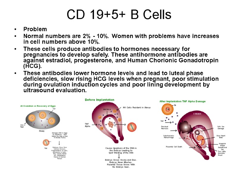
 CD 19+5+ B Cells Problem Normal numbers are 2% - 10%. Women with problems have increases in cell numbers above 10%. These cells produce antibodies to hormones necessary for pregnancies to develop safely. These antihormone antibodies are against estradiol, progesterone, and Human Chorionic Gonadotropin (HCG). These antibodies lower hormone levels and lead to luteal phase deficiencies, slow rising HCG levels when pregnant, poor stimulation during ovulation induction cycles and poor lining development by ultrasound evaluation.
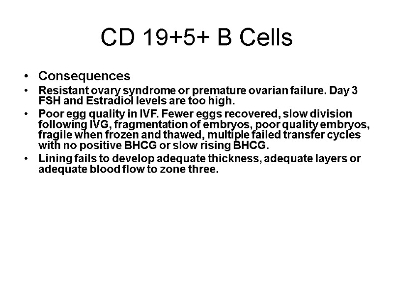
 CD 19+5+ B Cells Consequences Resistant ovary syndrome or premature ovarian failure. Day 3 FSH and Estradiol levels are too high. Poor egg quality in IVF. Fewer eggs recovered, slow division following IVG, fragmentation of embryos, poor quality embryos, fragile when frozen and thawed, multiple failed transfer cycles with no positive BHCG or slow rising BHCG. Lining fails to develop adequate thickness, adequate layers or adequate blood flow to zone three.
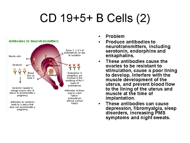
 CD 19+5+ B Cells (2) Problem Produce antibodies to neurotransmitters, including serotonin, endorphins and enkaphalins. These antibodies cause the ovaries to be resistant to stimulation, cause a poor lining to develop, interfere with the muscle development of the uterus, and prevent blood flow to the lining of the uterus and muscle at the time of implantation. These antibodies can cause depression, fibromyalgia, sleep disorders, increasing PMS symptoms and night sweats.
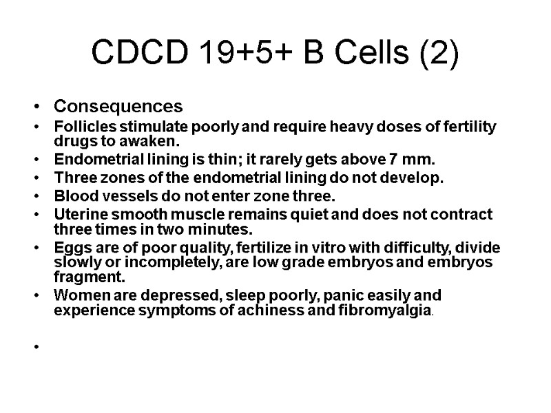
 CDCD 19+5+ B Cells (2) Consequences Follicles stimulate poorly and require heavy doses of fertility drugs to awaken. Endometrial lining is thin; it rarely gets above 7 mm. Three zones of the endometrial lining do not develop. Blood vessels do not enter zone three. Uterine smooth muscle remains quiet and does not contract three times in two minutes. Eggs are of poor quality, fertilize in vitro with difficulty, divide slowly or incompletely, are low grade embryos and embryos fragment. Women are depressed, sleep poorly, panic easily and experience symptoms of achiness and fibromyalgia.

