daae04d19344a21d3466eed34a19bbea.ppt
- Количество слайдов: 74


Il contenuto intracellulare di una cellula batterica è diviso tra due principali compartimenti nucleoide nel quale è situato il genoma citoplasma contenete l’apparato di sintesi proteica Quali sono i costituenti del nucleoide Genoma batterico RNA polimerasi Topoisomerasi Proteine basiche (definite Proteine Associate al Nucleoide NAP o Proteine Istone -simili)

IL NUCLEOIDE • la molecola di DNA cromosomico di E. coli è lunga circa 1. 6 mm • è contenuta in una cellula di 2 mm di lunghezza e 1 mm di larghezza • Un compattamento casuale della molecola determinerebbe un volume di circa 200 mm 3 circa 400 volte superiore al volume del nucleoide • Il volume del nucleoide è di circa di E. coli 0. 5 mm • Il cromosoma è quindi estremamente organizzato in anse topologicamente indipendenti circa 100 da 50 kb

Organizzazione del cromosoma batterico ad anse Il cromosoma potrebbe essere organizzato in domini topologicamante indipendenti , superavvolti negativamente. Analizzando il numero di tagli necessari per rilassare completamente il DNA è stato possibile valutare la quantità di topodomini presenti in E. coli (100). Tenendo conto che il cromosoma di E. coli è di 4. 600 kb la taglia media dei domini è valutata di circa 50 kb
 in vivo l’RNA")
Ancoraggi nel nucleoide batterico Componenti degli ancoraggi: • RNA? - 1) in vivo l’RNA nascente si osserva solo alla periferia del nucleoide + 2) in vitro l’RNasi rilassa il nucleoide (aumento di viscosità, diminuzione del coeff. di sedimentaz. ) + 3) Gli inbitori della trascrizione non influenzano il numero di domini cromosomici, ma i nucleoidi si rilassano più facilmente • Parete -Membrana? (replicazione) • Proteine istone-simili? (HU, H-NS, …) • DNA ancorato? Rilevanza funzionale in vivo?

Il cromosoma batterico è organizzato in numerosi domini superavvolti stabilizzati dal legame con proteine specifiche alla base dell’ansa. In Escherichia coli si calcolano circa 100 domini

Bacterial chromatin organization Plectonemic condition FIS IHF RNA polimerasi Toroidal condition H-NS HU Co-existence of plectonemic and toroidal conditions
 Anche nei batteri esistono proteine implicate nell’organizzazione")
Le proteine associate al nucleoide (NAP proteins) Anche nei batteri esistono proteine implicate nell’organizzazione strutturale del DNA. L’isolamento e la caratterizzazione delle proteine istone simili è risultato complesso • perchè in molti casi mostrano una stretta associazione con altre proteine prevalentemente quelle ribosomiali • perché il loro legame è meno sensibile all’aumento della forza ionica HU IHF FIS H-NS Dps Lrp Principali proteine associate all nucleoide
 Definite anche HLP (Histone-Like")
Struttura ed organizzazione delle prinicipali proteine associate al nucleoide (NAP) Definite anche HLP (Histone-Like Proteins)

La proteina HU Caratteristiche • proteina basica • molto abbondante 30000 copie/cellula • la più abbondante tra le proteine del nucleoide • nessuna sequenza consenso di legame al DNA Struttura p. M geni eterodimero Hua 9. 2 k. Da hup. A (90. 4 min) Hub 9. 5 k. Da hup. B ( 9. 9 min)

La proteina HU Funzioni • compatta il DNA in strutture nucleosoma-simili • induce curvatura nel DNA • riconosce il DNA curvo • interviene nella ricombinazione generale Isolata nella maggior parte delle specie batteriche proteine simili ad HU conservate anche negli Archea Anticorpi anti HU si legano sulla superficie del nucleoide piuttosto che al suo interno supportando l’ipotesi che HU si associ anche all’RNA e svolga un ruolo anche nella trascrizione e traduzione dei m. RNA
 adapted from Ussery et al. , 2001 N-domain (protein oligomerization Nucleoid")
C-domain (DNA binding) adapted from Ussery et al. , 2001 N-domain (protein oligomerization Nucleoid proteins are homo- or heterodimers

HU lega il DNA e lo ripiega

Superavvolgimento mediato da HU. I dimeri di HU si legano in vitro a distanza di circa 9 bp. In vitro associazione di 8 -10 dimeri con frammenti di 275 -290 bp Il legame di HU ogni 9 bp prevede interazioni con i dimeri adiacenti con formazione di una struttura solenoidale che si avvolge in senso sinistrorso. Ciascun avvolgimento toroidale avverrebbe intorno a 6 dimeri di HU

Le proteine che ripiegano il DNA

La proteina IHF Integration Host factor Caratteristiche • proteina basica • 5 -10 volte meno abbondante di HU • abbondante in fase stazionaria • debole specificità di sequenza per il legame al DNA (YAANNNNTTGATW) Struttura Eterodimero p. M geni IHFa 11. 2 k. Da him. A (38. 6 min) IHFb 10. 5 k. Da him. D ( 25 min)
")
IHF ; Caratteristiche funzionali • Induce forte curvatura nel DNA ( fino a 140°) • Interviene nella ricombinazione sito specifica • interviene nella trasposizione Si lega al solco minore del DNA utilizzando due foglietti B che in opposizione l’uno all’altro prendono contatto con il DNA • Isolata come fattore dell’ospite per l’integrazione del fago l è coinvolta in molte funzioni cellulari. • Forte ruolo strutturale; curvando il DNA fino a 140°C permette a siti di DNA distanti di trovarsi ravvicinati favorendo cosi sia processi di trascrizione che di regolazione

La curvatura mediata da IHF può facilitare il legame dellla Proteina Int con siti di DNA distanti tra loro.
3 - 6")
Intrinsically curved DNA Roll r Wedge model Tilt t e. g. GC(A/T)3 - 6 GCTCG(A/T)3 - 6 GCG Twist Junction model W

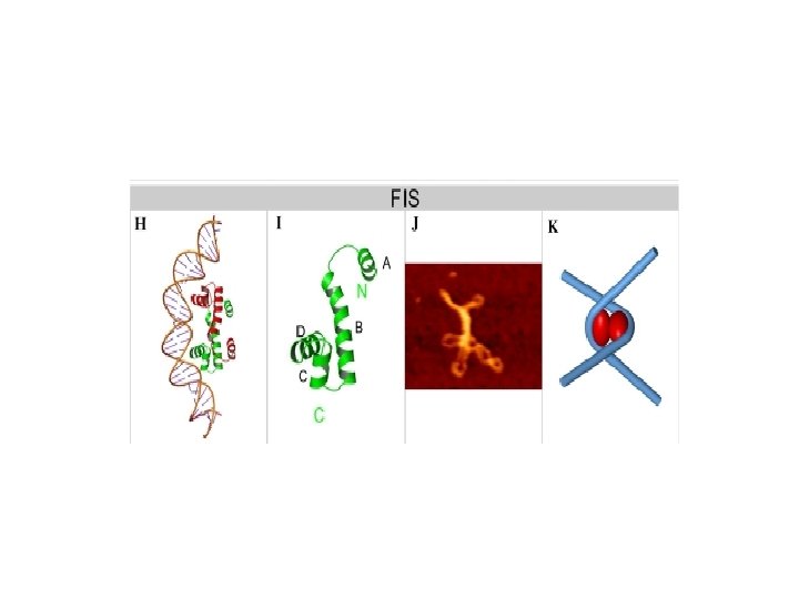
FIS Factor for Inversion Stimulation Caratteristiche • Proteina basica • abbondante in fase esponenziale • 10. 000 -60. 000 copie • scarsa specificità di sequenza (KNNYRNNWNNYRNNM) Struttura p. M geni OMODIMERO FIS 2 x 11. 5 k. Da fis ( 27. 4 min) W R K Y TA GA GT CT

L’inversione di fase in Salmonella è mediata dalla proteina Hin e dalla proteina Fis dell’ospite

Proteine del nucleoide come regolatori globali Potenziali siti dilegame per IHF Potenziali siti di legame per FIS

Espressione di IHF e FIS in funzione della curva di crescita La concentrazione di IHF aumenta in funzione della curva di crescita e raggiunge il massimo all’inizio della fase stazionaria La proteina FIS è invece molto abbondante all’inizio della fase esponenziale
 • Abundant peptide (~ 20000 copies/cell) • Small (136")
H-NS (Histone-like Nucleoid Structuring protein) • Abundant peptide (~ 20000 copies/cell) • Small (136 aminoacids, 15. 5 k. D), non-basic peptide • Mainly acts as a homodimer or tetramer • Able to form heterodimers with Stp. A or Hh. A • In vitro binding to DNA is non-specific; induces high DNA compaction • Higher affinity for intrinsically curved DNA; able to bend DNA in vitro • Global regulator: controls 5 % of the whole E. coli protein coding sequences • Generally acts as a transcriptional repressor of virulence genes outside the host

La proteina H-NS riconosce sequenze di DNA curvo ed è in grado di indurre curvatura nel DNA

Le proteine NAP che oltre a ripiegare il DNA formano ponti : un esempio H-NS viene considerata una proteine in grado di formare ponti A) B) C) D) Strutture proposte per il dimero di H-NS Analisi dei monomeri di H-NS Formazione delle anse mediate da H-NS 2 diversi modelli di interazione nella formazione complessi Altre proteine NAP che formano ponti su LRP e Mu. KB

Struttura della proteina H-NS La proteina H-NS è costituita da soli 137 AA Il dominio di oligomerizzazione è localizzato al N terminale ed è costituito da brevi sequenze di AA ( 1 -8, 12 -19, e 23 -47) capaci di formare 3 strutture ad alfa elica. I linker flesibili che separano le 3 a eliche permettono alle eliche 1 e 2 di ripiegarsi facilitando la formazione di oligomeri tra i diversi dimeri

H-NS ed il silenziamento di regione geniche acquisite per HGT Il legame di H-NS a regioni di DNA esogeno ricche in AT silenzia l’espressione genica. L’eventuale presenza di una proteina regolatrice sequenza specifica con un elevata affinità per il DNA può competere con H-NS per eliminare il silenziamento e permettere nuovamente l’espressione genica in condizioni specifiche. In questo modo, la cellula ospite può tollerare la presenza di sequenze di DNA estraneo e in seguito inserire la sequenza in un network di regolazione preesistente

H-NS e il silenziamento dei geni acquisiti tramite HGT

Concentrazione delle proteine del nucleoide in funzione della curva di crescita
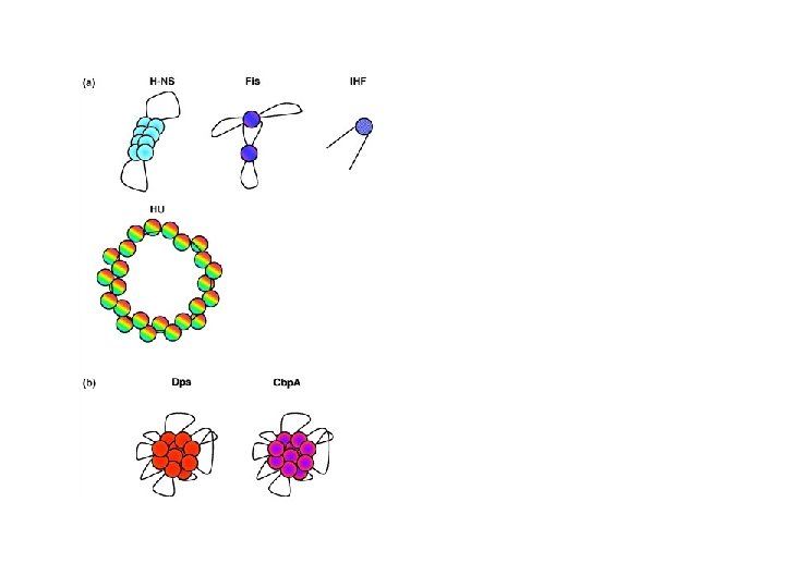

Bacterial chromatin organization FIS IHF H-NS HU H-NS • Small (136 aminoacids, 15. 5 k. D), abundant peptide (~ 20000 copies/cell) • Mainly acts as a homodimer or tetramer • Higher affinity for intrinsically curved DNA; able to bend DNA in vitro • Global regulator: controls 5 % of the whole E. coli protein coding sequences

Shigella • is a Gram negative, facultative anaerobe • is an intracellular pathogen • is the etiological agent of bacillary dysentery, an acute diarrheal disease • causes 160 million of episodes, determining 1. 1 million deaths/year in children and infants in developing countries. Infection is spread via fecal-oral route Subgrouped in four “species”: • Shigella flexneri • Shigella dysenteriae • Shigella boydii • Shigella sonnei

Model for Shigella invasion of the colonic mucosa Bacteria Enterocytes M cells Inflammatory cytokines cascade Polymorphonuclear leucocytes

Ipa. D Proteins involved in the invasion process are encoded by a virulence plasmid (p. INV) Ipa. B, Ipa. C Ics. A (vir. G) Ics. B Actin filaments nucleus

Genetic organization of p. INV vir. F is. . . • … located on a “desert island” • … the first positive activator of p. INV virulence genes vir. B is. . . • … located within the main Pathogenicity Island … the second positive regulator of the plasmid virulence regulons IS elements virulence genes

H-NS controls the virulence regulon in Shigella and in E. coli EIEC bacterial chromosome hns H-NS vir. G vir. B ipa mxi spa Virulence plasmid (p. INV, 220 -260 kb) vir. F

The expression “cascade” of virulence genes FIS Vir. F is. . . • … expressed at 37°C • … is controlled antagonistically by two nucleoid proteins H-NS (repressor) and FIS (activator) By which mechanism is the vir. F gene allowed to be expressed only at the host temperature?

H-NS is able to bind to and repress vir. F only at low temperature …more precisely: vir. F transcription is inhibited by H-NS only below 32°C

DNase. I footprinting of the vir. F promoter region by H-NS

The vir. F promoter region +1 H-NS box II H-NS box I

Curvature in H-NS regulated vir promoters vir. F vir. B vir. G bp 500 vir. F vir. B vir. G 400 60°C gel 4°C gel 300 200 Computer-generated models vir. F vir. B vir. G

Within the vir. F promoter region H-NS recognizes two sites separated by a region endowed with signficant intrinsic curvature -140 -345 DNA bending : the wedge model for sequence mediated curvature H-NS site II -278 / -274 to -234 / -218 +1 H-NS site I -54 / -46 to +33 / +46 +104

4°C 500 400 300 200 100 28°C 32°C 40°C 60°C hns vir. F B vir. F AB vir. F A hns vir. F B vir. F A bp 900 800 700 600 20°C hns vir. F B vir. F A Temperature-dependent curvature of the vir. F promoter Acrilamide separations

The curvature of the vir. F promoter is strongly temperature-dependent White symbols: Mg++ Black symbols: no Mg++

Temperature-dependent curvature of the vir. F promoter TGGE separation 700 600 500 400 700 300 600 500 200 400 100 bp 300 39°C 36°C 33. 5°C 31°C 29. 5°C 27°C 24. 5°C 22. 5°C 20°C 17°C 39°C 17°C 200 bp

Temperature-dependent vir. F expression Working model 37°C 30°C H-NS RNA polymerase

By which mechanism is the vir. F gene allowed to be expressed only at the host temperature? Small RNAs are emerging as key regulators of virulence gene expression in bacteria. Is this true also in Shigella?

The circular permutation assay Rationale Step 1 B C A Identification of unique restriction sites (A, B, C, D) in putative bent region Step 2 B C A D D A Dimerization B C D Step 3 A B C A D B C A Restriction A C A D D A B B B Restriction B D A B Restriction C C D D A A D B C Restriction D B C D

Circular permutation assay on the vir. F promoter region The bending centre maps halfway between the H-NS boxes and is located ~140 bp upstream the transcription start site (+1)

Molecular dissection of the vir. F promoter: mutagenesis of the b-region and shifting the b-region by ~½helix turn HI Bg l. I m Ba HI m Ba Ec I o. R ) ite a NS (H Is I Ba b m. H I +4 /+ 6 bp g( Bg HN S II s ite 8 -oxo-d. GTP d. PTP ) l. II

Mutagenesis of the bent region Mutagens used 8 -oxo-d. GTP d. PTP

Mutagenesis of the bent region Strategy for the construction of b-region mutants b b’ vir. F promoter PCR in the presence of d. PTP and 8 -oxod. GTP a PCR g p. Flacag lac. Z Transform E. coli DH 10 b and select on Ap-Xgal b’’
 of b-region mutants")
Acrilamide separation (4°C) of b-region mutants

Correlation between intrinsic curvature and thermoregulated expression of the vir. F promoter H-NS box I bending centre F 19 F 22 F 38 F 40 Computer predictions F 53 H-NS box II

DNAse. I footprints reveal that H-NS: • recognizes a wider site I in the strongly bent mutant (F 53) • is unable to recognize site II in the Fb+6 mutant F 53 X Fb+6 X

The vir. F gene is positively regulated by FIS, another nucleoid protein FIS (Factor for Inversion Stimulation) • Basic • homodimer (2 x 11. 5 k. Da) • abundant in exponential phase • very weak DNA sequence specificity • transcriptional regulator • participates in site-specific recombination and transposition

FIS has four binding sites within the vir. F promoter … … and alleviates H-NS-mediated repression of the vir. F promoter at 31°C In vitro transcription in the presence of both, H-NS and FIS

Identification of FIS binding sites on the vir. F promoter region

Circular permutation assay on the vir. F promoter region Effect of temperature Curvature is reduced as temperature increases Do temperature changes alter the position of the bending centre?

… yes, the bending centre of the vir. F promoter shifts considerably with temperature • The displacement of the bending centre is not a linear function of temperature. • The maximum displacement occurs between 28°C and 32°C.

The thermodependent expression of vir. F is mediated by changes in DNA bending of its promoter < 28°C 28 - 32 °C H-NS and its binding sites FIS and its binding sites RNA polymerase Bending center > 32°C

Does DNA curvature regulate virulence genes as a function of host temperature also in other bacterial pathogens? The Yersinia enterocolitica virulence plasmid contains DNA bends which melt at 37°C (Rohde et al. ) In pathogenic E. coli the plasmid hemolysin operon is regulated by temperature-dependent binding of H-NS to curved DNA (Madrid et al. )

From pathogenic bacteria to E. coli : How far does intrinsic DNA curvature sustain bacterial transcription?

The experimental approach The 211 bp fragment from the kinetoplast DNA of Crithidia fasciculata, known to be endowed with strong curvature, has been randomly mutagenized in order to obtain a spectrum of fragments covering a wide curvature range. Then, mutagenized fragments were cloned upstream (-45) a reporter gene and … k= Apparent size Real size

… their temperature-dependent transcription profiles were analyzed in vitro. . . UNBENT Real-time florescence assay REFERENCE

In short: Curved DNA regions are frequently located upstream bacterial promoters. Their marked temperature-sensitivity makes them excellent candidates as transcriptional modulators responding to environmental stimuli. A narrow range of curvature is able to sustain bacterial transcription in vitro.

It is known that H-NS does not. . . • … undergo chemical modifications; • … undergo structural transitions; • … display temperature-dependent dimerization or tetramerization kinetics.

30°C 37°C wt 30°C 37°C hns 118 37°C 30°C 37°C hns 2 37°C 30°C Distribution of p. MYSH 6504 topoisomers in hns + and hns strains at 30°C and at 37°C 30°C

In vivo and in vitro effect of FIS on the expression of vir. F at 37°C

Influence of FIS on the H-NS mediated repression of the vir. F promoter at 31°C

L’intuizione di Carl Woese di utilizzare il gene che codifica per RNA 16 S per la costruzione degli alberi filogenetici è ancora valida e resta un ‘analisi necessaria per stabilire la filogenesi tra gruppi tassonomici. La struttura dell’RNA ribosomiale è tale che alcune regioni implicate nella formazione delle strutture tridimensionali complesse sono estremamente conservate e le loro sequenze si sono evolute lentamente differenziandosi molto poco tra gruppi filogeneticamente distanti

daae04d19344a21d3466eed34a19bbea.ppt