0a7e1dabe2bc0489ef9c55fb50db70ea.ppt
- Количество слайдов: 64
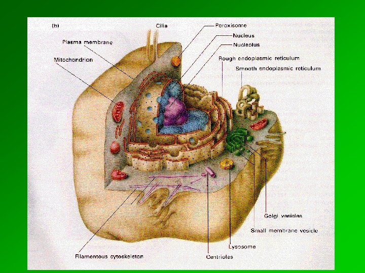
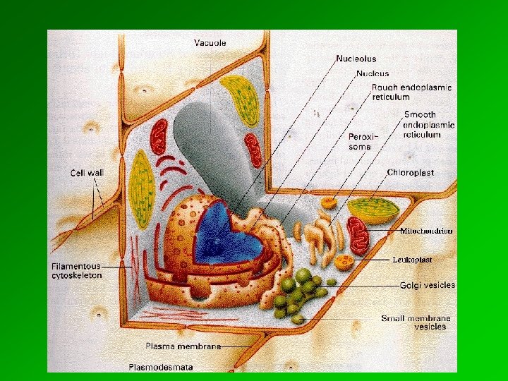
 II. INTERNAL ORGANIZATION OF EUKARYOTIC CELLS


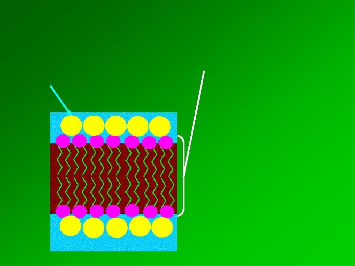
 Micelle Liposome Bilayer sheet

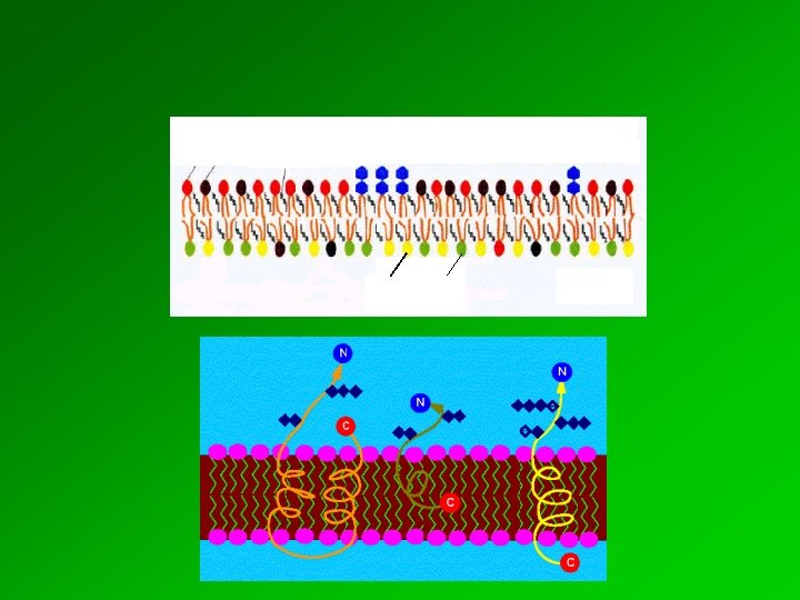
 Extracellular space Cytosol
 a. Transmembrane helices b. association by covalently attached lipid c. Interaction with transmembrane protein

 Plasma membrane Intercellular space

 Transport proteins Anchoring proteins Receptors Enzymes

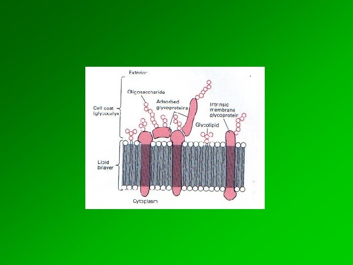
 Plasma membrane Attachment site of the glycocalyx on the plasma membrane The glycocalyx is exceptionally well developed on the surface of the intestinal epithelium.

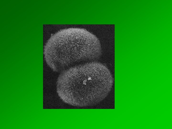
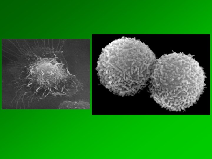
 Sensory epithelium

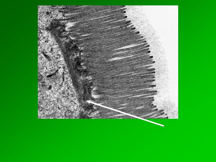
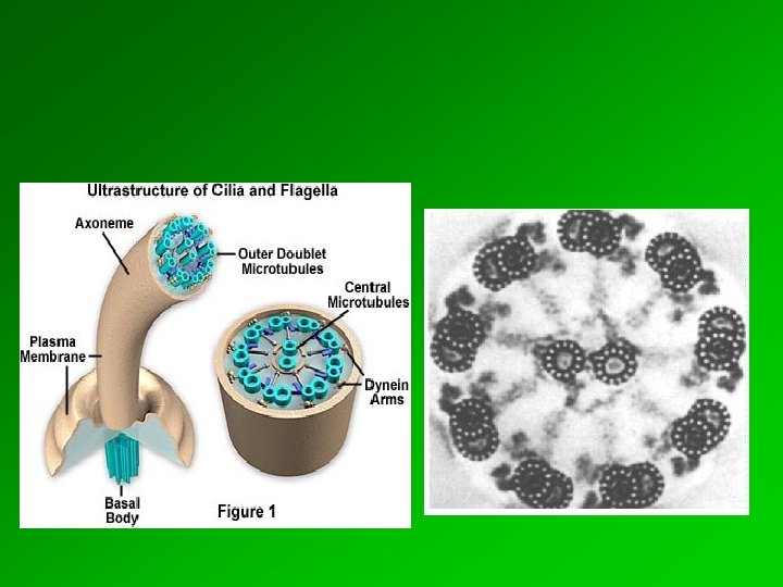
 basal body kinocilia and microvilli Cilia are parallelly oriented, motile, finger-like protrusions of cell membrane (Ø 300 nm, length 7 - 10 µm), connected to an electron dense basal body.


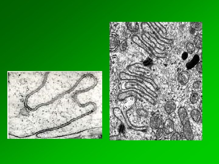
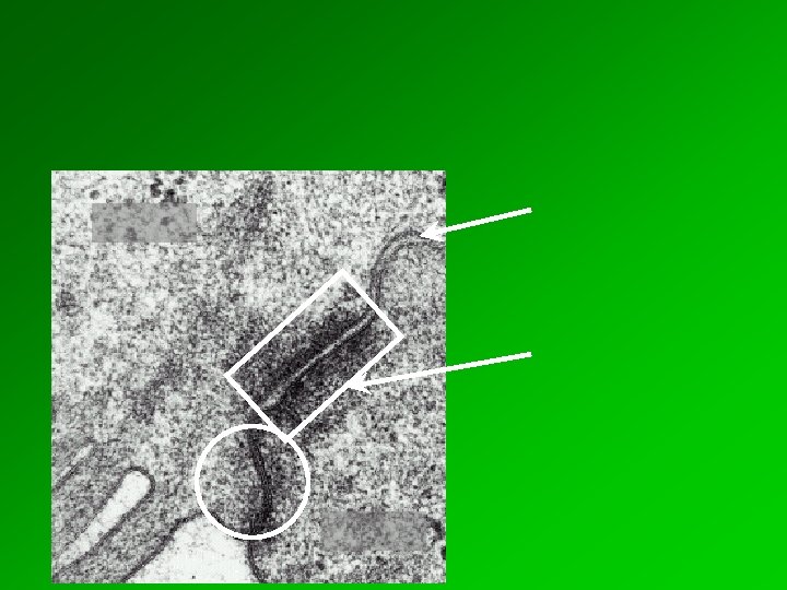
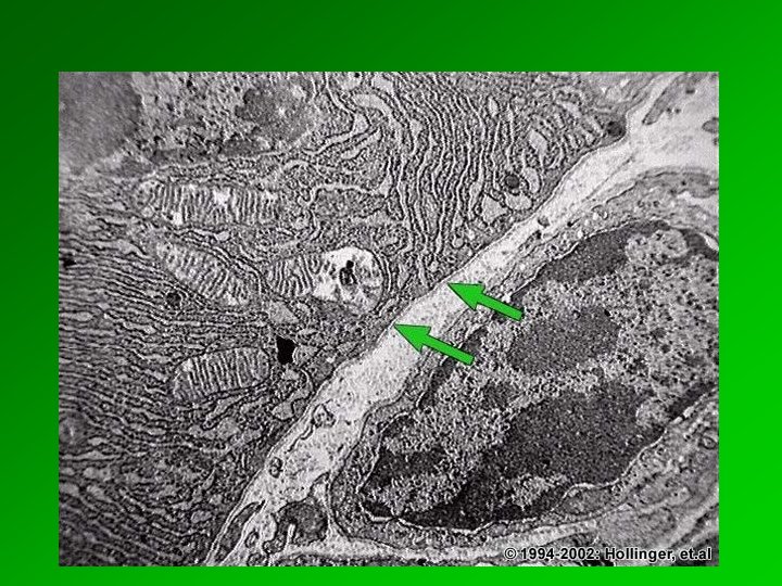
 Zonula occludens") Intercellular space (Spatium intercellulare) Zonula occludens
Intercellular space (Spatium intercellulare) Zonula occludens
 Integral membrane proteins of neigbouring cell membranes fuse with each other.
 Cell 2 Actin cytoskeleton Cadherin Cell 1
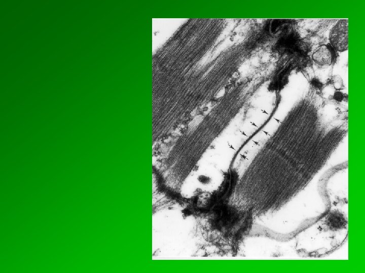
 Spot desmosome is a disk-like structure. Electron-dense material is present in the extracellular space, that often forms a central line. It can be found between epithelial cells, cardiac muscle cells and several other cell types. Intercellular space
 Cadherin EM Intermediary filaments Plaques
 Plasma membranes
 form connexins a channel (connexon). Connexons of two connexon adjacent") Plasma membrane proteins (connexins) form connexins a channel (connexon). Connexons of two connexon adjacent cell membranes join together in the intercellular space to constitute a continuous hidrophilic channel, the nexus
Plasma membrane proteins (connexins) form connexins a channel (connexon). Connexons of two connexon adjacent cell membranes join together in the intercellular space to constitute a continuous hidrophilic channel, the nexus
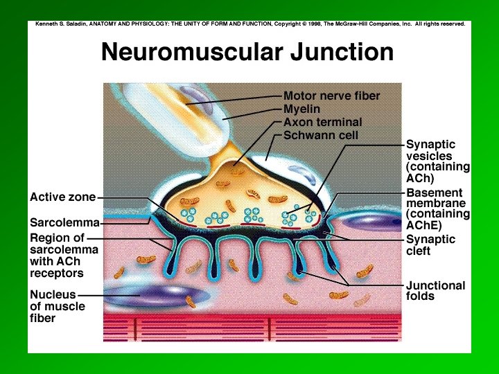
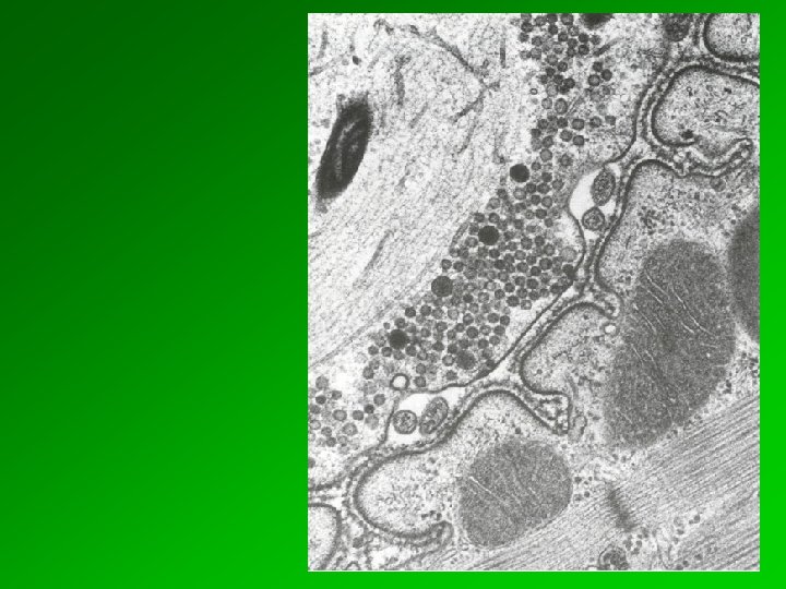
 vesicles presynaptic membrane synaptic cleft postsynaptic membrane thickening") Presynaptic site (neurotransmitter containing) vesicles presynaptic membrane synaptic cleft postsynaptic membrane thickening
Presynaptic site (neurotransmitter containing) vesicles presynaptic membrane synaptic cleft postsynaptic membrane thickening
 plasma membrane basal laminae filaments of the") cytoskeletal filaments hemidesmosome (consists of 2 plaques) plasma membrane basal laminae filaments of the ecm
cytoskeletal filaments hemidesmosome (consists of 2 plaques) plasma membrane basal laminae filaments of the ecm


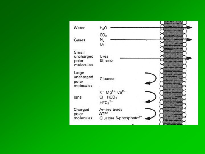
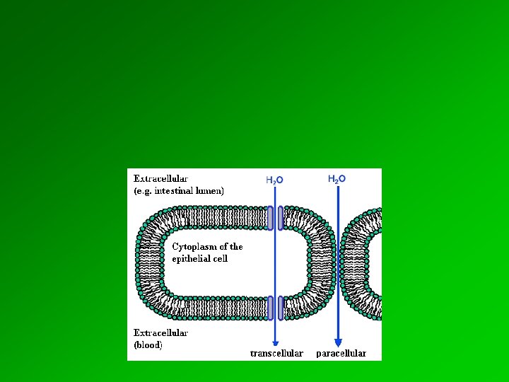
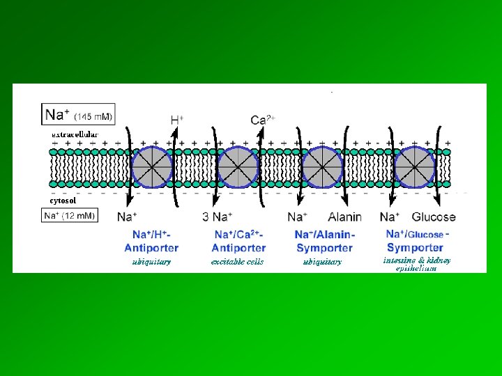
chemical gradient of an ion or a molecule through the membrane") Definition: (electro)chemical gradient of an ion or a molecule through the membrane
Definition: (electro)chemical gradient of an ion or a molecule through the membrane
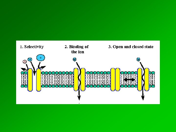
 2. Conformational change of the protein enables the substrate to cross the membrane 3. Dissociation of the substrate molecule


 Sig na l r ec cellular response
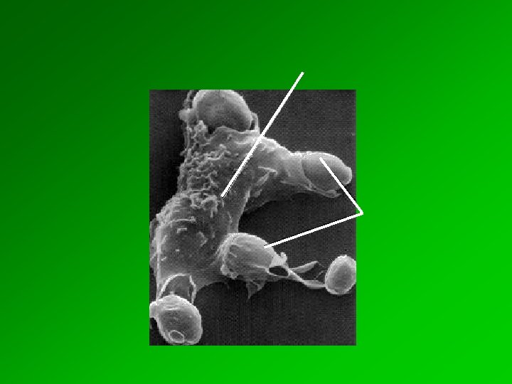
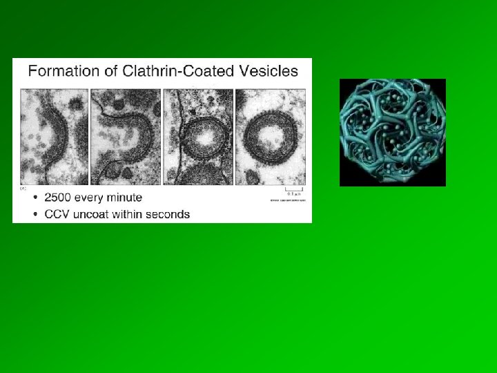
 Receptor mediated endocytosis