

Cell Membrane Fluid Mosaic.pptx
- Количество слайдов: 56
 Ideally, we need a scale we can see directly alongside the cells we are observing:
 The Cell Membrane
 At the end of this lesson, you should be able to: • Describe the function of the plasma membrane. • Describe the fluid mosaic model of membrane structure. • Explain how hydrophobic interactions determine membrane structure and function. • Describe how proteins are arranged in membranes and how they contribute to membrane functioning.
 Fig. 8. 6 Copyright © 2002 Pearson Education, Inc. , publishing as Benjamin Cummings
 Overview • The functions of the cell membrane depend on its structure. • The different components/structures determine the cell membrane’s various functions. • The fluid-mosaic model is the widely recognized and accepted model of the cell membrane.
 What’s in it? What are the different components of the cell membrane?
 Membrane is a collage of proteins & other molecules embedded in the fluid matrix of the lipid bilayer Glycoprotein Extracellular fluid Glycolipid Phospholipids Cholesterol Peripheral protein Cytoplasm Transmembrane proteins Filaments of cytoskeleton
 What are the different components of the cell membrane? • lipids • proteins • carbohydrates
 Amphipathic = has both hydrophilic and hydrophobic parts
 Phospholipids • Fatty acid tails Phosphate – hydrophobic • Phosphate group head – hydrophilic • arranged as a bilayer Fatty acid Aaaah, one of those structure–function examples
 Phospholipid bilayer polar hydrophilic heads nonpolar hydrophobic tails polar hydrophilic heads
 Behavior: • fluid • mobile
 Behavior: • form vesicles rather than free ends • can reseal to form intact membranes
 Behavior:
 Membrane Fat Composition Varies! • % unsaturated fatty acids keep the bilipid layer fluid • The number of unsaturated fatty acids in increases in autumn for cold-adapted organisms.
 Cholesterol makes the bilipid layer more fluid.
 More than lipids… • In 1972, S. J. Singer & G. Nicolson proposed that membrane proteins are inserted into the phospholipid bilayer It’s like a fluid… It’s like a mosaic… It’s the Fluid Mosaic Model!
 Why are proteins the perfect molecule to build structures in the cell membrane? 2007 -2008
 Membrane Proteins • Proteins determine membrane’s specific functions – cell membrane & organelle membranes each have unique collections of proteins • Membrane proteins: – peripheral proteins • loosely bound to surface of membrane • cell surface identity marker (antigens) – integral proteins • penetrate lipid bilayer, usually across whole membrane • transmembrane protein • transport proteins – channels, permeases (pumps)
 Many Functions of Membrane Proteins Outside Plasma membrane Inside Transporter Enzyme activity Cell surface receptor Cell surface identity marker Cell adhesion Attachment to the cytoskeleton
 • The proteins in the plasma membrane may provide a variety of major cell functions. Fig. 8. 9
 Classes of amino acids What do these amino acids have in common? nonpolar & hydrophobic
 Classes of amino acids What do these amino acids have in common? I like the polar ones the best! polar & hydrophilic
 Proteins domains anchor molecule • Within membrane Polar areas of protein – nonpolar amino acids • hydrophobic • anchors protein into membrane • On outer surfaces of membrane – polar amino acids • hydrophilic • extend into extracellular fluid & into cytosol Nonpolar areas of protein
 Examples Retinal chromophore H+ NH 2 water channel in bacteria Porin monomer b-pleated sheets Bacterial outer membrane Nonpolar (hydrophobic) a-helices in the cell membrane COOH H+ Cytoplasm proton pump channel in photosynthetic bacteria function through conformational change = shape change
 Membrane carbohydrates • Play a key role in cell-cell recognition – ability of a cell to distinguish one cell from another • antigens – important in organ & tissue development – basis for rejection of foreign cells by immune system
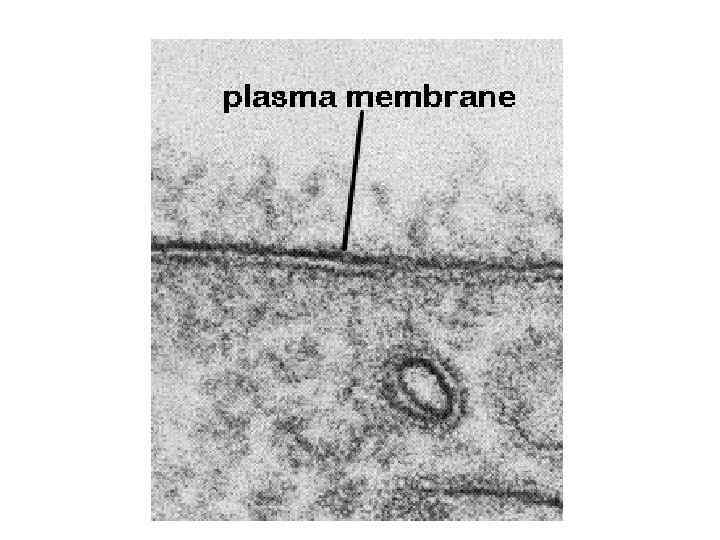
 Summary • Cell membrane separates living cell from nonliving surroundings – thin barrier = 8 nm thick • Controls traffic in & out of the cell – selectively permeable – allows some substances to cross more easily than others • hydrophobic vs. hydrophilic • Made of phospholipids, proteins& other macromolecules
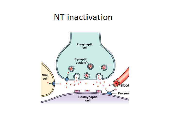
 Functions of the plasma membrane: • acts like the “skin of the cell” • separates the intracellular components from the cell’s environment (extracellular fluid) • controls the traffic of substances in and out of the cell (semi-permeable) • participates in signal transduction • provides an ID to the cell (cell recognition)
 Any Questions? ?
 Movement across the Cell Membrane 2007 -2008
 Diffusion • 2 nd Law of Thermodynamics governs biological systems – universe tends towards disorder (entropy) § Diffusion u movement from high low concentration
 Diffusion • Move from HIGH to LOW concentration – “passive transport” – no energy needed diffusion movement of water osmosis
 Diffusion across cell membrane • Cell membrane is the boundary between inside & outside… – separates cell from its environment Can it be an impenetrable boundary? OUT IN food carbohydrates sugars, proteins amino acids lipids salts, O 2, H 2 O NO! OUT IN cell needs materials in& products or waste out waste ammonia salts CO 2 H 2 O products
 Diffusion through phospholipid bilayer • What molecules can get through directly? – fats & other lipids § What molecules can NOT lipid inside cell salt NH 3 get through directly? u polar molecules § H 2 O u ions § salts, ammonia u outside cell sugar aa H 2 O large molecules § starches, proteins
 Channels through cell membrane • Membrane becomes semi-permeable with protein channels – specific channels allow specific material across cell membrane inside cell NH 3 H 2 O salt aa sugar outside cell
 Facilitated Diffusion • Diffusion through protein channels – channels move specific molecules across cell membrane facilitated = with help – no energy needed open channel = fast transport high low “The Bouncer”
 Active Transport • Cells may need to move molecules against concentration gradient – shape change transports solute from one side of membrane to other – protein “pump” conformationalchange – “costs” energy = ATP low ATP high “The Doorman”
 Active transport • Many models & mechanisms ATP antiport symport
 Getting through cell membrane • Passive Transport – Simple diffusion • diffusion of nonpolar, hydrophobic molecules – lipids – high low concentration gradient – Facilitated transport • diffusion of polar, hydrophilic molecules • through a protein channel – high low concentration gradient • Active transport – diffusion against concentration gradient • low high – uses a protein pump – requires ATP
 Transport summary simple diffusion facilitated diffusion active transport ATP
 How about large molecules? • Moving large molecules into & out of cell – through vesicles & vacuoles – endocytosis • phagocytosis = “cellular eating” • pinocytosis = “cellular drinking” – exocytosis
 Endocytosis phagocytosis pinocytosis receptor-mediated endocytosis fuse with lysosome for digestion non-specific process triggered by molecular signal
 The Special Case of Water Movement of water across the cell membrane 2007 -2008
 Osmosis is diffusion of water • Water is very important to life, so we talk about water separately • Diffusion of water from high concentration of water to low concentration of water – across a semi-permeable membrane
 Concentration of water • Direction of osmosis is determined by comparing total solute concentrations – Hypertonic - more solute, less water – Hypotonic - less solute, more water – Isotonic - equal solute, equal water hypotonic hypertonic net movement of water
 Managing water balance • Cell survival depends on balancing water uptake & loss freshwater balanced saltwater
 Managing water balance • Isotonic – animal cell immersed in mild salt solution • example: blood cells in blood plasma • problem: none – no net movement of water » flows across membrane equally, in both directions – volume of cell is stable balanced
 Managing water balance • Hypotonic – a cell in fresh water • example: Paramecium • problem: gains water, swells & can burst – water continually enters Paramecium cell ATP • solution: contractile vacuole – pumps water out of cell – ATP – plant cells • turgid freshwater
 Water regulation • Contractile vacuole in Paramecium ATP
 Managing water balance • Hypertonic – a cell in salt water • example: shellfish • problem: lose water & die • solution: take up water or pump out salt – plant cells • plasmolysis= wilt saltwater
 Aquaporins 1991 | 2003 • Water moves rapidly into & out of cells – evidence that there water channels Peter Agre John Hopkins Roderick Mac. Kinnon Rockefeller
 hypertonic or hypotonic") Osmosis… . 05 M . 03 M Cell (compared to beaker) hypertonic or hypotonic Beaker (compared to cell) hypertonic or hypotonic Which way does the water flow? in or out of cell
Osmosis… . 05 M . 03 M Cell (compared to beaker) hypertonic or hypotonic Beaker (compared to cell) hypertonic or hypotonic Which way does the water flow? in or out of cell
 Any Questions? ?