6ee94ef77a687d6d0f37dfe82400eb7f.ppt
- Количество слайдов: 112


Human Physiology: Cell Structure and Function BY DR BOOMINATHAN Ph. D. M. Sc. , (Med. Bio, JIPMER), M. Sc. , (FGS, Israel), Ph. D (NUS, SINGAPORE) PONDICHERRY UNIVERSITY Source: Collected from different sources on the internet-http: //koning. ecsu. ctstateu. edu/cell. html

Anatomy, physiology, … • Anatomy is the science of the structure • Physiology is the science of the function • Anatomy and physiology are closely linked, in particular physiology cannot be understood without anatomy • In many respects, both are ‘closed sciences’

Physiology • Some important moments: • 17 th century: William Harvey first describes the closed circulation • 19 th century: Claude Bernard formulates the modern version of homeostasis – the constancy of the internal milieu • 19 th century: Johannes Muller formulates the ‘law of specific nerve energy’

Physiology • Some important moments: • 17 th century: William Harvey first describes the closed circulation • 19 th century: Claude Bernard formulates the modern version of homeostasis – the constancy of the internal milieu • 19 th century: Johannes Muller formulates the ‘law of specific nerve energy’ • In general, a slow development of our modern view of the function of the body

Systems physiology: Missing from the scheme: Structure and motion: • Skeletal system • Muscles Integratory systems: • Nervous system • Hormones

Cell Structure & Function Source: http: //koning. ecsu. ctstateu. edu/cell. html

Unit-I Outline • Levels of Cellular Organization & function. Organelles, tissues, organs & systems. • Cell theory • Properties common to all cells • Cell size and shape – why are cells so small? • Prokaryotic cells • Eukaryotic cells – Organelles and structure in all eukaryotic cell – Organelles in plant cells but not animal • Cell junctions

History of Cell Theory • mid 1600 s – Anton van Leeuwenhoek – Improved microscope, observed many living cells • mid 1600 s – Robert Hooke – Observed many cells • 1850 – Rudolf Virchow – Proposed that all cells come from existing cells

Cell Theory Cells were discovered in 1665 by Robert Hooke. Early studies of cells were conducted by - Mathias Schleiden (1838) - Theodor Schwann (1839) Schleiden and Schwann proposed the Cell Theory. 9

Cell Theory 1. All organisms consist of 1 or more cells. 2. Cell is the smallest unit of life. 3. All cells come from pre-existing cells.

Cell Theory • All living things are made up of cells. • Cells are the smallest working units of all living things. • All cells come from preexisting cells through cell division.

Cell Theory 1. All organisms are composed of cells. 2. Cells are the smallest living things. 3. Cells arise only from pre-existing cells. All cells today represent a continuous line of descent from the first living cells. 12

Cell Theory Cell size is limited. -As cell size increases, it takes longer for material to diffuse from the cell membrane to the interior of the cell. Surface area-to-volume ratio: as a cell increases in size, the volume increases 10 x faster than the surface area 13

Cell Theory 14

Cell Theory Microscopes are required to visualize cells. Light microscopes can resolve structures that are 200 nm apart. Electron microscopes can resolve structures that are 0. 2 nm apart. 15

Cell Theory All cells have certain structures in common. 1. genetic material – in a nucleoid or nucleus 2. cytoplasm – a semifluid matrix 3. plasma membrane – a phospholipid bilayer 16

Definition of Cell A cell is the smallest unit that is capable of performing life functions.
 • Light microscope – Can observe living cells in true")
Observing Cells (4. 1) • Light microscope – Can observe living cells in true color – Magnification of up to ~1000 x – Resolution ~ 0. 2 microns – 0. 5 microns
 • Electron Microscopes – Images are black and white –")
Observing Cells (4. 1) • Electron Microscopes – Images are black and white – may be colorized – Magnifcation up to ~100, 000 • Transmission electron microscope (TEM) – 2 -D image • Scanning electron microscope (SEM) – 3 -D image

SEM TEM

Examples of Cells Amoeba Proteus Plant Stem Bacteria Red Blood Cell Nerve Cell

Two Types of Cells • Prokaryotic • Eukaryotic

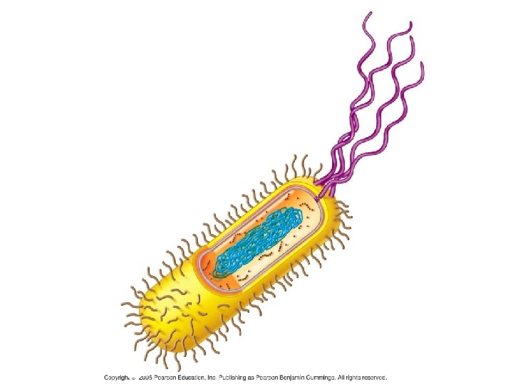
Prokaryotic • Do not have structures surrounded by membranes • Few internal structures • One-celled organisms, Bacteria http: //library. thinkquest. org/C 004535/prokaryotic_cells. html

Prokaryotic Cells Prokaryotic cells lack a membrane-bound nucleus. -genetic material is present in the nucleoid Two types of prokaryotes: -archaea -bacteria 24

Prokaryotic Cells Prokaryotic cells possess -genetic material in the nucleoid -cytoplasm -plasma membrane -cell wall -ribosomes -no membrane-bound organelles 25

Prokaryotic Cells 26

Prokaryotic Cells Prokaryotic cell walls -protect the cell and maintain cell shape Bacterial cell walls -may be composed of peptidoglycan -may be Gram positive or Gram negative Archaean cell walls lack peptidoglycan. 27

Prokaryotic Cells Flagella -present in some prokaryotic cells -used for locomotion -rotary motion propels the cell 28

Prokaryotic Cell Structure • Prokaryotic Cells are smaller and simpler in structure than eukaryotic cells. – Typical prokaryotic cell is _____ – Prokaryotic cells do NOT have: • Nucleus • Membrane bound organelles

Prokaryotic Cell

TEM Prokaryotic Cell

Eukaryotic • Contain organelles surrounded by membranes • Most living organisms Plant http: //library. thinkquest. org/C 004535/eukaryotic_cells. html Animal

“Typical” Animal Cell http: //web. jjay. cuny. edu/~acarpi/NSC/images/cell. gif

Plant Cell http: //waynesword. palomar. edu/images/plant 3. gif

Eukaryotic Cells Eukaryotic cells -possess a membrane-bound nucleus -are more complex than prokaryotic cells -compartmentalize many cellular functions within organelles and the endomembrane system -possess a cytoskeleton for support and to maintain cellular structure 36

Eukaryotic Cells 37

Eukaryotic Cells 38

Eukaryotic Cells Nucleus -stores the genetic material of the cell in the form of multiple, linear chromosomes -surrounded by a nuclear envelope composed of 2 phospholipid bilayers -in chromosomes – DNA is organized with proteins to form chromatin 39

Eukaryotic Cells 40

Eukaryotic Cells Ribosomes -the site of protein synthesis in the cell -composed of ribosomal RNA and proteins -found within the cytosol of the cytoplasm and attached to internal membranes 41

Cell Structure • All Cells have: – an outermost plasma membrane – genetic material in the form of DNA – cytoplasm with ribosomes

Cell Parts Organelles

Surrounding the Cell

Cell Membrane • Outer membrane of cell that controls movement in and out of the cell • Double layer http: //library. thinkquest. org/12413/structures. html

Cell Wall • Most commonly found in plant cells & bacteria • Supports & protects cells http: //library. thinkquest. org/12413/structures. html

Inside the Cell

Nucleus • Directs cell activities • Separated from cytoplasm by nuclear membrane • Contains genetic material - DNA

Nuclear Membrane • Surrounds nucleus • Made of two layers • Openings allow material to enter and leave nucleus http: //library. thinkquest. org/12413/structures. html

Chromosomes • In nucleus • Made of DNA • Contain instructions for traits & characteristics http: //library. thinkquest. org/12413/structures. html

Nucleolus • Inside nucleus • Contains RNA to build proteins http: //library. thinkquest. org/12413/structures. html

Cytoplasm • Gel-like mixture • Surrounded by cell membrane • Contains hereditary material

Endoplasmic Reticulum • Moves materials around in cell • Smooth type: lacks ribosomes • Rough type (pictured): ribosomes embedded in surface http: //library. thinkquest. org/12413/structures. html

Ribosomes • Each cell contains thousands • Make proteins • Found on ribosomes & floating throughout the cell http: //library. thinkquest. org/12413/structures. html

Mitochondria • Produces energy through chemical reactions – breaking down fats & carbohydrates • Controls level of water and other materials in cell • Recycles and decomposes proteins, fats, and carbohydrates http: //library. thinkquest. org/12413/structures. html

Golgi Bodies • Protein 'packaging plant' • Move materials within the cell • Move materials out of the cell http: //library. thinkquest. org/12413/structures. html

Lysosome • Digestive 'plant' for proteins, fats, and carbohydrates • Transports undigested material to cell membrane for removal • Cell breaks down if lysosome structure is disrupted. http: //library. thinkquest. org/12413/structures. html

Vacuoles • Membrane-bound sacs for storage, digestion, and waste removal • Contains water solution • Help plants maintain shape http: //library. thinkquest. org/12413/structures. html

Chloroplast • Usually found in plant cells • Contains green chlorophyll • Where photosynthesis takes place http: //library. thinkquest. org/12413/structures. html

1. Plasma Membrane • All membranes are phospholipid bilayers with embedded proteins • The outer plasma membrane – isolates cell contents – controls what gets in and out of the cell – receives signals

2. Genetic material in the form of DNA – Prokaryotes – no membrane around the DNA (no nucleus) – Eukaryotes – DNA is within a membrane (there is nucleus)

3. Cytoplasm with ribosomes – Cytoplasm – fluid area inside outer plasma membrane and outside DNA region – Ribosomes – make proteins

Cell Structure • All Cells have: – an outermost plasma membrane – genetic material in the form of DNA – cytoplasm with ribosomes
 • Cells need sufficient surface area to")
Why Are Cells So Small? (4. 2) • Cells need sufficient surface area to allow adequate transport of nutrients in and wastes out. • As cell volume increases, so does the need for the transporting of nutrients and wastes.

Why Are Cells So Small? • However, as cell volume increases the surface area of the cell does not expand as quickly. – If the cell’s volume gets too large it cannot transport enough wastes out or nutrients in. • Thus, surface area limits cell volume/size.

Why Are Cells So Small? • Strategies for increasing surface area, so cell can be larger: – “Frilly” edged……. – Long and narrow…. . • Round cells will always be small.

Eukaryotic Cells • Structures in all eukaryotic cells – Nucleus – Ribosomes – Endomembrane System • Endoplasmic reticulum – smooth and rough • Golgi apparatus • Vesicles – Mitochondria – Cytoskeleton

NUCLEUS CYTOSKELETON RIBOSOMES ROUGH ER MITOCHONDRION CYTOPLASM SMOOTH ER CENTRIOLES GOLGI BODY PLASMA MEMBRANE LYSOSOME VESICLE Fig. 4 -15 b, p. 59
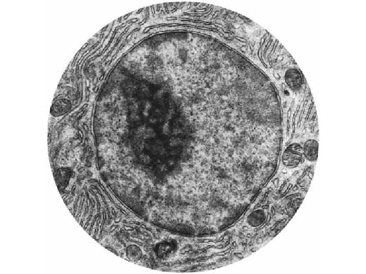
 • Function – isolates the cell’s genetic material, DNA – DNA")
Nucleus (4. 5) • Function – isolates the cell’s genetic material, DNA – DNA directs/controls the activities of the cell • DNA determines which types of RNA are made • The RNA leaves the nucleus and directs the synthesis of proteins in the cytoplasm at a _______

Nucleus • Structure – Nuclear envelope • Two Phospholipid bilayers with protein lined pores – Each pore is a ring of 8 proteins with an opening in the center of the ring – Nucleoplasm – fluid of the nucleus

Nuclear pore bilayer facing cytoplasm Nuclear envelope bilayer facing nucleoplasm Fig. 4 -17, p. 61

Nucleus • DNA is arranged in chromosomes – Chromosome – fiber of DNA with proteins attached – Chromatin – all of the cell’s DNA and the associated proteins

Nucleus • Structure, continued – Nucleolus • Area of condensed DNA • Where ribosomal subunits are made – Subunits exit the nucleus via nuclear pores

ADD THE LABELS
 • Series of organelles responsible for: –")
Endomembrane System (4. 6 – 4. 9) • Series of organelles responsible for: – Modifying protein chains into their final form – Synthesizing of lipids – Packaging of fully modified proteins and lipids into vesicles for export or use in the cell – And more that we will not cover!
 – Continuous with the outer")
Structures of the Endomembrane System • Endoplasmic Reticulum (ER) – Continuous with the outer membrane of the nuclear envelope – Two forms - smooth and rough • Transport vesicles • Golgi apparatus
 – The ER is continuous with the outer membrane of the")
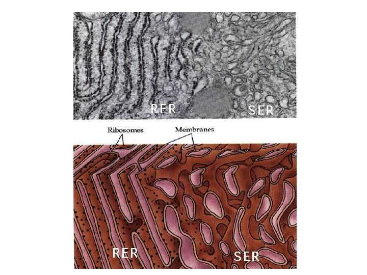
Endoplasmic Reticulum (ER) – The ER is continuous with the outer membrane of the nuclear envelope – There are 2 types of ER: • Rough ER – has ribosomes attached • Smooth ER – no ribosomes attached
 • Network of flattened membrane sacs create")
Endoplasmic Reticulum • Rough Endoplasmic Reticulum (RER) • Network of flattened membrane sacs create a “maze” – RER contains enzymes that recognize and modify proteins • Ribosomes are attached to the outside of the RER and make it appear rough

Endoplasmic Reticulum • Function RER • Proteins are modified as they move through the RER • Once modified, the proteins are packaged in transport vesicles for transport to the Golgi body
 – Tubular membrane structure – Continuous with RER")
Endomembrane System • Smooth ER (SER) – Tubular membrane structure – Continuous with RER – No ribosomes attached • Function SER – Lipids are made inside the SER • fatty acids, phospholipids, sterols. . – Lipids are packaged in transport vesicles and sent to the Golgi

Endomembrane System Vacuoles -membrane-bound structures with various functions depending on the cell type There are different types of vacuoles: -central vacuole in plant cells -contractile vacuole of some protists -vacuoles for storage 82

Endomembrane System Endomembrane system -a series of membranes throughout the cytoplasm -divides cell into compartments where different cellular functions occur 1. endoplasmic reticulum 2. Golgi apparatus 3. lysosomes 83
 -membranes that create a network of channels throughout")
Endomembrane System Rough endoplasmic reticulum (RER) -membranes that create a network of channels throughout the cytoplasm -attachment of ribosomes to the membrane gives a rough appearance -synthesis of proteins to be secreted, sent to lysosomes or plasma membrane 84
 -relatively few ribosomes attached -functions: -synthesis of membrane")
Endomembrane System Smooth endoplasmic reticulum (SER) -relatively few ribosomes attached -functions: -synthesis of membrane lipids -calcium storage -detoxification of foreign substances 85

Endomembrane System

Endomembrane System Golgi apparatus -flattened stacks of interconnected membranes -packaging and distribution of materials to different parts of the cell -synthesis of cell wall components 87

88

Endomembrane System Lysosomes -membrane bound vesicles containing digestive enzymes to break down macromolecules -destroy cells or foreign matter that the cell has engulfed by phagocytosis 89

90

Endomembrane System Microbodies -membrane bound vesicles -contain enzymes -not part of the endomembrane system -glyoxysomes in plants contain enzymes for converting fats to carbohydrates -peroxisomes contain oxidative enzymes and catalase 91

Golgi Apparatus • Golgi Apparatus – Stack of flattened membrane sacs • Function Golgi apparatus – Completes the processing substances received from the ER – Sorts, tags and packages fully processed proteins and lipids in vesicles

Golgi Apparatus • Golgi apparatus receives transport vesicles from the ER on one side of the organelle – Vesicle binds to the first layer of the Golgi and its contents enter the Golgi

Golgi Apparatus – The proteins and lipids are modified as they pass through layers of the Golgi – Molecular tags are added to the fully modified substances • These tags allow the substances to be sorted and packaged appropriately. • Tags also indicate where the substance is to be shipped.

Golgi Apparatus

Transport Vesicles • Transport Vesicles – Vesicle = small membrane bound sac – Transport modified proteins and lipids from the ER to the Golgi apparatus (and from Golgi to final destination)

Endomembrane System • Putting it all together – DNA directs RNA synthesis RNA exits nucleus through a nuclear pore ribosome protein is made proteins with proper code enter RER proteins are modified in RER and lipids are made in SER vesicles containing the proteins and lipids bud off from the ER

Endomembrane System • Putting it all together ER vesicles merge with Golgi body proteins and lipids enter Golgi each is fully modified as it passes through layers of Golgi modified products are tagged, sorted and bud off in Golgi vesicles …

Endomembrane System • Putting it all together Golgi vesicles either merge with the plasma membrane and release their contents OR remain in the cell and serve a purpose

Vesicles • Vesicles - small membrane bound sacs – Examples • Golgi and ER transport vesicles • Peroxisome – Where fatty acids are metabolized – Where hydrogen peroxide is detoxified • Lysosome – contains digestive enzymes – Digests unwanted cell parts and other wastes
 • The lysosome is an example of an organelle made at")
Lysosomes (4. 10) • The lysosome is an example of an organelle made at the Golgi apparatus. – Golgi packages digestive enzymes in a vesicle. The vesicle remains in the cell and: • Digests unwanted or damaged cell parts • Merges with food vacuoles and digest the contents • Figure 4. 10 A
 • Tay-Sachs disease occurs when the lysosome is missing the enzyme")
Lysosomes (4. 11) • Tay-Sachs disease occurs when the lysosome is missing the enzyme needed to digest a lipid found in nerve cells. – As a result the lipid accumulates and nerve cells are damaged as the lysosome swells with undigested lipid.
 • Function – synthesis of ATP – 3 major pathways involved")
Mitochondria (4. 15) • Function – synthesis of ATP – 3 major pathways involved in ATP production 1. Glycolysis 2. Krebs Cycle 3. Electron transport system (ETS)

Mitochondria • Structure: – ~1 -5 microns – Two membranes • Outer membrane • Inner membrane - Highly folded – Folds called cristae – Intermembrane space (or outer compartment) – Matrix • DNA and ribosomes in matrix

Mitochondria
 • Function – synthesis of ATP – 3 major pathways involved")
Mitochondria (4. 15) • Function – synthesis of ATP – 3 major pathways involved in ATP production 1. Glycolysis - cytoplasm 2. Krebs Cycle - matrix 3. Electron transport system (ETS) - intermembrane space

Mitochondria TEM

Mitochondria -organelles present in all types of eukaryotic cells -contain oxidative metabolism enzymes for transferring the energy within macromolecules to ATP -found in all types of eukaryotic cells 110

Mitochondria -surrounded by 2 membranes -smooth outer membrane -folded inner membrane with layers called cristae -matrix is within the inner membrane -intermembrane space is located between the two membranes -contain their own DNA 111

Mitochondria 112
6ee94ef77a687d6d0f37dfe82400eb7f.ppt