

CIRCULATORY SYSTEM.pptx
- Количество слайдов: 74
 HUMAN CIRCULATORY SYSTEM
 Functions of human circulatory system The human circulatory system functions like a network of highways. It transports materials around the body. SOME TRANSPORTED MATERIALS • Oxygen • Carbon dioxide • Digested food • Hormones • Waste chemicals - urea • Heat
 The Human Circulatory System It consists of: • HEART • BLOOD VESSELS • BLOOD
 THE HEART
 HEART FACTS: About 250 -340 grams, In your life time, pumps about 300 million liter of blood, It contracts about 2. 5 billion times.
 MAIN STRUCTURE OF THE HEART The heart is made of a special type of muscle called cardiac muscle which contracts and relaxes rhythmically for a lifetime. The heart is located in the chest cavity and is surrounded by a membrane called the pericardium. The blood vessels which supply food and oxygen to heart are called as coronary arteries.
 EXTERNAL STRUCTURE
 INTERNAL STRUCTURE
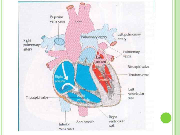
 INTERNAL STRUCTURE OF THE HEART The heart consists of four chambers : The two upper chambers = ATRIA The two lower chambers = VENTRICLES Between atria and ventricle there are valves, preventing the blood coming back to the atria when the ventricles contract. The valve on the left is BICUSPID VALVE The valve on the right is TRICUSPID VALVE The lub-dub heart sound is generated by valves.

 VALVES
 SEMILUNAR VALVES Semilunar valves are found between the arteries and the ventricles. They prevent the blood entering the arteries when the ventricle contract. Between left ventricle and aorta there is aortic valve Between right ventricle and pulmonary artery there is pulmonary valve
 VALVES
 C A R D I A C C Y C L E
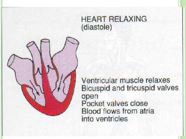
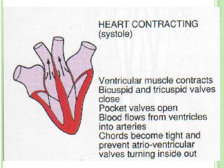
 The Cardiac activity heart pumps blood into the body. Relaxation of heart is known as diastole. Contraction of heart is known as systole. Blood is pumped into the ventricles by atrial contraction, and blood is pumped into the vessels by ventricular contraction.

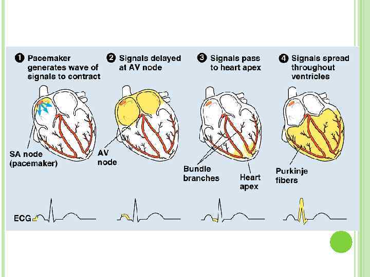
 Control of HEART Heartbeat is controlled by autonomic nervous system. The autonomic nervous system stimulates the sinoatrial node and atrioventricular node for initiation of a contraction. The atria and ventricles contract as a result. SA node sends impulses to heart every 0. 85 seconds
 HEART RATE Parasympathetic nerves reduces the heart rate. Sympathetic nervs speed up the heart rate. Acetylcholine reduces the heart rate. Adrenaline speed up the heart rate. CO 2 reduces the heart rate. High temperature increases the heart rate.
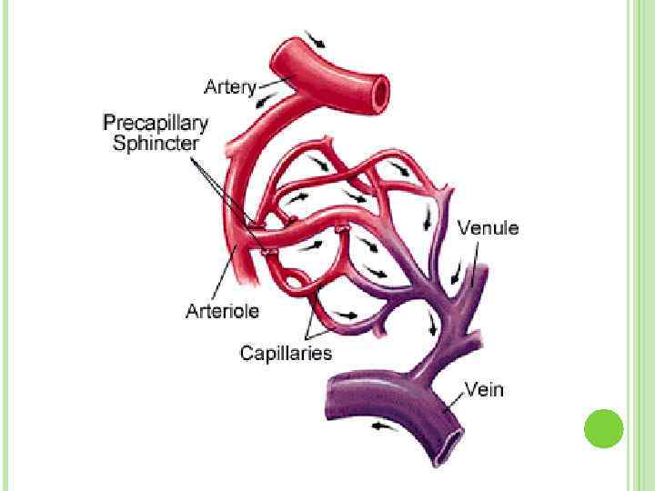
 BLOOD VESSELS There are 3 types of vessels in our body. These are; ARTERIES VEINS CAPILLARIES

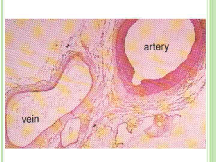
 Arteries 1. ARTERIES carry blood away from heart to the different tissues of the body. Artery walls are stronger and thicker and more elastic than the veins. The pulse is the rhythmic contraction and relaxation of arteries which are parallel to the contraction of the heart. Branches of arteries are called as arteriole. They carry mainly oxygenated blood
 2. VEINS o Veins carry blood to heart o Their walls are much thinner than the walls of arteries. o Veins are farther from the heart and exposed to lower pressures. o Veins are larger in diameter than arteries. o Most veins have one-way valves. A valve is a flap of tissue that ensures blood passes through but does not flow backwards. o Branches of veins are called as venules o Veins mainly carry deoxygenated blood
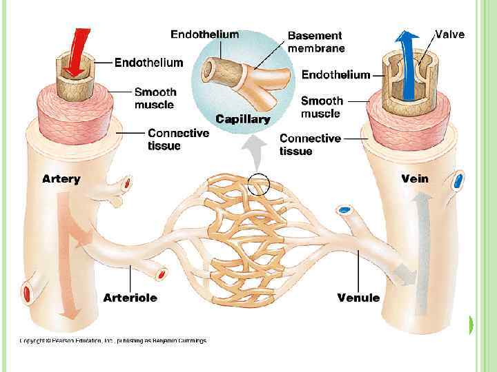

 3. CAPILLARIES Capillary walls are only one cell thick. Gas and nutrient molecules pass easily through their thin walls. They are nonmuscular in structure. Capillaries connect arteries to the veins.
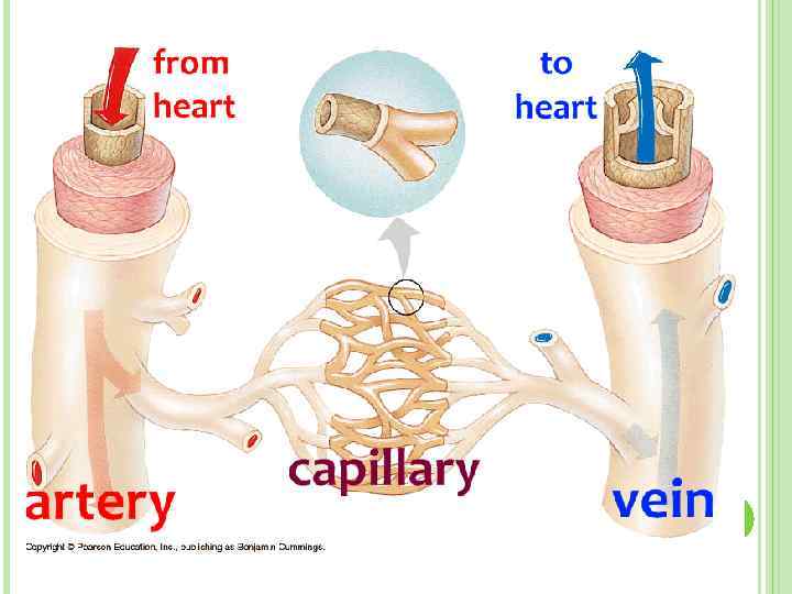
 artery vein arteriole venule capillary
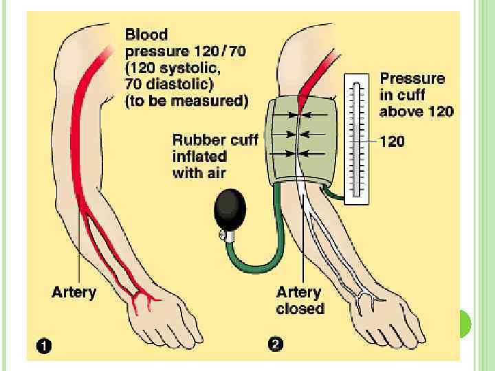
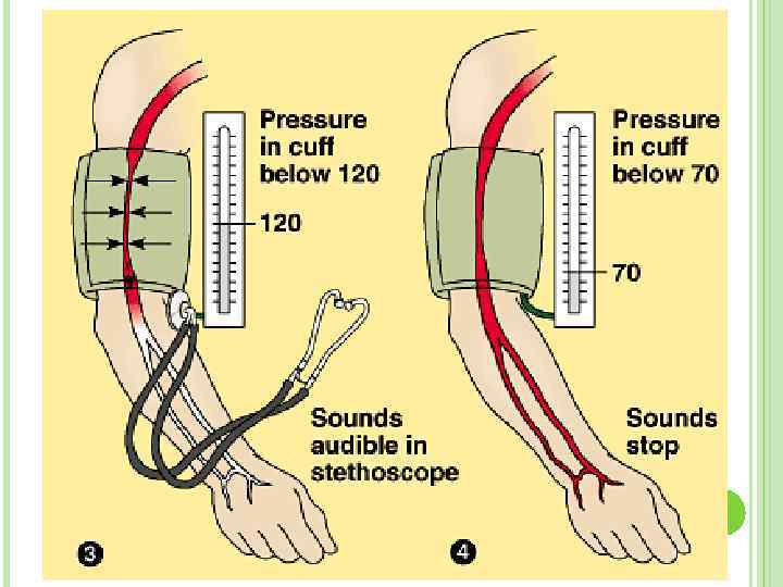
 BLOOD PRESSURE Blood exerts pressure on the walls of vessels during circulation Blood pressure increases when the ventricles contract (systole) and decreases when the ventricles relax (diastole) In normal healthy human systolic pressure is 120 mm Hg and diastolic is 70 mm Hg (120/70) The blood pressure increases during physical work, and decreases during rest and sleep Abnormal increase of blood pressure is known as hypertension Abnormal decrease – hypotension
 Measuring Blood Pressure
 BLOOD CIRCULATION There two types of circulation in human body: 1. Pulmonary Circulation: Oxygen poor blood is pumped into lungs. And oxygen rich one is brought back to the heart. 2. Systemic Circulation: Oxygen rich blood is pumped into body parts. And contaminated blood is brought back to the lungs.
 Pulmonary circulation Systemic circulation
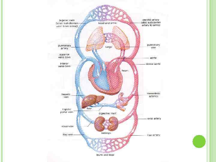
 Left BLOOD MOVEMENT ventricle pumps oxygenated blood to body, that’s why it’s walls are thicker Right ventricle pumps deoxygenated blood to lungs All arteries except pulmonary artery carry oxygenated blood All veins except pulmonary vein carry deoxygenated blood
 Left ventricle Left atrium aorta Pulmonary vein Other arteries arterioles Lungs Pulmonary artery Capillaries Right ventricle venules Right atrium Veins
 INTERNAL STRUCTURE
 Blood BLOOD is a type of tissue that formed by mesoderm layer of embryo. An adult Human body has approximately 5, 5 liters of blood.
 FUNCTIONS OF BLOOD Transport of materials Hormone transport Homeostasis Immune response Blood Clotting
 BLOOD COMPONENTS Blood contain 2 main parts. These are: Blood Plasma Blood cells
 and dissolved proteins.") PLASMA Plasma is liquid part of blood. It includes water (90%) and dissolved proteins. It also contains salts, glucose, aminoacids, fatty acids, vitamins, hormones and cellular wastes.
PLASMA Plasma is liquid part of blood. It includes water (90%) and dissolved proteins. It also contains salts, glucose, aminoacids, fatty acids, vitamins, hormones and cellular wastes.
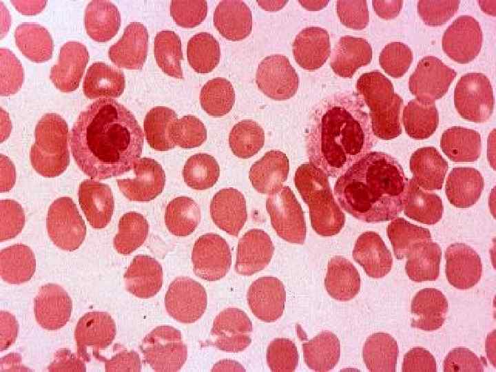
 Leucocytes") Blood Cells There are three types of blood cells: Erythrocytes (=Red Blood Cells) Leucocytes (=White Blood Cells) Thrombocytes (=Platelets)
Blood Cells There are three types of blood cells: Erythrocytes (=Red Blood Cells) Leucocytes (=White Blood Cells) Thrombocytes (=Platelets)
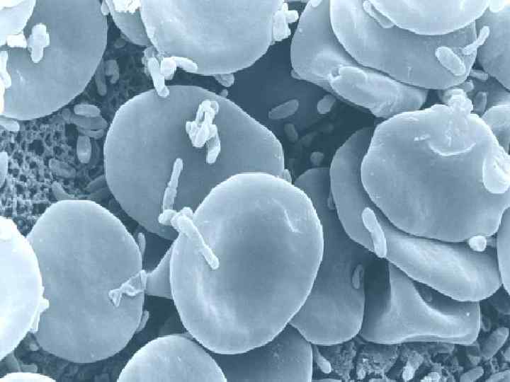
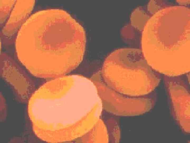
 There approximately 5 to 5, 5 million of erythrocytes per cubic millimeter of blood. The major function of erythrocytes is to transport oxygen from lungs to tissues and transport CO 2 from body tissues to lungs.
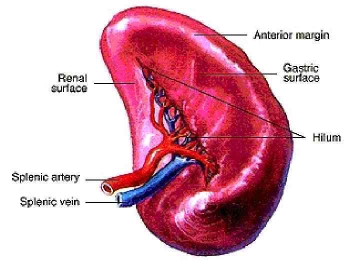
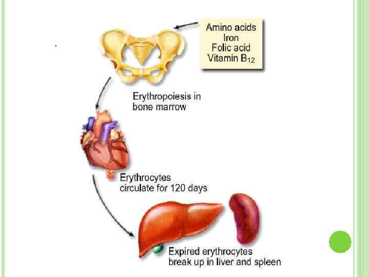
 stage. They are produced by") Mammalian erythrocytes have no nucleus at adult (maturation) stage. They are produced by red bone marrow. Erythrocytes live(!) for 120 days Erythrocytes are broken down by Reticulo-Endothelial System in spleen, liver and lymph nodes.
Mammalian erythrocytes have no nucleus at adult (maturation) stage. They are produced by red bone marrow. Erythrocytes live(!) for 120 days Erythrocytes are broken down by Reticulo-Endothelial System in spleen, liver and lymph nodes.
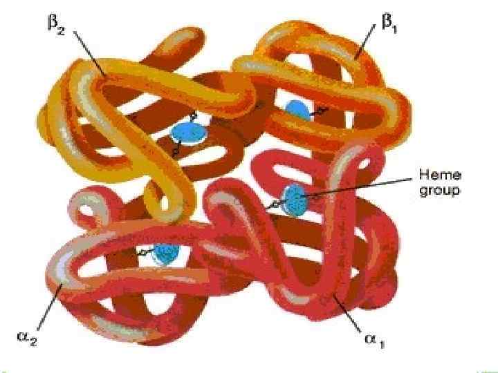
 containing pigment. It gives") HEMOGLOBIN Erythrocytes are filled with hemoglobin. Hemoglobin is iron (Fe) containing pigment. It gives red color to blood. Hemoglobin carries oxygen. Erythrocytes live(!) for 120 days
HEMOGLOBIN Erythrocytes are filled with hemoglobin. Hemoglobin is iron (Fe) containing pigment. It gives red color to blood. Hemoglobin carries oxygen. Erythrocytes live(!) for 120 days
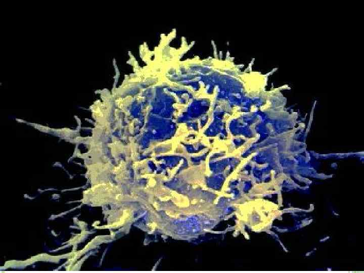
 LEUCOCYTES Leucocytes protect the body from infections. They are produced by red bone marrow and lymph nodes. They can move through the tissue.
 Normally there are only 6000 to 8000 leucocytes per cubic millimeter of blood. When there is an infection in the body, number of leucocytes may increase to 30000 per cubic millimeter.
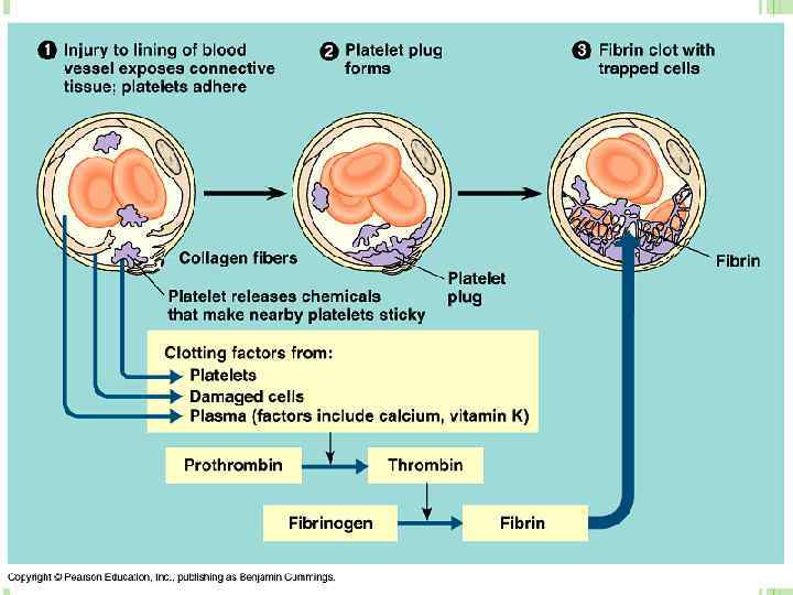
 PLATELETS Platelets are produced by bone marrow. They play major role in blood clotting. Blood clotting is the solidification of blood in order to stop bleeding.
 Vitamin K Thrombogen Thrombocytes + O") THE MECHANISM OF BLOOD CLOTTING Prothrombin (In liver) Vitamin K Thrombogen Thrombocytes + O 2 Thrombokinase Fibrinogen Ca ions Thrombin Cloth Platelets + Fibrin
THE MECHANISM OF BLOOD CLOTTING Prothrombin (In liver) Vitamin K Thrombogen Thrombocytes + O 2 Thrombokinase Fibrinogen Ca ions Thrombin Cloth Platelets + Fibrin
 DISEASES RELATED TO CIRCULATORY SYSTEM Anemia Leukemia Arteriosclerosis
 Anemia
 When Arteriosclerosis blood vessels become narrow and lose their elasticity Fats and Ca++ ions adhere to the walls of blood vessels, and by this stroke and heart attack may occur o This disease occurs as a result of eating disorders o Is seen mainly in men and women over the age 40
 Heart attack, infarcts
 STROKE