History n Mr X is a 74 year old Caucasian male n PC: Acute SOB n HPC: SOB episode began at 10 -11 am this morning gradually worsening, but suddenly became acutely worse. Noticed by neighbour. Ambulance blue lighted to AE resus
 ‘A short of breath radio operator’ Dr Antonios
 PMH n n n AHA diagnosed 1988 MI 1999 Fibrosing alveolitis diagnosed 1993 Hypercholestolaemia 1993 Multiple admissions for SOB, 6 times in the last 6/12
 DH n n n n n Salbutamol 100 mg 2 puffs qds Ipratropium Bromide tds 2 puffs Beconase nasal spray 2 puffs Bumetanide 1 md od Simvastatin 20 mg Asprin 75 mg od Azatioprine 50 mg Folic acid 5 mg Lansoprazole 15 mg Calcichew 500 mg tds
 SH n Ex radio operator for the army n Lives alone in a flat n Not married, no children n Smoked for most of life n Ex- heavy alcohol drinker.
 CVS n Ankle oedema 4 months ago R > L n. Resp n n n SOB for 15 years getting worse, can’t complete sentences SOB on 1 flight of stairs No PND 2 pillow orthopnia Same throughout the year, no variation to day/night or climate change.
 n Sputum produced throughout the year, difficult to clear, White in colour Autoimmune Haemolytic anaemia diagnosed 15 years ago drug controlled by Azathioprine and folic acid, Predisolone was discontinued 2 years ago n Cryptogenic Fibrosing alveolitis for 10 years n
 Examination n n n Anxious, gasping for breath, holding arms on trolley, Abdominal muscles used for breathing Central cyanosis Pulse 106 bmp regular HS I + II + 0 Apex beat laterally displaced Pitting right ankle oedema No sacral oedema
 Respiratory R n n n L Resp rate 22 pm Trachea central Uneven lung expansion Hyperinflated upper chest Hyperresonant percussion Exp wheezing Insp crackles
 Impressions n Pneumothorax n COPD/Fibrosing alveolitis n AHA n Congestive heart failure n Newly diagnosed Diabetes mellitus
 Pneumothorax. Tension: due to trauma Primary Risk factors: • Age< 40 yrs • Smoking • Height (in males) Spontaneous: due to rupture of bleb Secondary Risk Factors: • Age> 40 yrs • Pulmonary fibrosis • Emphysema
 Epidemiology. n Incidence - 1. 2 -6/100, 000 women - 18 -28/100, 000 men n Hospital admission rates 5. 8/100, 000 per year for women n 16. 7/100, 000 per year for men n n Mortality rates 0. 62/million per year for women n 1. 26/million per year for men n (between 1991 -1995)
- few signs n Large") Signs and Symptoms. n Sudden SOB n Small (<2 cm)- few signs n Large (>2 cm) Pallor n Tachycardia n chest wall movement n breath sounds n
Signs and Symptoms. n Sudden SOB n Small (<2 cm)- few signs n Large (>2 cm) Pallor n Tachycardia n chest wall movement n breath sounds n
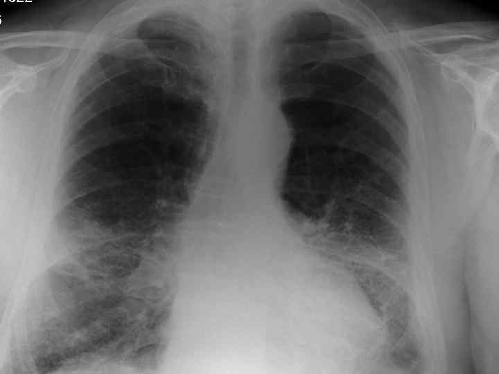
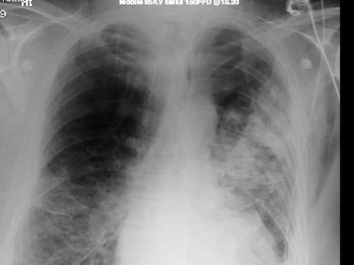
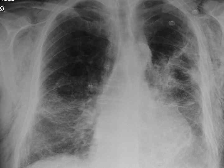
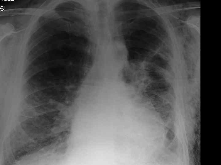
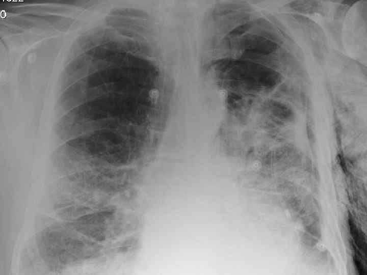
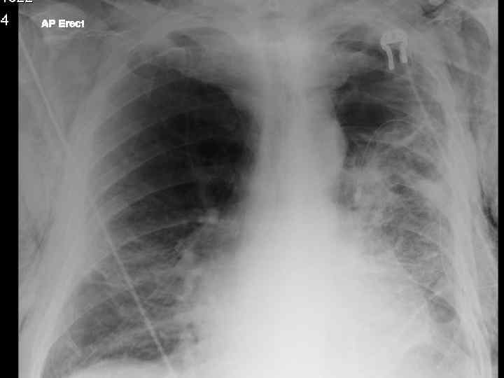
 Investigations. n CXR n CT n Arterial blood gases
 Treatment of spontaneous pneumothorax. n Depends on: Age n Levels of dyspnoea n Results of CXR n
 Treatment. Primary pneumothorax NO Breathless and/or rim of air>2 cm on CXR? YES Aspiration successful? NO Repeat aspiration, successful? NO Intercostal drainage, successful? YES Remove 24 hrs after full re-expansion NO Refer to chest physician within 48 hrs Consider discharge BTS Guidelines
 Treatment. Secondary pneumothorax NO Breathless + age> 50 years + rim of air>2 cm on CXR? Aspiration successful? YES NO Intercostal drainage, successful? YES Admit to hospital for 24 hours NO Refer to chest physician within 48 hrs YES Remove 24 hrs after full re-expansion Consider discharge BTS Guidelines
 Simple aspiration.
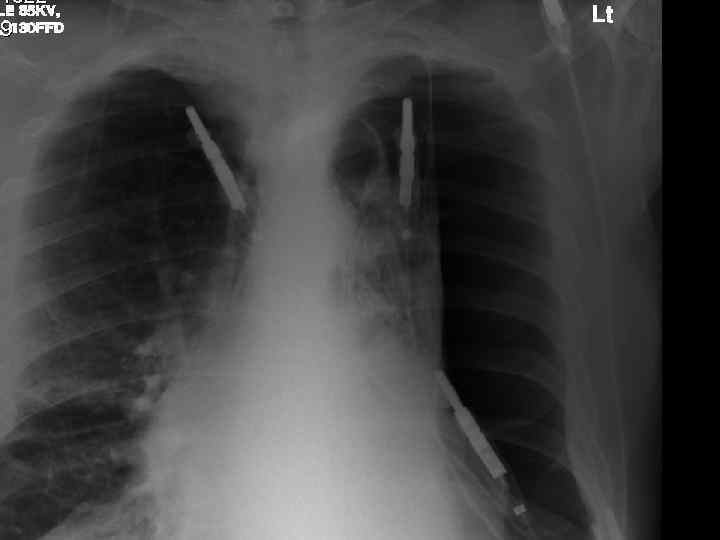
 Intercostal drainage n Chest drain into triangle marked by: Apex of axilla n Nipple (4 th ics, mid clav. line) n Base of scapula n n Complications: Penetration of major organs n Pleural infection n Surgical emphysema n
 Surgical emphysema n Air filled space in subcutaneous tissue n Felt on skin as bubble wrap n Severe respiratory compromise n Rx- skin incision
 Follow up n Unsuccessful referral to resp. physician n Successful discharge with: Chest clinic appointment (after 7 -10 days) n No air travel for 3 mths n Back to hospital if symptoms worsen n
 Fibrosing alveolitis n Affects less than 3 in 10, 000 n Onset usually after 50 years of age n Affects twice as many men as women n Twice as common in smokers than nonsmokers
 Pathogenesis of pulmonary fibrosis in CFA
 Histological features of CFA n Cellular infiltration and thickening of alveolar walls n Alveolitis – increased number of cells within the alveolar space
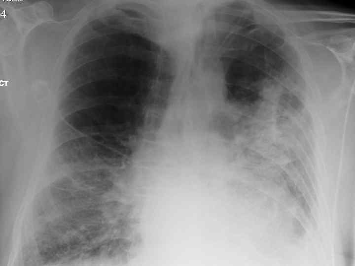
 Clinical features n Progressive breathlessness n Cyanosis n Pulmonary hypertension/cor pulmonale n Clubbing n Bilateral fine end insp. Crackles n X-ray shows ground glass appearance
 Investigations n Chest X-ray n High res. CT scan n Respiratory function tests n Blood gasses n ANF, RF, ESR n Bronchoalveolar lavage n Biopsy
 Differential diagnosis n Extrinsic allergic alveolitis n Bronchiectasis n Chronic LHF n Sarcoidosis n Industrial lung disease n Lymphangitis carcinomatosa
 Treatment n Corticosteroids: Prednisolone n Other immunosuppresants: Azathioprine n Oxygen therapy n Single lung transplant