d8516f1dfd96a7cc3dbf51d6514e342d.ppt
- Количество слайдов: 151
 Heart Sounds, ECG & Fractals
 The Heart ECG Wave Heart Sounds Abnormal Sounds Audicor’s Solution Fractal Dimension Sound Analysis The heart is a 2 -step mechanical pump that is coordinated by precisely timed electrical impulses. Fractal Results The END Lets Go! 2
 The Heart ECG Wave The heart is a pulsating pump that composes of four chambers and Abnormal Sounds four heart valves. Audicor’s Solution The upper chambers are the right Fractal Dimension atrium (RA) and left atrium (LA) The lower chambers are the right Sound Analysis ventricle (RV) and left ventricle Fractal Results (LV). Heart sounds The END Lets Go! 3
 Electrophysiology of cardiac conduction The Heart ECG Wave Heart Sounds Abnormal Sounds Audicor’s Solution Fractal Dimension Sound Analysis Fractal Results The END Lets Go! 4
 Heart Valves The Heart ECG Wave Heart Sounds Abnormal Sounds Audicor’s Solution Fractal Dimension Sound Analysis Fractal Results The END Lets Go! 5
 Events occurring during the cardiac cycle The Heart ECG Wave Heart Sounds Abnormal Sounds Audicor’s Solution Fractal Dimension Sound Analysis Fractal Results The cardiac cycle consists of two basic components: A period of ventricular diastole during which the ventricles are filled with blood. A period of ventricular systole during which blood is propelled out of the heart. The END Lets Go! 6
 Events occurring during the cardiac cycle The Heart ECG Wave Heart Sounds Abnormal Sounds Audicor’s Solution Fractal Dimension Sound Analysis Fractal Results Clinically, systole is taken as the interval between the first and the second heart sound. Diastole is considered to be the interval between second heart sound and the first heart sound. The END Lets Go! 7
 The Electrical system The Heart ECG Wave Heart Sounds Abnormal Sounds Audicor’s Solution Fractal Dimension Sound Analysis Fractal Results The END Lets Go! 8
 What is measured on the ECG The Heart ECG Wave Heart Sounds Abnormal Sounds Audicor’s Solution Rate and rhythm of the heart. Evidence of heart enlargement. Evidence of damage to the heart Fractal Dimension Impaired blood flow to the heart Sound Analysis Heart rhythm problems Fractal Results Electrolyte imbalance The END Lets Go! 9
 What are the limitations of the ECG Electrophysiology ECG Wave Heart Sounds Abnormal Sounds Audicor’s Solution Fractal Dimension Sound Analysis Fractal Results The ECG is a static picture Many heart attacks cannot be detected by ECG. Many abnormal patterns on an ECG may be non-specific. The ECG may be normal despite the presence of a cardiac condition The END Lets Go! 10
 ECG wave Electrophysiology ECG Wave Heart Sounds Abnormal Sounds ECG tracings show a pattern of electrical impulses that are generated by the heart. Audicor’s Solution P wave: the sequential activation (depolarization) of the right and left atria Fractal Dimension QRS complex: right and left ventricular depolarization Sound Analysis ST segmet: ventricular repolarization. Fractal Results The END The T wave corresponds to electrical relaxation and preparation for their next muscle contraction. Lets Go! 11
 ECG wave Electrophysiology ECG Wave Heart sounds Abnormal Sounds Audicor’s Solution Fractal Dimension Sound Analysis Fractal Results ML Model Lets Go! 12
 Heart Sounds The Heart ECG Wave Heart Sounds Abnormal Sounds Audicor’s Solution Fractal Dimension Sound Analysis Fractal Results The END The auscultation of the heart may reveal different phenomena called heart sounds and murmurs. Heart sounds are a prolonged series of vibrations of both high and low frequency The murmurs are a longer series of vibrations, mostly of either high or low frequency. Lets Go! 13
 Heart Sounds The Heart ECG Wave Heart Sounds Abnormal Sounds Audicor’s Solution Fractal Dimension Sound Analysis Fractal Results The END The sounds heard during auscultation are called the first (S 1) and second (S 2) heart sounds respectively, with respect to their temporal relationship, and are systolic sounds. Phonocardiography often yields third (S 3) and fourth (S 4) heart sounds especially in children and in cases of heart disease. These are diastolic sounds. Lets Go! 14
 Heart Sounds Genesis The Heart ECG Wave Heart Sounds Abnormal Sounds Audicor’s Solution Fractal Dimension Sound Analysis Fractal Results The END Many hypotheses have been suggested to explain the origin of these sounds. Some being controversial at the time. With the advent of echocardiography the movement of intracardiac structures could be monitored with virtually no time delay. Concerning S 1, S 2, and S 4 these controversies have largely been resolved. However there still exists controversy regarding S 3. Lets Go! 15
 S 1 & S 2 Electrophysiology ECG Measurements Heart Sounds Abnormal Sounds Audicor’s Solution S 1 occurs when the mitral and tricuspid valves close at the beginning of systole. Fractal Dimension Sound Analysis Fractal Results S 2 results from closure of the aortic and pulmonic valves at the end of systole. The END Lets Go! 16
 S 3 The Heart ECG Wave Heart Sounds Abnormal sounds low frequency sound: 0 - 70 Hz 50% in the 0 -15 Hz band Audicor’s Solution occurring in early ventricular diastole Fractal Dimension due to over-distention of the ventricle during the rapid early filling phase Sound Analysis Fractal Results occurs 0. 12 – 0. 20 secs after S 2 The END Lets Go! 17
 The physiological cause and effect of S 3 Electrophysiology ECG Measurements Heart Sounds Abnormal Sounds Audicor’s Solution Fractal Dimension Sound Analysis Fractal Results The END The third heart sound (S 3) occurs 0. 12 to 0. 20 seconds after S 2 in early Diastole. Of the many proposed theories, the most likely explanation is that excessive rapid filling of the ventricle is suddenly halted, causing vibrations that are audible as S 3. Pathologic states where an S 3 is encountered include anemia, thyrotoxicosis, mitral regurgitation, hypertrophic cardiomyopathy, aortic and tricuspid regurgitation and left ventricular dysfunction. Lets Go! 18
 S 4 The Heart ECG Wave Heart Sounds Abnormal Sounds Audicor’s Solution Fractal Dimension Sound Analysis Fractal Results low frequency sound: 0 - 70 Hz occuring at the late diastolic filling phase at when the atria contract Ventricles have decreased compliance, or receive increased diastolic volume occurs just before S 1 70 ms after onset of ECG P wave The END Lets Go! 19
 The physiological cause and effect of S 4 Electrophysiology ECG Measurements Heart Sounds Abnormal Sounds Audicor’s Solution Fractal Dimension Sound Analysis Fractal Results The END The S 4 occurs just before the first heart sound in the cardiac cycle. It is produced in late diastole as a result of atrial contraction causing vibrations of the LV muscle, mitral valve apparatus, and LV blood mass. Disease processes that produce an S 4 include hypertension, aortic stenosis and regurgitation, severe mitral regurgitation, cardiomyopathy, and ischemic heart disease. Lets Go! 20
 The physiological cause and effect of S 3 & S 4 Electrophysiology ECG Measurements Heart Sounds Abnormal Sounds Audicor’s Solution Fractal Dimension Sound Analysis Fractal Results In 1856, Potain first described "gallop rhythm" as an audible phenomenon in which a tripling or quadrupling of heart sounds resembles the canter of a horse. That term is still used to describe a third or fourth heart sound. The END Lets Go! 21
 The physiological cause and effect of S 3 & S 4 Electrophysiology ECG Measurements Heart Sounds Abnormal Sounds Audicor’s Solution Fractal Dimension Sound Analysis Gallops are diastolic events and seem to be related to 2 periods of filling of the ventricles: 1. The rapid filling phase (ventricular diastolic 2. gallop or S 3) 2. The presystolic filling phase related to atrial systole (atrial gallop or S 4) Fractal Results The END Lets Go! 22
 The physiological cause and effect of S 3 & S 4 Electrophysiology ECG Measurements Heart Sounds Abnormal Sounds Audicor’s Solution Fractal Dimension Sound Analysis Fractal Results The END Experimental evidence in both humans and animal models suggests that abnormal compliance of the left ventricle is often associated with an S 4 and/or a pathological S 3. In the early diastolic phase of the cardiac cycle, the left ventricle relaxes and the intraventricular blood pressure falls below that of the left atrium. Therefore, blood flows from the atrium into the ventricle. Lets Go! 23
 The physiological cause and effect of S 3 & S 4 Electrophysiology ECG Measurements Heart Sounds Abnormal Sounds Audicor’s Solution Fractal Dimension Sound Analysis Fractal Results The END This continues until the intraventricular pressure equals the pressure in the atrium and the flow of blood into the ventricle therefore stops. This deceleration of the blood early in diastole produces vibrations inside the ventricle, which can result in an S 3 if the vibrations have sufficient energy. The steep left ventricular pressure increase in early diastole causes a reversal of the transmitral pressure gradient and hence a more rapid deceleration of inflow. Lets Go! 24
 The physiological cause and effect of S 3 & S 4 Electrophysiology ECG Measurements Heart Sounds Abnormal Sounds Audicor’s Solution Fractal Dimension Sound Analysis Fractal Results The END Since the vibrations of S 3 occur during deceleration of inflow, a conversion of kinetic into vibratory energy is likely. These vibrations are audible if transmitted with enough intensity. The higher the inflow rate (valve regurgitation) and the steeper the rapid filling wave (high filling rates), the greater the deceleration and more likely an S 3 will occur. Lets Go! 25
 The physiological cause and effect of S 3 & S 4 Electrophysiology ECG Measurements Heart Sounds Abnormal Sounds Audicor’s Solution Fractal Dimension Sound Analysis Fractal Results S 3 is produced when the rapidly distending ventricle reaches a point when its distention is checked by the resistance of its wall and the ensuing vibrations are audible as the third heart sound. The END Lets Go! 26
 The physiological cause and effect of S 3 & S 4 Electrophysiology ECG Measurements Heart Sounds Abnormal Sounds Audicor’s Solution Fractal Dimension Sound Analysis Fractal Results The END The remainder of the filling of the ventricle occurs late in diastole because of active contraction of the atrium. The deceleration of the blood later in diastole also produces vibrations inside the ventricle. If the atrial contraction that produced the late diastolic filling was sufficiently strong and the ventricle is relatively stiff, these vibrations may have enough energy to produce an S 4. Lets Go! 27
 Heart Sounds Characteristics The Heart http: //www. cardiologysite. com/auscultation/html/s 3_gallop. h tml ECG Wave Heart Sounds Abnormal Sounds Audicor’s Solution Fractal Dimension Sound Analysis Fractal Results The END Lets Go! 28
 Heart Sounds The Heart http: //depts. washington. edu/physdx/heart/tech 2. html ECG Wave Heart Sounds Abnormal Sounds Audicor’s Solution Fractal Dimension Sound Analysis Fractal Results The END Lets Go! 29
 PCG against ECG The Heart ECG Wave Heart Sounds Abnormal Sounds Audicor’s Solution Fractal Dimension Sound Analysis Fractal Results The END Lets Go! 30
 PCG against ECG The Heart ECG Wave Heart Sounds Abnormal Sounds Audicor’s Solution Fractal Dimension Sound Analysis Fractal Results The END Lets Go! 31
 The relationship between heart sounds and cardiac events The Heart ECG Wave Heart Sounds Abnormal Sounds Audicor’s Solution Fractal Dimension Sound Analysis Fractal Results The END Lets Go! 32
 How do S 3 & S 4 Help? The Heart ECG Wave Heart Sounds Abnormal Sounds Audicor’s Solution Fractal Dimension Sound Analysis Fractal Results The END Experimental evidence suggests that abnormal compliance of the left ventricle is often associated with an S 4 and/or a pathological S 3. The S 3 may be a normal finding in patients less than 30 years old. However, in older patients, the S 3 is usually evidence of impaired ability of the ventricle to contract during systole. The prevalence of the S 4 increases with age and usually indicates an abnormal increase in ventricular stiffness Lets Go! 33
 Clinical significance of S 3 The Heart ECG Wave Heart Sounds Abnormal Sounds Audicor’s Solution Fractal Dimension Sound Analysis Fractal Results The END The presence of S 3 may be the earliest clue to left ventricular failure. The presence of heart disease and may offer valuable information about diagnosis, prognosis, and treatment. The most useful clinical importance of S 3 is in detecting left-sided heart failure, especially in the early stages when other signs may be normal. More recently, S 3 was the best predictor of response to digoxin in CHF patients. Lets Go! 34
 Usefulness of S 3, S 4 & ECG in assisting early detection The Heart ECG Wave Heart Sounds Abnormal Sounds Audicor’s Solution Fractal Dimension Sound Analysis Fractal Results The END Several types of cardiac disease have characteristic electrical and hemodynamic manifestations. For example, acute myocardial ischemia is typically associated both with displacement of the ST segments of the ECG and with alterations of the mechanical properties of the left ventricle. The latter changes may produce pathological heart sounds – S 3 and/or S 4. Lets Go! 35
 S 3 In Children The Heart ECG Wave The genesis of S 3 has been clearly associated with the rapid filling phase of diastole. Heart Sounds Present work has shown that S 3 occurs earlier in the cardiac cycle with increase in Abnormal Sounds age of child subjects. Audicor’s Solution Fractal Dimension Sound Analysis Fractal Results The END This supports the hypothesis that S 3 is due to L. V. reaching it’s elastic limit during diastole. This notion is supported further by the finding of the spectral energy of S 3 is distributed more towards the high frequency of the end of the spectrum with age. Lets Go! 36
 S 3 In Children The Heart ECG Wave Heart Sounds Abnormal Sounds Audicor’s Solution Fractal Dimension Sound Analysis Fractal Results The END This is consistent with an increase in stiffness of the L. V. with age. The resonant frequencies of L. V. increase with stiffness. higher frequencies are more attenuated by passage through body tissue than lower frequencies. As the frequency distribution of S 3 is shifted to higher frequencies as the child becomes older, it would be expected that the energy in S 3 would decrease with age. Thus S 3 usually disappears around adulthood, but may reoccur with cardiac pathology. Lets Go! 37
 Combining ECG & Heart Sounds The Heart Sounds ECG Wave Audicor uses an advanced new technology called correlated audioelectric cardiography (COR). This technology builds on the traditional findings of the standard, 12 -lead resting Audicor’s Solution ECG, augmenting it by simultaneously Fractal Dimension acquiring acoustical signals from both the V 3 and V 4 lead positions. Sound Analysis Abnormal Sounds Fractal Results The END (Two acoustic sensors replace the V 3 and V 4 ECG electrodes of a standard 12 -Lead ECG ) Lets Go! 38
 Combining ECG & Heart Sounds The Heart Sounds Audicor CE combines the detection of heart sounds with ECG data in order to ECG Wave provide physicians with additional Abnormal Sounds information that is valuable in Audicor’s Solution assessing: S 3 and S 4 heart sounds that may be Fractal Dimension indicative of acute coronary syndrome or heart failure Sound Analysis Acute and prior (age-undetermined) Fractal Results myocardial infarction (MI) The END Ischemia Left ventricular hypertrophy (LVH) Lets Go! 39
 Combining ECG & Heart Sounds The Heart ECG Wave Heart Sounds Abnormal Sounds The S 3 heart sound is often very difficult to detect by auscultation due to its low frequency and intensity. Audicor’s Solution Noisy clinical environments further complicate Fractal Dimension Sound Analysis Fractal Results The END this difficulty. To improve the detection of the S 3, Inovise Medical, Inc. has developed AUDICOR®, a device that records and algorithmically interprets simultaneous 12 -lead ECG and electronic cardiac sound recording Lets Go! 40
 Audicor Heart Sound Algorithm The Heart ECG Wave Heart Sounds Audicor’s Solution The AUDICOR heart sounds algorithm receives Fractal Dimension Sound Analysis three synchronous inputs: 1. A standard ECG signal 2. Two single-channel sound signals. Fractal Results The END Lets Go! 41
 Processing The Sound Data The Heart ECG Wave Heart Sounds Abnormal Sounds The sound data for each channel is then processed by removing offsets, prescaling, and filtering it into narrow frequency bands to optimize the detection of each S 1 through S 4 heart sound. Audicor’s Solution Using the ECG as a reference, the S 1 and S 2 Fractal Dimension Sound Analysis Fractal Results The END detection time windows are identified for each beat. Utilizing a threshold adaptively computed from a moving window root mean square for each frequency band, the location of each S 1 and S 2 is determined within the computed detection window. Lets Go! 42
 Detecting S 3 The Heart ECG Wave Heart Sounds Abnormal Sounds Audicor’s Solution The S 3 detection time windows are located using information within the ECG and the computed position of the S 2 offset. The energy content is determined within the S 3 Fractal Dimension detection time window. Sound Analysis Fractal Results The END Using a set of rules based on frequency and amplitude measurements, possible S 3 s are detected within the S 3 windows. Lets Go! 43
 Detecting S 4 The Heart ECG Wave Heart Sounds Abnormal Sounds Audicor’s Solution Fractal Dimension Sound Analysis Fractal Results The S 4 detection time windows are located based on PQ intervals and Q-wave onset positions. Further processing on S 4 s is similar to that described before for S 3 s. The END Lets Go! 44
 ECG Diagnostic Algorithm The Heart Sounds Superior diagnostic performance up to 84% more sensitive than current systems in acute MI detection, particularly in women was achieved in the following ways : Abnormal Sounds • Developed the computerized ECG diagnostic ECG Wave Audicor’s Solution algorithms using very large clinically correlated databases of over 100, 000 ECGs. Fractal Dimension Sound Analysis Fractal Results The END • Divided the data into demographically balanced learning and test sets to help ensure that it was not overtraining the algorithms using limited sets of data. Lets Go! 45
 ECG Diagnostic Algorithm The Heart ECG Wave Heart sounds Abnormal Sounds Audicor’s Solution Fractal Dimension Sound Analysis Fractal Results • Accounted for differences in gender and age, emphasizing features that discriminate between prior and acute MI. • Avoided circularity in developing and testing the algorithms by selecting all cases and non-cases of various diseases using criteria independent of the ECG The END Lets Go! 46
 ECG Diagnostic Algorithm The Heart ECG Wave Heart Sounds Abnormal Sounds Audicor’s Solution Fractal Dimension Sound Analysis Fractal Results The END • Criteria for IMI is based upon the relationships between portions of the vectorcardiographic (VCG) QRS loop in the frontal plane and the corresponding portions of the ECG QRS complexes recorded in leads II and III. • Commercial ECG algorithms for detection of prior myocardial infarction (MI) predominantly rely on QRS criteria and on established qualitative ST and T changes. Lets Go! 47
 ECG Diagnostic Algorithm The Heart ECG Wave • Two distinct new approaches for quantifying Heart Sounds Abnormal Sounds Audicor’s Solution Fractal Dimension Sound Analysis Fractal Results The END ST and T changes to assist with the detection of prior MI. 1. The first method uses the mean axes of vectorcardiographic T-loops taken from the inverse Dower transform of the 12 lead ECG to indicate ischemic regions of the left ventricular wall. 2. The second method establishes regional scores for residual ST elevation supportive of ischemia or infarction. Lets Go! 48
 ECG Diagnostic Algorithm The Heart ECG Wave Heart Sounds Abnormal Sounds Audicor’s Solution Fractal Dimension Sound Analysis • These 2 ST-T measures qualify borderline QRS infarct criteria, resulting in composite criteria having higher sensitivities and specificities than QRS criteria alone. Fractal Results The END Lets Go! 49
 ECG Diagnostic Algorithm The Heart ECG Wave Heart Sounds Abnormal Sounds Audicor’s Solution Fractal Dimension Sound Analysis Fractal Results The END LVH is defined as values of the ECG left ventricular mass index (LVMI) >116 g/m(2) in men or >104 g/m(2) in women. Univariate linear regression was performed separately on the male and female subjects in the Learning Set to find all the ECG parameters that correlated significantly with LVMI. Multivariate linear regression (MLR) was applied to these parameters to identify the 4 variables for each sex that discriminated best between the subjects with and without LVH. Lets Go! 50
 Diagnostic Performance of a Computerized Algorithm for Augmenting the ECG with Acoustical Data The Heart ECG Wave Heart Sounds Results: Abnormal Sounds The following data show the ability of the Audicor’s Solution computerized acoustical algorithm to detect an S 3 or an S 4 in patients in a variety of clinical settings. Fractal Dimension Sound Analysis Fractal Results The e. ND The performance of the algorithm is compared to a consensus of 2 experienced cardiologists concerning the audibility of the recorded S 3 or the S 4 in the each of the same patients. Lets Go! 51
 Diagnostic Performance of a Computerized Algorithm for Augmenting the ECG with Acoustical Data The Heart ECG Wave Heart Sounds Abnormal Sounds Audicor’s Solution Fractal Dimension Sound Analysis Fractal Results The END Lets Go! 52
 Audicor Analysis The Heart ECG Wave Heart Sounds Abnormal Sounds Audicor’s Solution Fractal Dimension Sound Analysis Fractal Results The END Lets Go! 53
 Audicor Analysis The Heart ECG Wave Heart Sounds Abnormal Sounds Audicor’s Solution Fractal Dimension Sound Analysis Fractal Results The END Lets Go! 54
 The AUDICOR Decision Pathway – EMS The Heart ECG Wave Heart Sounds Abnormal Sounds Audicor’s Solution Fractal Dimension Sound Analysis Fractal Results The END Lets Go! 55
 ECG, Hemodynamic & Acoustical Findings: Experimental Model of Myocardial Ischemia The Heart ECG Wave Heart Sounds Abnormal Sounds Audicor’s Solution Fractal Dimension Sound Analysis Fractal Results The END Acute myocardial ischemia often displaces the ST segments in the ECG. However, since the specificity and sensitivity for ischemia of ST segment displacement are imperfect Echocardiography and radionuclide studies are often used to augment the ECG in evaluating patients for ischemia. Lets Go! 56
 ECG, Hemodynamic & Acoustical Findings: Experimental Model of Myocardial Ischemia The Heart ECG Wave Heart Sounds Abnormal Sounds Audicor’s Solution Fractal Dimension Sound Analysis Ischemia also has hemodynamic effects that include reduced left ventricular (LV) contractility and compliance. These hemodynamic changes are typically associated with a third and fourth heart sound (S 3 and S 4), respectively. Fractal Results The END Lets Go! 57
 ECG, Hemodynamic & Acoustical Findings: Experimental Model of Myocardial Ischemia The Heart ECG Wave Heart Sounds Conclusion: Abnormal Sounds Detecting and recording heart sounds may improve the identification of acute myocardial ischemia as the cause of ST segment abnormalities. Audicor’s Solution Fractal Dimension Sound Analysis Fractal Results The END Lets Go! 58
 ECG & Acoustical Data in the detection of Left Ventricular Enlargement The Heart ECG Wave Heart Sounds The presence of 3 rd and 4 th heart sounds was Abnormal Sounds associated mainly with relative prolongation of Audicor’s Solution the PR interval and with flattening or negativity of T waves in multiple leads. Fractal Dimension Sound Analysis Fractal Results The END Conversely these sounds were not associated with the abnormalities of QRS voltage traditionally attributed to increased left ventricular mass. Lets Go! 59
 ECG & Acoustical Data in the detection of Left Ventricular Enlargement The Heart ECG Wave Heart Sounds Abnormal Sounds Audicor’s Solution Fractal Dimension Sound Analysis Fractal Results Conclusion: ECG and acoustical data can detect abnormalities of ventricular function that the cardiac diseases responsible for LVE produce. The END Lets Go! 60
 Detecting Hemodynamic Abnormalities Using ECG and Cardiac Acoustical Data The Heart ECG Wave Heart Sounds Background: Hemodynamic abnormalities can produce ECG changes. Abnormal Sounds For example, the ECG evidence of left Audicor’s Solution ventricular hypertrophy (LVH) is a consequence of the hemodynamic Fractal Dimension abnormalities that produced the LVH. Sound Analysis Fractal Results The END However they hypothesized that abnormal hemodynamics are more likely to predict the presence of a third heart sound (S 3) than of ECG findings. Lets Go! 61
 Detecting Hemodynamic Abnormalities Using ECG and Cardiac Acoustical Data The Heart ECG Wave Heart Sounds Conclusions: The electronically recorded S 3 is associated with a wider range of Audicor’s Solution hemodynamic abnormalities than is Fractal Dimension ECG evidence of LVH, ST-T or prior MI and can therefore Sound Analysis augment the diagnostic capabilities of the ECG. Fractal Results Abnormal Sounds The END Lets Go! 62
 Using Simultaneous ECG and Acoustical Data to Evaluate and Monitor Patients with Cardiac Disease The Heart ECG Wave Heart Sounds Background: Acute myocardial ischemia is associated with hemodynamic as well as ECG abnormalities. Abnormal Sounds For example, impaired left ventricular (LV) Audicor’s Solution systolic function can produce a third heart sound (S 3) that previous research, as Fractal Dimension reflected in the ACC/AHA Practice Sound Analysis Guidelines, has shown to be associated with increased clinical risk. Fractal Results The END Lets Go! 63
 Using Simultaneous ECG and Acoustical Data to Evaluate and Monitor Patients with Cardiac Disease The Heart ECG Wave Heart Sounds Results: In the 89 pre-cath patients, the recorded S 3 Abnormal Sounds had a sensitivity /specificity for detecting Audicor’s Solution an LV ejection fraction <50% and LV enddiastolic pressure >15 mm. Hg of 13/21 (sens, 62%); 60/68 (spec, 88%). Fractal Dimension Sound Analysis Fractal Results The END In the acute MI patient, acoustical changes preceded ECG changes and a new S 3 appeared shortly after the onset of the acute MI. Lets Go! 64
 Using Simultaneous ECG and Acoustical Data to Evaluate and Monitor Patients with Cardiac Disease The Heart ECG Wave Heart Sounds Abnormal Sounds Audicor’s Solution Fractal Dimension Sound Analysis Fractal Results Conclusions: Electronically recorded S 3 identifies patients with impaired LV systolic function and recorded heart sounds can be added to multi-parameter monitoring of patients with suspected acute MI. The END Lets Go! 65
 Automatic Segmentation of Heart Sound Signals Using Hidden Markov Models The Heart ECG Wave Heart Sounds Abnormal Sounds HMM Models Fractal Dimension Sound Analysis Fractal Results The END Segmentation of heart sounds into their component segments, using Hidden Markov Models. The heart sounds data is preprocessed into feature vectors, where the feature vectors are comprised of the average Shannon energy of the heart sound signal, the delta Shannon energy, and the delta-delta Shannon energy. Lets Go! 66
 Pre-processing The Heart ECG Wave Heart Sounds Abnormal Sounds HMM Models The system filters the original heart sound signal using a band-pass filter with cutoff frequencies at 30 Hz and 200 Hz. Next, the signal is normalized according to: Fractal Dimension Sound Analysis Fractal Results The END Then, it calculates the average Shannon energy In continuous 0. 04 -second segments, with 0. 02 seconds of overlap per segment. Lets Go! 67
 Pre-processing The Heart ECG Wave Heart Sounds Abnormal Sounds HMM Models Fractal Dimension Sound Analysis Shannon energy emphasizes the medium intensity signals and attenuates the high intensity signals. This tends to make medium and high intensity signals similar in amplitude. The system calculates the average Shannon energy of each frame, where Xnorm is the normalized heart signal, using: Fractal Results The END Lets Go! 68
 Pre-processing The Heart ECG Wave Heart Sounds Abnormal Sounds HMM Models Then, the system normalizes the average Shannon energy over all of the frames, where is the average Shannon energy for frame t the mean value the standard deviation Fractal Dimension Sound Analysis Fractal Results The END Lets Go! 69
 Mel-spaced filterbanks The Heart ECG Wave Heart Sounds Abnormal Sounds HMM Models Fractal Dimension Sound Analysis Fractal Results The END Next, the system extracts the spectral Characteristics from the heart sound signal. Since the average duration of the S 1 sound is 0. 16 seconds (empirical), the system divides the signal into 0. 15 -second frames, with 0. 02 seconds of overlap for each frame. The frequency spectrum of S 1 contains peaks in the 10 to 50 Hz range and the 50 to 140 Hz range, while the frequency spectrum of S 2 Contains peaks in the 10 to 80 Hz range, the 80 to 200 Hz range, and the 220 to 400 Hz range. As a result, this study limits the spectral feature extraction between the frequencies of 10 Hz and 430 Hz. Lets Go! 70
 Mel-spaced filterbanks The Heart ECG Wave Heart Sounds Abnormal Sounds HMM Models Fractal Dimension Mel-Spaced filter banks provide a simple method for extracting spectral characteristics from an acoustic signal. This method involves creating a set of triangular filter banks across the spectrum. The filterbanks are equally spaced along the mel-scale, as defined in: Sound Analysis Fractal Results The END Lets Go! 71
 Mel-spaced filterbanks The Heart ECG Wave Heart Sounds Abnormal Sounds HMM Models Fractal Dimension Sound Analysis Fractal Results The END Equal spacing on the mel-scale provides non. Linear spacing on the normal frequency axis. This non-linear spacing means that there are numerous, small banks at the lower frequencies and sparse, large banks at the Higher frequencies. Since most of the energy of the heart sounds is in the lower frequency ranges, using a melscale matches the frequency spectrum of the heart sounds. Lets Go! 72
 Mel-spaced filterbanks The Heart ECG Wave Heart Sounds Abnormal Sounds HMM Models Fractal Dimension Sound Analysis Fractal Results Each triangular filter is multiplied by the Discrete Fourier transfer of the heart sound frame and summed. This creates a set of frequency bins, where each bin represents a portion of the frequency spectrum. The END Lets Go! 73
 Regression coefficients The Heart ECG Wave Heart Sounds Abnormal Sounds HMM Models Fractal Dimension The final feature extraction step is to calculate a set of regression coefficients. Regression coefficients are used to represent the changes in each feature over time. The system computes the first order regression (delta coefficients) and the second order Coefficients (delta-delta coefficients) using the following regression formula: Sound Analysis Fractal Results The END Lets Go! 74
 Regression coefficients The Heart ECG Wave Heart Sounds Abnormal Sounds HMM Models Fractal Dimension Sound Analysis Fractal Results The system combines the Shannon energy, the Spectral features, and the regression coefficients into a single feature vector per frame. It stores these feature vectors for later use in the training and testing of the heart sound HMM. The END Lets Go! 75
 Heart sound Hidden Markov Model The Heart ECG Wave Heart Sounds Abnormal Sounds HMM Models Fractal Dimension Sound Analysis Fractal Results The END One can model the phonocardiogram signal as a four state HMM: The first state corresponds to S 1. The second state corresponds to the silence during the systolic period. The third state corresponds to S 2. The fourth state corresponds to the silence during the diastolic period. Lets Go! 76
 Heart sound Hidden Markov Model The Heart ECG Wave Heart Sounds Abnormal Sounds HMM Models Fractal Dimension Sound Analysis Fractal Results The END Lets Go! 77
 Heart sound Hidden Markov Model The Heart ECG Wave Heart Sounds Abnormal Sounds HMM Models Fractal Dimension Sound Analysis This model ignores the possibility of the S 3 and S 4 heart sounds, because these sounds are difficult to hear and record; therefore, they are most likely noticeable in the heart sound data. Fractal Results The END Lets Go! 78
 Heart sound Hidden Markov Model The Heart ECG Wave Heart Sounds Abnormal Sounds HMM Models Fractal Dimension Sound Analysis Fractal Results The END This four state HMM is useful for modeling the sequence of symbols (or labels) of the phonocardiogram; However, it is too simple to accurately model The transitions between sound and silence. One solution is to embed another HMM inside of each of the heart sound symbol states. The embedded HMM models the signal as it traverses a specific labeled region of the signal. Lets Go! 79
 Heart sound Hidden Markov Model The Heart ECG Wave Heart Sounds Abnormal Sounds HMM Models Fractal Dimension Sound Analysis Fractal Results The END Using this combined approach, we can model both the high-level state sequence of our signal (S 1 -sil-S 2 -sil) and the continuous transitions of the signal. This type of model is similar to how a speech processing system has a high-level probabilistic grammar to model the transition of words or phonemes, and an embedded HMM for each Phoneme. Lets Go! 80
 Discussion The Heart ECG Wave Heart Sounds Abnormal Sounds HMM Models Fractal Dimension Sound Analysis Fractal Results The END Shannon Energy features with and without the Melspaced filterbank features are nearly identical in performance. Shannon Energy features are better suited for lowering the frame error rate while Mel-spaced filterbanks are better suited for lowering the model error rate. Mel-spaced filterbanks are marginally better as features for noisy PCGs than Clean PCGs. Lets Go! 81
 FFT S 3 Analysis The Heart ECG Wave The PCG and ECG data were sampled at a rate of 2042 Hz (this gave a Nyquist frequency of about 1000 Hz). The signals are then digitized by means of a two channel, 8 bit analogue to digital converter controlled by an Intel 8085 microprocessor based system (sdk 85) with 8 k memory. Each sampled datum was represented as an unsigned hexadecimal number. These files are then simultaneously plotted by means of a graphics terminal. Heart Sounds Abnormal Sounds FFT Fractal Dimension Sound Analysis Fractal Results The END Lets Go! 82
 FFT S 3 Analysis The Heart ECG Wave Heart Sounds The ECG was used as a time reference for the PCG plot, which aided in obtaining the starting and end points of S 3. Each PCG file was multiplied by a file containing a hamming window (0. 54 + 0. 46 cosθ) co-positioned with the S 3, but zero everywhere else. This had the effect of extracting the S 3 from the PCG file and multiplying it by a window function. A conventional FFT is then applied to these files to produce the S 3 spectra. Abnormal Sounds FFT Fractal Dimension Sound Analysis Fractal Results The END Lets Go! 83
 Why FFT does not work for S 3 The Heart ECG Wave Heart Sounds Abnormal Sounds FFT Fractal Dimension Sound Analysis Fractal Results The END When applying it to the short duration S 3, the FFT suffers from a fundamental limitation in frequency resolution determined by the window size. The FFT gives poor resolution for S 3 spectral analysis. The time duration of S 3 is relatively short (50 ms). This short observation time, combined with the spectral blurring effects of the window function accounts for the poor resolution of the FFT method. Lets Go! 84
 Why FFT does not work for S 3 The Heart ECG Wave Heart Sounds Abnormal Sounds FFT Fractal Dimension Sound Analysis Fractal Results The major limitation of the FFT approach to spectral analysis is that of frequency resolution, i. e. the capability of distinguishing between closely spaced spectral peaks. The FFT resolution is about 1/T, where T is the available data time. Hence, when dealing with short data lengths the resolution is restricted. The END Lets Go! 85
 Why FFT does not work for S 3 The Heart ECG Wave Heart Sounds Abnormal Sounds FFT Fractal Dimension Sound Analysis Fractal Results The END Another problem inherent to the FFT method is the effect of spectral “leakage”. In FFT analysis a real signal represents a truncated function, which is equivalent to multiplying it by a “window” function. The resultant FFT spectrum contains energy due to both the signal itself and the window function. The result is the spectrum of the convolution of the signal and window functions. This leakage can be reduced by appropriate design of the window function, but this always results in reduced frequency resolution. Lets Go! 86
 Maximum Entropy Spectral Analysis The Heart ECG Wave Heart Sounds Abnormal Sounds MESA Fractal Dimension Sound Analysis Fractal Results The END Classical spectral analysis requires the assumptions, about the signal under analysis, of long samples of data and of stationarity. However in real applications of biomedical spectral analysis both these assumptions are violated. In the case of the spectral analysis of S 3, the time duration is short enough to consider it stationary; but the assumption of a long signal history is obviously erroneous. Lets Go! 87
 Maximum Entropy Spectral Analysis The Heart ECG Wave Heart Sounds Abnormal Sounds MESA Fractal Dimension Sound Analysis Fractal Results The END 88 The MESA technique has been demonstrated to produce superior spectral resolution when compared with more traditional methods, especially for short data lengths [Burg 1967, Kay 1981, Ulrych 1975]. Another advantage of MESA is that one can use a simple rectangular window as there is no spectral “leakage”. Studies have shown that FFT is incapable of satisfactorily resolving the frequency peaks of in S 3 and introduces unwanted leakage. However the Maximum Entropy Method is capable of satisfactory resolution with no Lets Go! leakage.
 Conclusions The Heart ECG Wave Heart Sounds Abnormal Sounds Research has shown that S 3 can significantly enhance heart disease analysis. The normal range of human hearing lies within the range of 20 Hz – 20000 Hz, with maximum sensitivity lying in the speech range; about 1000 Hz to 3000 Hz. In order to be heard, low frequency sounds such as S 3, must attain energy levels thousands of times greater than those needed by vibrations within the speech range. Future Research Fractal Dimension Sound Analysis Fractal Results The END Lets Go! 89
 Conclusions The Heart ECG Wave Heart Sounds Abnormal Sounds Thus the extraction of S 3 & S 4 is almost entirely dependant on the development of accurate automated methods. Following a review of the literature it is apparent that a truly successful S 3 & S 4 detection algorithm has yet to be developed. Future Research Fractal Dimension Sound Analysis Fractal Results The END Lets Go! 90
 Future Research The Heart ECG Wave Heart Sounds Abnormal Sounds Future Research Fractal Dimension Sound Analysis My future research may be focused on developing an accurate S 3 & S 4 detection algorithm. Fractal Results The END Lets Go! 91
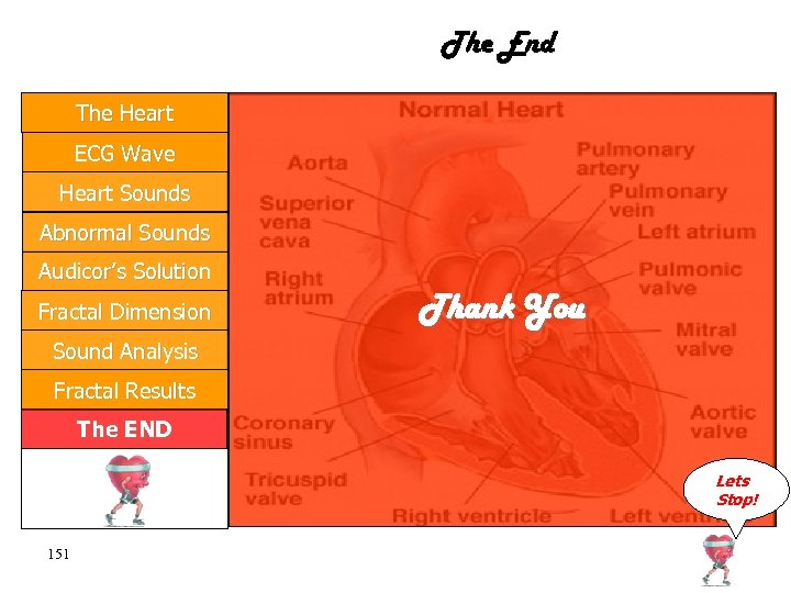
 The End The Heart ECG Wave Heart Sounds Abnormal Sounds Audicor’s Solution Fractal Dimension Thank You Sound Analysis Fractal Results The END Lets Stop! 92
 Fractal Definition The Heart ECG Wave Heart Sounds Abnormal sounds Audicor’s Solution An object which appears self-similar under varying degrees of magnification. Fractal Dimension In effect, possessing symmetry across scale, with each small part of the object replicating Sound Analysis Fractal Results the structure of the whole. The END Lets Go! 93
 Self-similarity The Heart ECG Wave Heart Sounds Abnormal sounds Audicor’s Solution Fractal Dimension Sound Analysis Fractal Results The END Lets Go! 94
 Fractal Dimension The Heart ECG Wave Heart Sounds Abnormal sounds All fractals are characterized by their own Audicor’s Solution dimension, which is usually a non-integer dimension, Fractal Dimension that is greater than their topological dimension and less than their Euclidean dimension. Sound Analysis Fractal Results The END Lets Go! 95
 Fractal Dimension The Heart ECG Wave Heart Sounds Abnormal sounds Audicor’s Solution This definition of fractal dimension is often used as an alternative definition of fractal objects. Fractal Dimension However, the fractal dimension may be estimated in numerous ways, such as the box. Sound Analysis Fractal Results counting dimension and the variance dimension. The END Lets Go! 96
 Chaotic dynamics The Heart ECG Wave Heart Sounds Abnormal sounds Audicor’s Solution A chaotic system refers to a system that never exactly repeats its behavior. Fractal Dimension Regardless of how long we let the model run for, we would never come across a repetition in Sound Analysis Fractal Results the waveform due to its aperiodic feature. The END Lets Go! 97
 Chaos The Heart ECG Wave Heart Sounds Abnormal sounds Audicor’s Solution Fractal Dimension Sound Analysis This behavior is known as chaos. By plotting the models variables against each other we receive a visualization of the dynamics of the system. Fractal Results The END Lets Go! 98
 Phase Portrait The Heart ECG Wave Heart Sounds Abnormal sounds Audicor’s Solution Fractal Dimension Sound Analysis Fractal Results The phase portrait will have the same form although the model is started with different initial conditions, the system will be attracted to this type of final solution. In two dimensions these plots are known as phase portraits. The END Lets Go! 99
 Strange Attractors The Heart ECG Wave Heart Sounds Abnormal sounds This plot is known as the attractor of the system. Audicor’s Solution The attractors for chaotic systems are termed Fractal Dimension strange attractors. Sound Analysis Fractal Results The END The fractal structures of these strange attractors may be classified by calculating their fractal dimensions. Lets Go! 100
 The Cantor Set The Heart ECG Wave Heart Sounds Abnormal sounds The cantor set consists of an infinite set of disappearing line segments in the unit interval. The set is generated by iteratively removing the middle third of line segments, resulting in a collection of infinitely many Fractal Dimension disappearing line segments lying on the unit Sound Analysis interval. Audicor’s Solution Fractal Results The END Both the line segments individual and combined length approach zero as the number of line segments approach infinity. Lets Go! 101
 The Cantor Set The Heart ECG Wave Heart Sounds Abnormal sounds Audicor’s Solution Fractal Dimension Sound Analysis Fractal Results The END Lets Go! 102
 The Koch curve The Heart ECG Wave Heart Sounds Abnormal sounds Audicor’s Solution Fractal Dimension Sound Analysis Fractal Results The END The iterative procedure used to construct the Koch curve begins, similar to the Cantor set, with the initiator of the set as the unit line segment. The generator is constructed by removing the middle third of the line segment and then replace it with two equal segments formed as two sides of a triangle. The process is repeated an infinite number of times to produce the Koch curve. Lets Go! 103
 The Koch curve The Heart ECG Wave Heart Sounds Abnormal sounds Audicor’s Solution Fractal Dimension Sound Analysis Fractal Results The END Lets Go! 104
 Regular Fractals The Heart ECG Wave Heart Sounds Abnormal sounds Audicor’s Solution Fractal Dimension Sound Analysis Fractal Results The END Regular fractals are fractal objects that possess exact self-similarity, objects with structures comprising of exact copies of themselves at all magnifications. The most commonly known regular fractals are possibly the Cantor set and the Koch curve, both simply constructed using an iterative procedure. Lets Go! 105
 Random Fractals The Heart ECG Wave Heart Sounds Random fractals are statistically self-similar. Each small part of a random fractal has the same statistical properties as the whole. Abnormal sounds Random fractals may be constructed mathematically by introducing a random Audicor’s Solution feature in the generating process of a regular fractal. Fractal Dimension Sound Analysis Fractal Results The END 106 For instance, when generating the Cantor set any third of the line segment is removed instead of the middle third. Many properties of natural objects and phenomena may be described using random Lets fractals. Go!
 Fractal boundaries The Heart ECG Wave Heart Sounds Abnormal sounds Audicor’s Solution Fractal Dimension Sound Analysis Fractal Results In order for a fractal curve to be classified as a fractal boundary it must meet two conditions: 1. The curve must be non-crossing, meaning that the fractal curve does not intersect itself 2. As the fractal curve is zoomed in it reveals more structure (details). The END Lets Go! 107
 Fractal boundaries The Heart ECG Wave Heart Sounds Abnormal sounds Audicor’s Solution Fractal Dimension Sound Analysis Fractal Results The END Lets Go! 108
 Fractal boundaries The Heart ECG Wave Heart Sounds Abnormal sounds Audicor’s Solution Dimension measurements are suitable in order to characterize and quantify the statistical selfsimilarity property of random fractal boundaries. Fractal Dimension As random fractals do not possess exact selfsimilarity the similarity dimension may not be Sound Analysis Fractal Results The END used. Instead we define estimates of the fractal dimension of random fractals, which do not require the exact self-similar property. Lets Go! 109
 The box counting dimension The Heart ECG Wave Heart Sounds Abnormal sounds Audicor’s Solution The box counting dimension, enables noninteger dimensions to be found for fractal curves. covers the object in self-similar boxes. In order to determine the box counting Fractal Dimension dimension of a fractal object, the object is covered with elements or boxes of side length ε. Sound Analysis Fractal Results The END The number of boxes, N, required to cover the object together with the side length ε is then used to determine the dimension. Lets Go! 110
 The box counting dimension The Heart ECG Wave Heart Sounds Abnormal sounds Audicor’s Solution Fractal Dimension Sound Analysis Fractal Results The END Lets Go! 111
 The box counting dimension The Heart ECG Wave Heart Sounds Abnormal sounds Audicor’s Solution The straight line is covered with elements of length ε, for simplicity we assume that the line is of unit length. Fractal Dimension In order to cover the line N elements are required regardless of the dimension of the Sound Analysis Fractal Results elements, here illustrated as cubes. The END Lets Go! 112
 The box counting dimension The Heart ECG Wave Heart Sounds Abnormal sounds Audicor’s Solution Fractal Dimension Sound Analysis To cover the unit line segment any elements with a dimension greater than or equal to the dimension of the line itself may be used, and still only require N of them. This leads to the following expression: Fractal Results The END Lets Go! 113
 The box counting dimension The Heart ECG Wave Heart Sounds Abnormal sounds Audicor’s Solution If, in stead, the same procedure would be applied to a plane of unit area, the expression received would be: Fractal Dimension Sound Analysis Fractal Results Similar reasoning with a 3 -dimensional object would lead to: The END Lets Go! 114
 The box counting dimension The Heart ECG Wave Heart Sounds Abnormal sounds Audicor’s Solution Fractal Dimension Sound Analysis In general, in order to cover an object of unit hypervolume the number of elements reuired are: In logarithmic form: Fractal Results The END Lets Go! 115
 The box counting dimension The Heart ECG Wave Heart Sounds Abnormal sounds By disregarding the assumption of unit hypervolume, a general expression of the box counting dimension may be received: Audicor’s Solution Fractal Dimension Sound Analysis Fractal Results where V is the hypervolume of the fractal object. The END Lets Go! 116
 The box counting dimension The Heart ECG Wave Heart Sounds Abnormal sounds Audicor’s Solution Fractal Dimension By rearranging the expression it is easy to see that it is an equation of the straight line, where the gradient of the line is the box counting dimension of the object, and by plotting log(N) against log(1/ε) for various elements with different side lengths d the box counting dimension may be determined: Sound Analysis Fractal Results The END Lets Go! 117
 The box counting dimension The Heart ECG Wave Heart Sounds To obtain a measure of the box counting dimension there are different methods of covering the fractal object. Three of them are illustrated below. Abnormal sounds Audicor’s Solution Fractal Dimension Sound Analysis Fractal Results The END Lets Go! 118
 The box counting dimension The Heart ECG Wave Heart Sounds Abnormal sounds The first method illustrated is covering the curve by placing boxes against each other in a way that a minimum number of boxes are being used. Audicor’s Solution Another method is to cover the fractal object with a grid of boxes and count the number of Fractal Dimension boxes that contain a part of the curve. Sound Analysis Fractal Results The END The last method illustrated is covering the curve with circles instead of boxes, placed in a similar way as with the boxes in the first method. Lets Go! 119
 The box counting dimension The Heart ECG Wave Heart Sounds Abnormal sounds Regardless of which method being used, the box counting dimension is still obtained from the derivate of the plot of log(N) against log(1/ε): Audicor’s Solution Fractal Dimension Sound Analysis Fractal Results The END Lets Go! 120
 The box counting dimension The Heart ECG Wave Heart Sounds Abnormal sounds Audicor’s Solution Fractal Dimension Sound Analysis Fractal Results The END Lets Go! 121
 The box counting dimension The Heart ECG Wave Heart Sounds Abnormal sounds Normally, in practical applications, the box counting dimension is estimated by selecting two points at small values of ε in the plot, resulting in an estimation given by: Audicor’s Solution Fractal Dimension Sound Analysis Fractal Results The END To receive a more accurate estimate of the box counting dimension a best fitted line may be drawn through the points at small values of ε. The slope, and consequently the box counting dimension, is then calculated from this best fitted line. Lets Go! 122
 Regular Brownian motion The Heart ECG Wave Heart Sounds Abnormal sounds Regular Brownian motion, or Brownian motion, is named after its discoverer Robert Brown. He observed that small particles floating in water underwent rapid irregular motions due Fractal Dimension to their bombardment by water molecules. Audicor’s Solution Sound Analysis Fractal Results The END If a group of particles is released at a certain location the bombarding molecules will cause the particles to spread out, diffuse, through time. Lets Go! 123
 Regular Brownian motion The Heart ECG Wave Heart Sounds Abnormal sounds Audicor’s Solution Fractal Dimension Sound Analysis Fractal Results The END The trajectories of particles undergoing Brownian motion in the plane may cross over themselves. Hence, Brownian motion may not be classified as a fractal boundary. Furthermore, as the Brownian trajectory is zoomed into, more structure is revealed, indicating that the statistical self-similarity features of the Brownian trajectory extends over all scales of magnification Lets Go! 124
 Regular Brownian motion The Heart ECG Wave Heart Sounds Abnormal sounds Audicor’s Solution Fractal Dimension Sound Analysis Fractal Results The END Lets Go! 125
 Regular Brownian motion The Heart ECG Wave Heart Sounds Abnormal sounds If the Brownian motion is sampled at intervals Audicor’s Solution of and the positions of the sampled points at time are denoted by , then the observed Fractal Dimension steps taken in the two coordinate directions and both follows a Sound Analysis Gaussian probability distribution. Fractal Results The END Lets Go! 126
 Regular Brownian motion The Heart ECG Wave Heart Sounds Abnormal sounds Audicor’s Solution Fractal Dimension Sound Analysis Fractal Results The END Lets Go! 127
 Regular Brownian motion The Heart ECG Wave Heart Sounds Abnormal sounds Audicor’s Solution Consequently, the step lengths between observed points also follows a Gaussian distribution: Fractal Dimension Sound Analysis Fractal Results The END Lets Go! 128
 Regular Brownian motion The Heart ECG Wave Heart Sounds The methods generally used to construct a Brownian motion in the plane are derived from these features. Abnormal sounds Hence, the motion is constructed by using Audicor’s Solution steps in the two coordinate directions, ∆x and ∆y randomly selected from a Gaussian Fractal Dimension distribution. Sound Analysis Fractal Results The END The step length r randomly selected from a Gaussian distribution T he step angel is randomly selected from a uniform distribution between 0 and. Lets Go! 129
 Regular Brownian motion The Heart ECG Wave Heart Sounds Abnormal sounds The time trace of Brownian motion, B(t), is equal to the time history of the coordinates of a Brownian trajectory, illustrating how the coordinate values vary in time. Audicor’s Solution The construction of a Brownian motion trace is derived from the property that successively Fractal Dimension increments the trace following a Guassian distribution: Sound Analysis Fractal Results The END Lets Go! 130
 Regular Brownian motion The Heart ECG Wave Heart Sounds Abnormal sounds Audicor’s Solution Thus, by sampling the Brownian motion trace at discrete times a discrete approximation may be constructed, by summarizing a series of random incremental steps, Fractal Dimension Thus Sound Analysis , is built up as an accumulated sum, Fractal Results The END Lets Go! 131
 Regular Brownian motion The Heart ECG Wave Heart Sounds Abnormal sounds Audicor’s Solution Fractal Dimension Sound Analysis Fractal Results The END Lets Go! 132
 Regular Brownian motion The Heart ECG Wave Heart Sounds Abnormal sounds Audicor’s Solution Fractal Dimension Sound Analysis Fractal Results The END For a continuous Brownian motion trace, B(t), the self-similar properties are apparent as zooming into it. Both the original trace and zoomed in traces displays the similar irregularity, as they are statistically self-similar. However, in order to retain the self-similar properties of the original trace the axes need to be scaled differently. Lets Go! 133
 Regular Brownian motion The Heart ECG Wave Heart Sounds Abnormal sounds Audicor’s Solution Fractal Dimension Sound Analysis Fractal Results The END Lets Go! 134
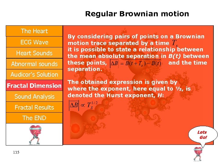 Regular Brownian motion The Heart ECG Wave Heart Sounds Abnormal sounds Audicor’s Solution By considering pairs of points on a Brownian motion trace separated by a time it is possible to state a relationship between the mean absolute separation in B(t) between these points, and the time separation. The obtained expression is given by Fractal Dimension where the exponent, here equal to ½, is denoted the Hurst exponent, H: Sound Analysis Fractal Results The END Lets Go! 135
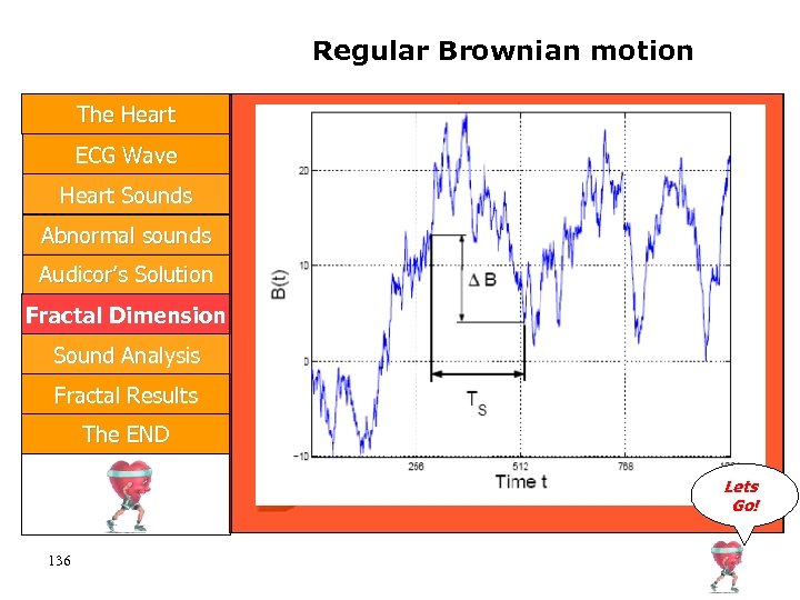 Regular Brownian motion The Heart ECG Wave Heart Sounds Abnormal sounds Audicor’s Solution Fractal Dimension Sound Analysis Fractal Results The END Lets Go! 136
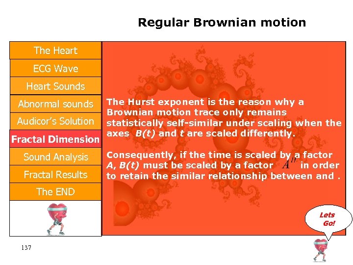 Regular Brownian motion The Heart ECG Wave Heart Sounds Abnormal sounds Audicor’s Solution Fractal Dimension Sound Analysis Fractal Results The Hurst exponent is the reason why a Brownian motion trace only remains statistically self-similar under scaling when the axes B(t) and t are scaled differently. Consequently, if the time is scaled by a factor A, B(t) must be scaled by a factor in order to retain the similar relationship between and. The END Lets Go! 137
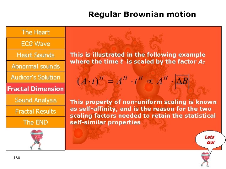 Regular Brownian motion The Heart ECG Wave Heart Sounds Abnormal sounds This is illustrated in the following example where the time t is scaled by the factor A: Audicor’s Solution Fractal Dimension Sound Analysis Fractal Results The END This property of non-uniform scaling is known as self-affinity, and is the reason for the two scaling factors needed to retain the statistical self-similar properties Lets Go! 138
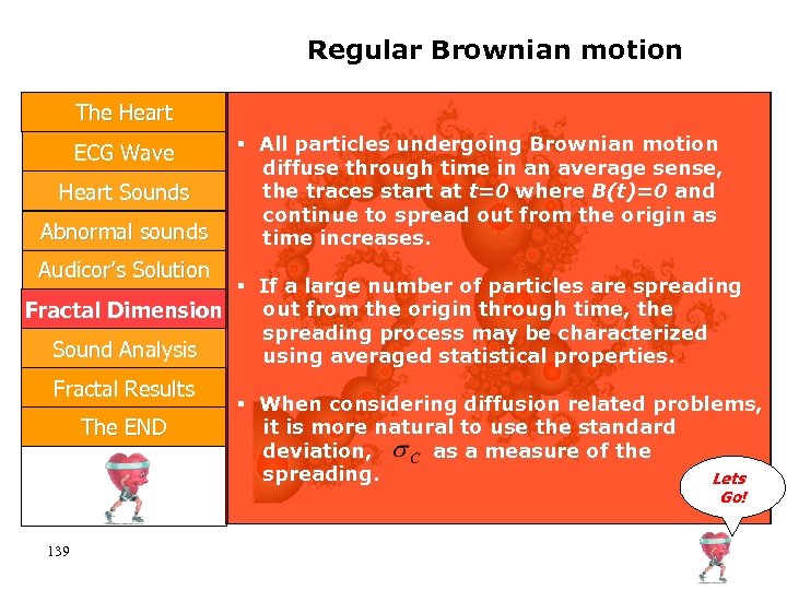 Regular Brownian motion The Heart ECG Wave Heart Sounds Abnormal sounds All particles undergoing Brownian motion diffuse through time in an average sense, the traces start at t=0 where B(t)=0 and continue to spread out from the origin as time increases. Audicor’s Solution If a large number of particles are spreading out from the origin through time, the Fractal Dimension spreading process may be characterized Sound Analysis using averaged statistical properties. Fractal Results The END When considering diffusion related problems, it is more natural to use the standard deviation, as a measure of the spreading. Lets Go! 139
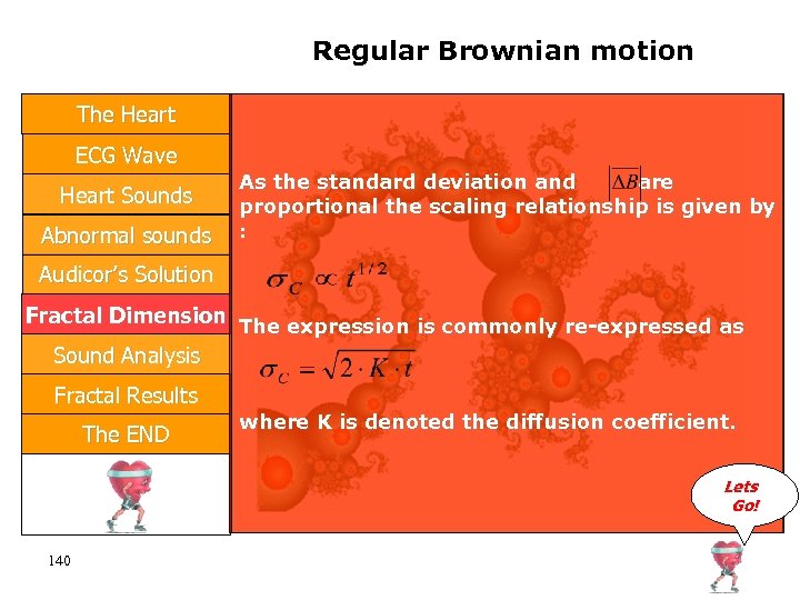 Regular Brownian motion The Heart ECG Wave Heart Sounds Abnormal sounds As the standard deviation and are proportional the scaling relationship is given by : Audicor’s Solution Fractal Dimension The expression is commonly re-expressed as Sound Analysis Fractal Results The END where K is denoted the diffusion coefficient. Lets Go! 140
 Regular Brownian motion The Heart ECG Wave Heart Sounds Abnormal sounds Audicor’s Solution Fractal Dimension Sound Analysis Fractal Results The END Lets Go! 141
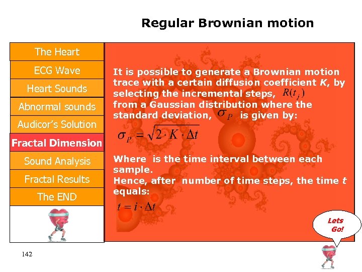 Regular Brownian motion The Heart ECG Wave Heart Sounds Abnormal sounds Audicor’s Solution It is possible to generate a Brownian motion trace with a certain diffusion coefficient K, by selecting the incremental steps, from a Gaussian distribution where the standard deviation, is given by: Fractal Dimension Sound Analysis Fractal Results The END Where is the time interval between each sample. Hence, after number of time steps, the time t equals: Lets Go! 142
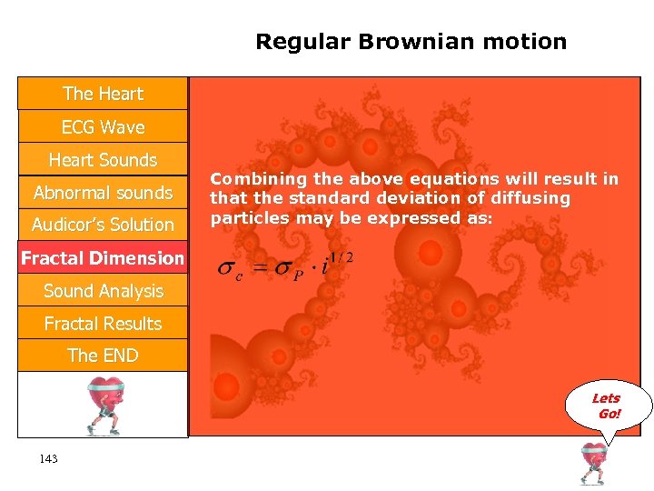 Regular Brownian motion The Heart ECG Wave Heart Sounds Abnormal sounds Audicor’s Solution Combining the above equations will result in that the standard deviation of diffusing particles may be expressed as: Fractal Dimension Sound Analysis Fractal Results The END Lets Go! 143
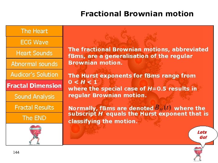 Fractional Brownian motion The Heart ECG Wave Heart Sounds Abnormal sounds Audicor’s Solution Fractal Dimension Sound Analysis Fractal Results The END The fractional Brownian motions, abbreviated f. Bms, are a generalisation of the regular Brownian motion. The Hurst exponents for f. Bms range from 0
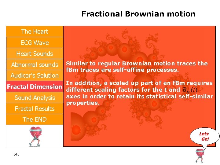 Fractional Brownian motion The Heart ECG Wave Heart Sounds Abnormal sounds Audicor’s Solution Similar to regular Brownian motion traces the f. Bm traces are self-affine processes. In addition, a scaled up part of an f. Bm requires Fractal Dimension different scaling factors for the t and axes in order to retain its statistical self-similar Sound Analysis Fractal Results properties. The END Lets Go! 145
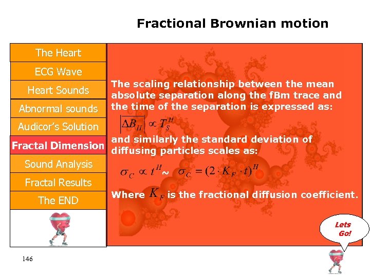 Fractional Brownian motion The Heart ECG Wave Heart Sounds Abnormal sounds The scaling relationship between the mean absolute separation along the f. Bm trace and the time of the separation is expressed as: Audicor’s Solution and similarly the standard deviation of Fractal Dimension diffusing particles scales as: Sound Analysis ~ Fractal Results The END Where is the fractional diffusion coefficient. Lets Go! 146
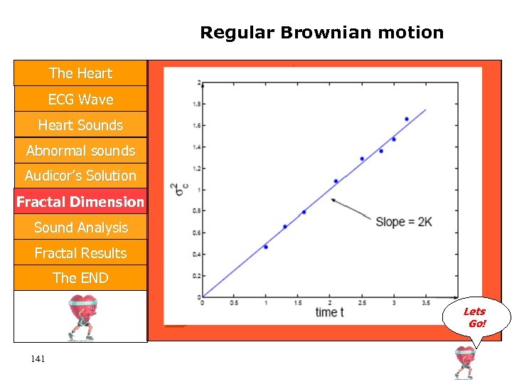
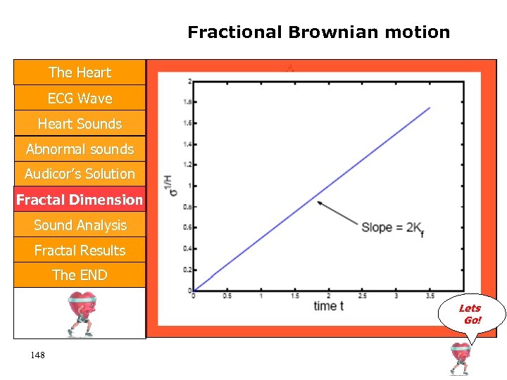
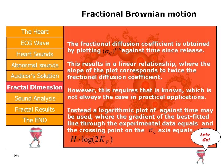 Fractional Brownian motion The Heart ECG Wave Heart Sounds Abnormal sounds Audicor’s Solution The fractional diffusion coefficient is obtained by plotting against time since release. This results in a linear relationship, where the slope of the plot corresponds to twice the fractional diffusion coefficient. Fractal Dimension However, this requires that is known, which is not always the case in practical applications. Sound Analysis Fractal Results The END Instead a logarithmic plot of against time may be used, where the gradient of the best-fitted line through the experimental data equals and the crossing point on the axis equals Lets Go! 147
 Fractional Brownian motion The Heart ECG Wave Heart Sounds Abnormal sounds Audicor’s Solution Fractal Dimension Sound Analysis Fractal Results The END Lets Go! 148
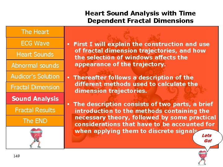 Heart Sound Analysis with Time Dependent Fractal Dimensions The Heart ECG Wave Heart Sounds Abnormal sounds Audicor’s Solution Fractal Dimension Sound Analysis Fractal Results The END First I will explain the construction and use of fractal dimension trajectories, and how the selection of windows affects the appearance of the trajectory. Thereafter follows a description of the different methods used to calculate the dimension trajectories. The description consists of two parts, a brief introduction to the methods containing the necessary theory, followed by some practical considerations that have to be accounted for when applying them to discrete signals. Lets Go! 149
 Heart Sound Analysis with Time Dependent Fractal Dimensions The Heart ECG Wave Heart Sounds Abnormal sounds Audicor’s Solution Fractal Dimension Sound Analysis Fractal Results The END First I will explain the construction and use of fractal dimension trajectories, and how the selection of windows affects the appearance of the trajectory. Thereafter follows a description of the different methods used to calculate the dimension trajectories. The description consists of two parts, a brief introduction to the methods containing the necessary theory, followed by some practical considerations that have to be accounted for when applying them to discrete signals. Lets Go! 150
 The End The Heart ECG Wave Heart Sounds Abnormal Sounds Audicor’s Solution Fractal Dimension Thank You Sound Analysis Fractal Results The END Lets Stop! 151


