
Heart ’’ s Anatomy and Physiology Stephen Jo


- Размер: 1.7 Mегабайта
- Количество слайдов: 27
Описание презентации Heart ’’ s Anatomy and Physiology Stephen Jo по слайдам
Heart ’’ s Anatomy and Physiology Stephen Jo T. Bonilla, RN. MD Department of Surgery St. Luke’s Medical Center
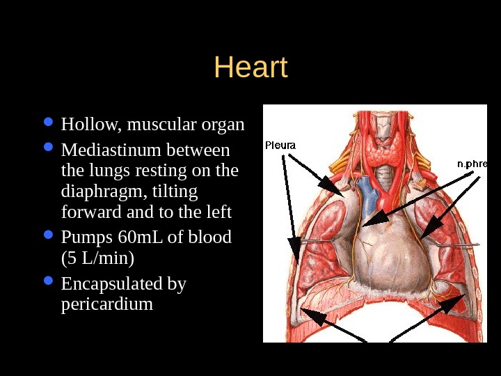
Heart Hollow, muscular organ Mediastinum between the lungs resting on the diaphragm, tilting forward and to the left Pumps 60 m. L of blood (5 L/min) Encapsulated by pericardium
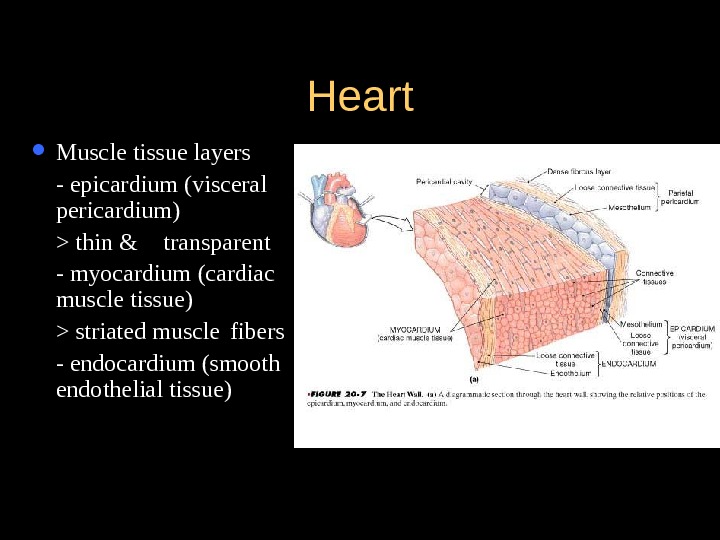
Heart Muscle tissue layers — epicardium (visceral pericardium) > thin & transparent — myocardium (cardiac muscle tissue) > striated muscle fibers — endocardium (smooth endothelial tissue)
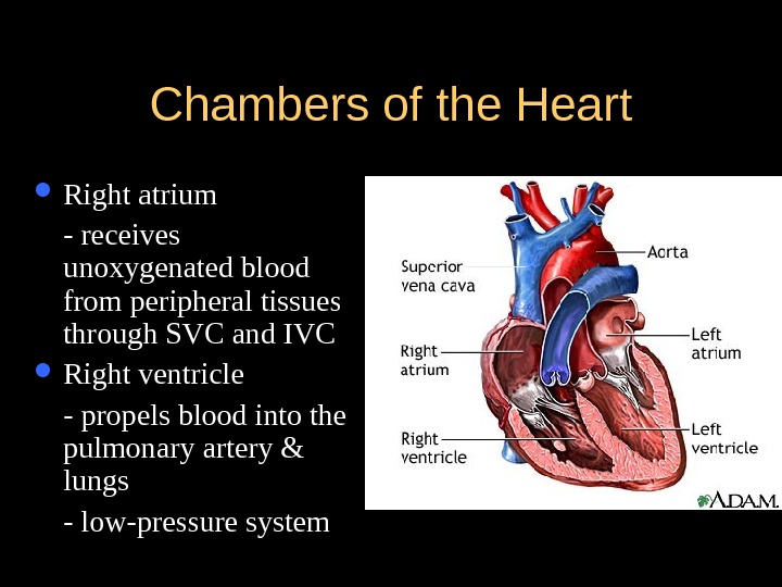
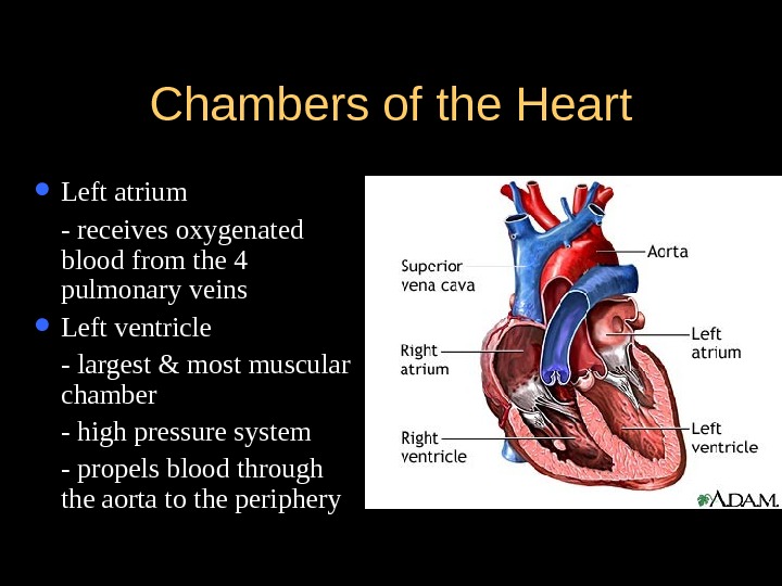
Chambers of the Heart Right atrium — receives unoxygenated blood from peripheral tissues through SVC and IVC Right ventricle — propels blood into the pulmonary artery & lungs — low-pressure system
Chambers of the Heart Left atrium — receives oxygenated blood from the 4 pulmonary veins Left ventricle — largest & most muscular chamber — high pressure system — propels blood through the aorta to the periphery
Heart Valves Atrioventricular valves — Tricuspid valve (3 leaflets) > separates RA from RV — Bicuspid valve (mitral valve) > separates LA from LV Semilunar valves — Pulmonic valve > separates RV from the pulmonary artery — Aortic valve > separates LV from the aorta
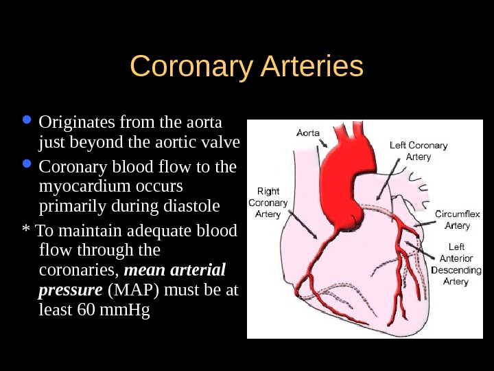
Coronary Arteries Originates from the aorta just beyond the aortic valve Coronary blood flow to the myocardium occurs primarily during diastole * To maintain adequate blood flow through the coronaries, mean arterial pressure (MAP) must be at least 60 mm. Hg
Coronary Arteries Left main coronary artery (LCA) — Left anterior descending artery (LAD) >descends toward the anterior wall & apex of LV > supplies LV, ventricular septum, chordae, papillary muscle & RV — Left circumflex artery (LXA) > descends toward the lateral wall of LV & apex > supplies LA, lateral & posterior LV surfaces *45% supplies SA node
Coronary Arteries Right main coronary artery (RCA) — descends toward the apex of RV — supplies the RA, RV, & inferior portions of LV
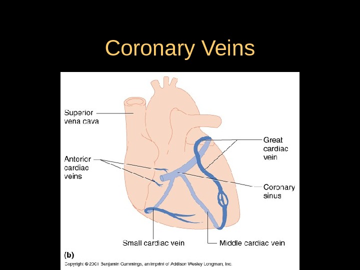
Coronary Veins
Physiology of the Heart Electrophysiologic properties (regulates heart rate & rhythm) — Automaticity – ability of all cardiac cells to initiate an impulse spontaneously & repetitively — Excitability – ability of cardiac cells to respond to stimulus by initiating an impulse (depolarization) — Conductivity – cardiac cells transmit the electrical impulses they receive — Contractility – cardiac cells contract in response to an impulse — Refractoriness – cardiac cells are unable to respond to a stimulus until they’ve recovered (repolarized)
Conduction System of the Heart Sinoatrial node (SA node) — pacemaker cells initiate impulses at 60 – 100 beats/min Atrioventricular node (AV node) — located in the junctional area — normal delay (allows the atria to contract completely before the ventricles are stimulated to contract) — 40 – 60 beats/min Bundle of His — located in the interventricular septum — Left and Right bundle branches Purkinje fibers — terminal branches that carry the impulse to RV & LV — 20 – 40 beats/min
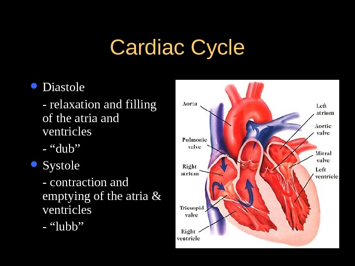
Cardiac Cycle Diastole — relaxation and filling of the atria and ventricles — “dub” Systole — contraction and emptying of the atria & ventricles — “lubb”
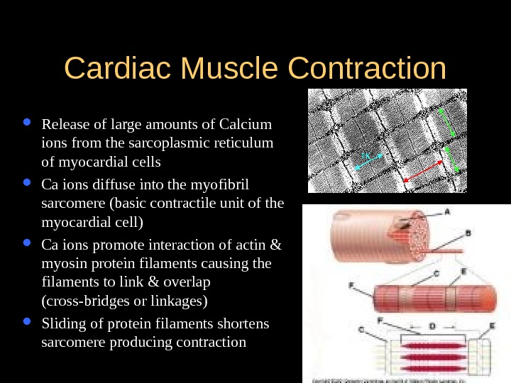
Cardiac Muscle Contraction Release of large amounts of Calcium ions from the sarcoplasmic reticulum of myocardial cells Ca ions diffuse into the myofibril sarcomere (basic contractile unit of the myocardial cell) Ca ions promote interaction of actin & myosin protein filaments causing the filaments to link & overlap (cross-bridges or linkages) Sliding of protein filaments shortens sarcomere producing contraction
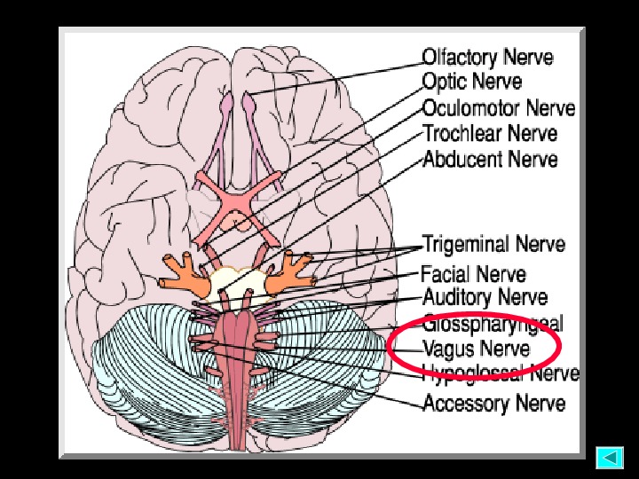
Mechanical Properties of the Heart Cardiac output — volume of blood (liters) ejected by the heart each minute — 4 – 7 L/min — heart rate x stroke volume Heart rate — number of times the ventricles contract each minute — 60 – 100 beats/min — controlled by the ANS via the vagus nerve Stroke volume — amount of blood ejected by the LV during each systole
Mechanical Properties of the Heart Preload — degree of myocardial stretch at the end of diastole & just before contraction — determined by the amount of blood returning to the heart from the venous ® & pulmonary system (L) – LVED *Starling’s law – the more the heart is filled during diastole, the more forcefully it contracts
Mechanical Properties of the Heart Afterload — pressure or resistance that the ventricles must overcome to eject blood through the semilunar valves & into the peripheral blood vessels — directly proportional to the BP & diameter of blood vessels Impedance (peripheral component of afterload) — pressure that the heart must overcome to open the aortic valve — depends on aortic compliance, blood viscosity & arteriolar constriction
Mechanical Properties of the Heart Myocardial contractility — force of cardiac contraction independent of preload — increased by sympathetic nervous stimulation, calcium release & positive inotropic drugs — decreased by hypoxemia & acidemia
Vascular System Provide conduits for blood to travel from the heart to nourish the body Carry cellular wastes to the excretory organs Allow lymphatic flow to drain tissue fluid back to the circulation Return blood to the heart for recirculation
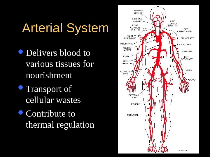
Arterial System Delivers blood to various tissues for nourishment Transport of cellular wastes Contribute to thermal regulation
Arterial System Blood pressure — force of blood exerted against the vessel walls = CO x peripheral vascular resistance — regulated by: > autonomic nervous system (ANS) > kidneys (renin-angiotensin-aldosterone) > endocrine system (catecholamines, kinins, serotonin, histamine)
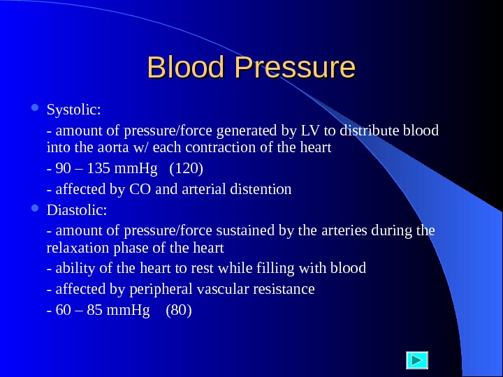
Blood Pressure Systolic: — amount of pressure/force generated by LV to distribute blood into the aorta w/ each contraction of the heart — 90 – 135 mm. Hg (120) — affected by CO and arterial distention Diastolic: — amount of pressure/force sustained by the arteries during the relaxation phase of the heart — ability of the heart to rest while filling with blood — affected by peripheral vascular resistance — 60 – 85 mm. Hg (80)
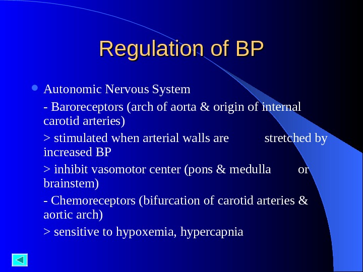
Regulation of BP Autonomic Nervous System — Baroreceptors (arch of aorta & origin of internal carotid arteries) > stimulated when arterial walls are stretched by increased BP > inhibit vasomotor center (pons & medulla or brainstem) — Chemoreceptors (bifurcation of carotid arteries & aortic arch) > sensitive to hypoxemia, hypercapnia
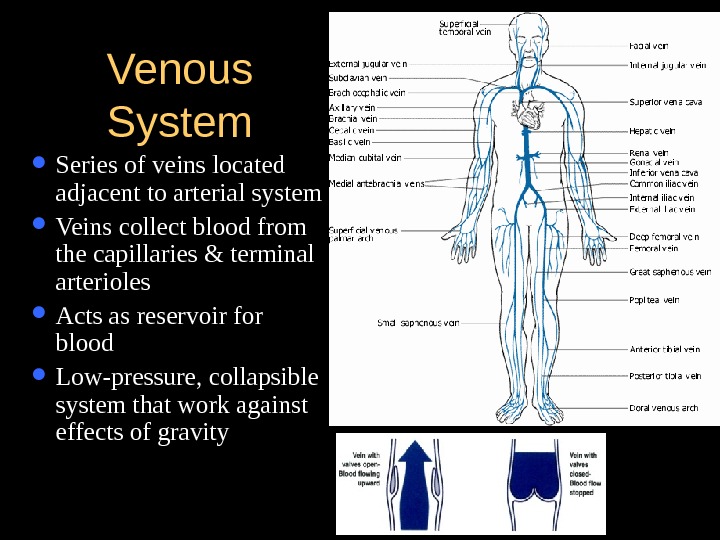
Venous System Series of veins located adjacent to arterial system Veins collect blood from the capillaries & terminal arterioles Acts as reservoir for blood Low-pressure, collapsible system that work against effects of gravity