Атеросклероз+ГБ.ppt
- Количество слайдов: 69


Гродненский Государственный Медицинский Университет Гродно-2013
")
Болезни сердечно-сосудистой системы (атеросклероз, артериальная гипертензия, ИБС)
 n эндокринный (сахарный диабет)")
Этиологические факторы атеросклероза n нервный n нарушение обмена веществ (гиперхолестеринемия) n эндокринный (сахарный диабет) n наследственность n гипертония

Макроскопические стадии атеросклероза: n Жировых пятен и полосок n Фиброзных бляшек n Осложненных поражений (изъязвление, кровоизлияние, тромбоз) n Атерокальциноза
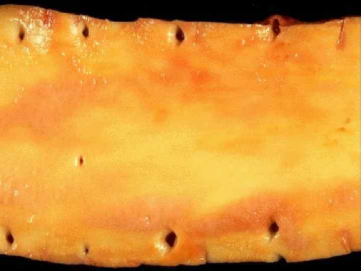

These three aortas demonstrate mild, moderate, and severe atherosclerosis from bottom to top. At the bottom, the mild atherosclerosis shows only scattered lipid plaques. The aorta in the middle shows many more larger plaques. The severe atherosclerosis in the aorta at the top shows extensive ulceration in the plaques.

Микроскопические стадии атеросклероза: n Долипидная n Липидоза n Липосклероза n Атероматоза n Изъязвления n Атерокальциноза

Изменения в долипидной стадии атеросклероза n Повышение проницаемости интимы n Распад эластических мембран n Накопление глюкозамингликанов (Но сохраняется высокая активность липолитических и протеолитических ферментов)

This microscopic cross section of the aorta shows a large overlying atheroma on the left. Cholesterol clefts are numerous in this atheroma. The surface on the far left shows ulceration and hemorrhage. Despite this ulceration, atheromatous emboli are rare (or at least, complications of them are rare).

This high magnification of the atheroma shows numerous foam cells and an occasional cholesterol cleft. A few dark blue inflammatory cells are scattered within the atheroma.

The coronary artery shown here has narrowing of the lumen due to build up of atherosclerotic plaque. Severe narrowing can lead to angina, ischemia, and infarction.

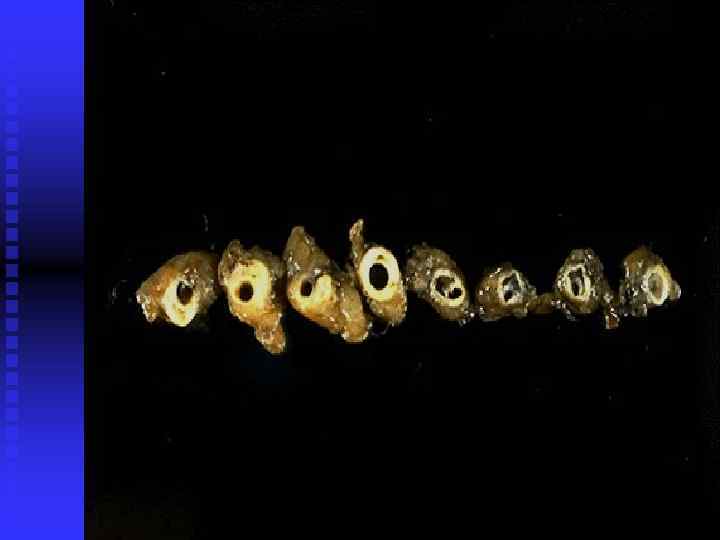
Клинико-анатомические формы атеросклероза: n Атеросклероз аорты n Артерий нижних конечностей n Брыжеечных артерий n Артерий почек n Артерий головного мозга n Артерий сердца
пристеночным тромбом с тромбоэмболией")
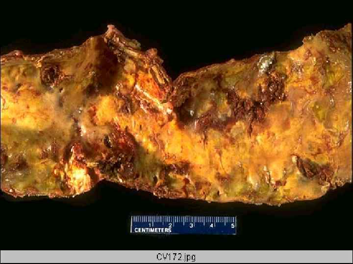
Атеросклероз аорты: -наиболее выражено поражение брюшного отдела, задней стенки -осложняется: а)пристеночным тромбом с тромбоэмболией б)эмболией атероматозными массами в)анеризмой (цилиндрической, мешковидной, грыжевидной)

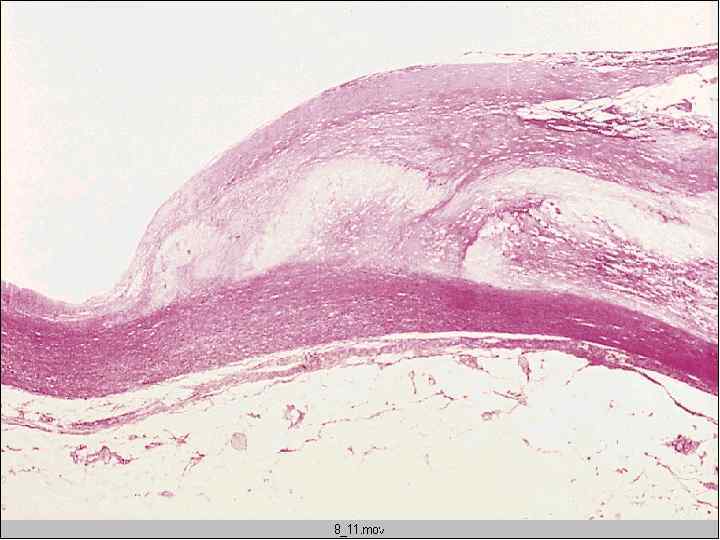
This aorta has been opened longitudinally to reveal an area of fairly limited dissection that is organizing. The red-brown thrombus can be seen in on both sides of the section as it extends around the aorta. The intimal tear would have been at the left. This creates a "double lumen" to the aorta. This aorta shows severe atherosclerosis which, along with cystic medial necrosis and hypertension, is a risk factor for dissection.

This microscopic cross section of the aorta demonstrates a red blood clot that is compressing the aortic lumen. This occurred as a result of aortic dissection in which there was a tear in the intima followed by dissection of blood at high pressure out through the muscular wall to the adventitia. This blood dissecting out can lead to sudden death from hemothorax.

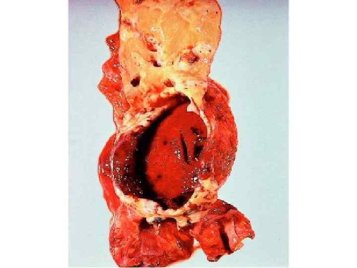
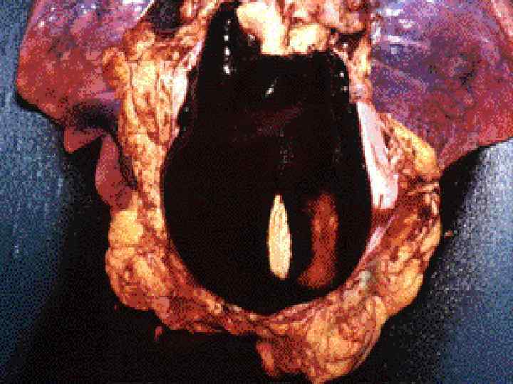
Atherosclerosis may weaken the wall of the aorta such that it bulges out to form an aneurysm. An atherosclerotic aortic aneurysm typically occurs in the abdominal portion below the renal arteries, as shown here. Aortic aneurysms that get bigger than 6 or 7 cm are likely to rupture.

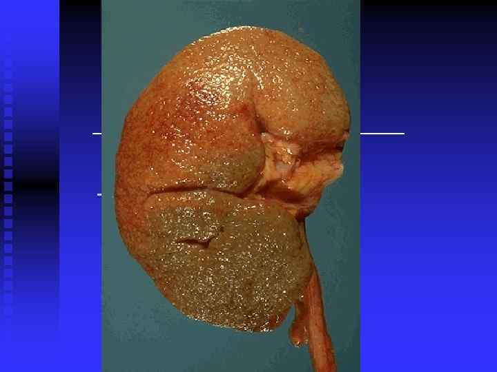
Атеросклероз артерий почек

Атеросклероз брыжеечных артерий

Атеросклероз артерий нижних конечностей

Симптомы перемежающейся хромоты n Боль в мышцах конечности при физической нагрузке n Похолодание конечности n Атрофия мышц конечности

Атеросклероз мозговых артерий

This is an intermediate to remote infarct in the distribution of the middle cerebral artery.

This cerebral infarction demonstrates the presence of many macrophages at the right which are cleaning up the lipid debris from the liquefactive necrosis.

In this case of Alzheimer's disease, there is more marked atrophy seen superiorly and laterally, with sparing of the occipital region.
 n Острая ИБС (инфаркт мио- карда, очаговая дистрофия миокарда) n")
Атеросклероз артерий сердца (ИБС) n Острая ИБС (инфаркт мио- карда, очаговая дистрофия миокарда) n Хроническая ИБС (диффуз- ный и очаговый кардиосклероз, хроническая аневризма)

Морфологические проявления и. М со стороны коронарных сосудов: Морфологические проявления спазма (гофрированность эластической мембраны, расположение эндотелия в виде частокола и т. д. ) n Плазматическое пропитывние стенки n Атеросклероз и тромбоз артерии n Изъязвление атеросклеротической бляшки или эмболия атероматозными массами n Кровоизлияние в атеросклеротическую бляшку n

This is coronary atherosclerosis with the complication of hemorrhage into atheromatous plaque, seen here in the center of the photograph. Such hemorrhage acutely may narrow the arterial lumen.

Despite the frequency of aortic atherosclerosis, cholesterol emboli are rare, or at least insignficant most of the time. Seen here in a renal artery branch are cholesterol clefts of such an embolus. This patient had severe ulcerative, friable atheromatous plaques and had undergone angiography, which increases the risk for such emboli.

Морфологические проявления ИМ со стороны миокарда n Пестрота миокарда не разрезе n Контрактурный тип поврежения миофибрилл n Исчезновение поперечной исчерченности n Эозинофильная или фуксинофильная дегенерация мышечных волокон n Глыбчатый распад миофибрилл n Внутриклеточный миоцитолиз с фрагментацией волокон n Накопление липидов, исчезновение гликогена n Венозное полнокровие и отек интерстиция, стазы, периваскулярные геморрагии n Мышечные волокна резко ШИК- положительные

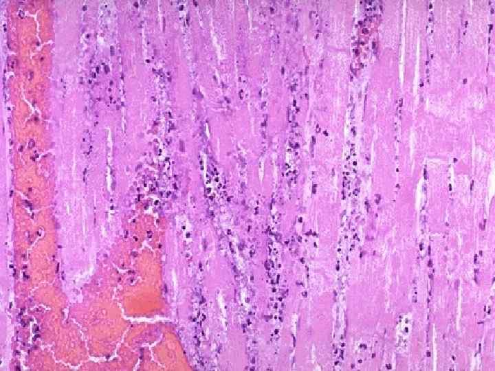
The earliest change histologically seen with acute myocardial infarction in the first day is contraction band necrosis. The myocardial fibers are beginning to lose cross striations and the nuclei are not clearly visible in most of the cells seen here. Note the many irregular darker pink wavy contraction bands extending across the fibers.

This high power microscopic view of the myocardium demonstrates an infarction of about 1 to 2 days in duration. The myocardial fibers have dark red contraction bands extending across them. The myocardial cell nuclei have almost all disappeared. There is beginning acute inflammation. Clinically, such an acute myocardial infarction is marked by changes in the electrocardiogram and by a rise in the MB fraction of creatine kinase.

In this microscopic view of a recent myocardial infarction, there is extensive hemorrhage along with myocardial fiber necrosis with contraction bands and loss of nuclei.

Причины инфаркта миокарда Спазм коронарной артерии n Тромбоз n Эмболия n Функциональное напряжение органа при недостаточном кровоснабжении n Наиболее частая локализация инфаркта миокарда Передняя стенка левого желудочка n Боковая стенка n Межжелудочковая перегородка n Верхушка сердца n

A thrombosis of a coronary artery is shown here in cross section. This acute thrombosis diminishes blood flow and leads to ischemia and/or infarction, marked clinically by the sudden onset of chest pain.

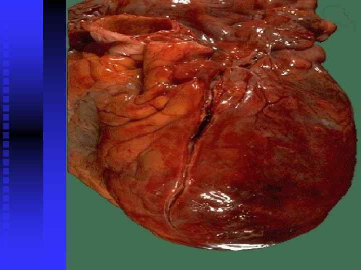
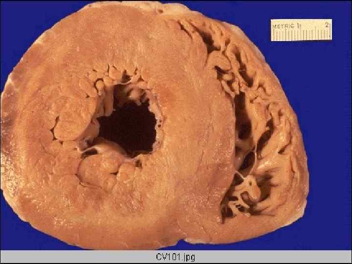
The interventricular septum of the heart has been sectioned to reveal an extensive acute myocardial infarction. The dead muscle is tan-yellow with a surrounding hyperemic border.

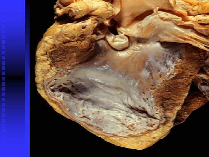
Разновидности инфаркта миокарда n Субэндокардиальный n Субэпикардиальный n Интрамуральный n Трансмуральный

Осложнения инфаркта миокарда Фибринозный перикардит n Пристеночный тромбоз с тромбоэмболией n Разрыв сердца с гемоперикардом и тампонадой n Асистолия n Фибрилляция желудочков n Острая сердечно-сосудистая недостаточность n Кардиогенный шок n

This is an intermediate myocardial infarction of 1 to 2 weeks in age. Note that there are remaining normal myocardial fibers at the top. Below these fibers are many macrophages along with numerous capillaries and little collagenization.

There is pale white collagen within the interstitium between myocardial fibers. This represents an area of remote infarction.
нервный фактор б)почечный")
Артериальная гипертензия: болезнь высокого артериального давления, болезнь «не отреагированных эмоций» Этиология: а)нервный фактор б)почечный в)рефлекторный г)наследственность д)эндокринные нарушения и т. д.

Причины симптоматической гипертонии Заболевания почек (острые и хронический нефриты, пиелонефриты, почечно-каменная болезнь и др. ) n Эндокринная патология (болезни гипофиза, надпочечников, шитовидной железы и др. ) n Органическое поражение ЦНС (опухоли, травмы, цистицеркоз и др. ) n Болезни сердечно-сосудистой системы (коарктация аорты, васкулиты, пороки сердца и др. ) n

Стадии АГ n Функциональная n Распространенных измене- ний артерий n Изменения органов вследствие поражения артерий

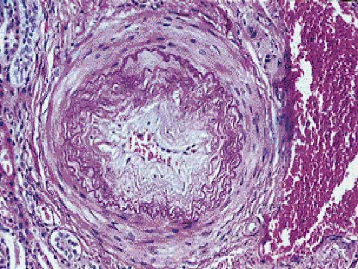
Изменения сосудов мелкого калибра при АГ n Плазматическое пропиты- вание n Артериолосклероз n Артериологиалиноз n Фибриноидный некроз стенки
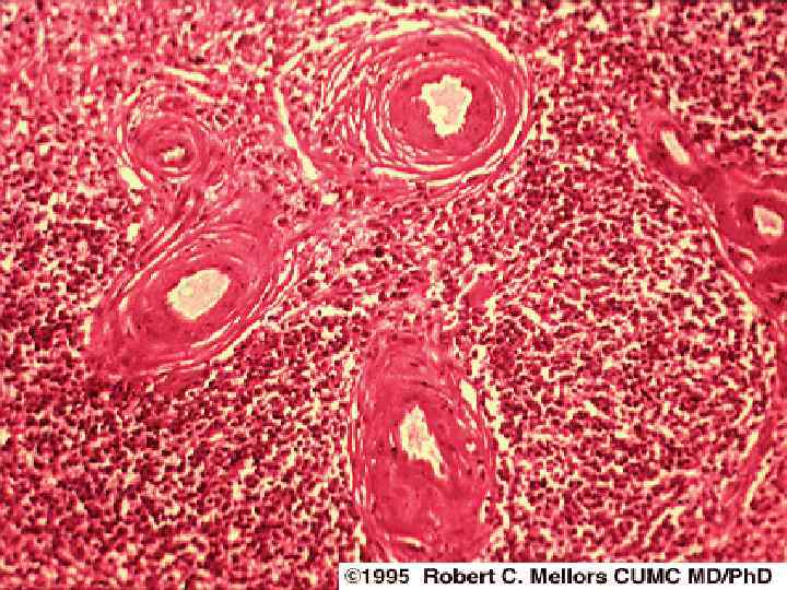

This is a different kind of arteriosclerosis. This is hyperplastic arteriolosclerosis, which most often appears in the kidney in patients with malignant hypertension. The arteriolar wall is markedly thickened and the lumen is narrowed.

One complication of hyperplastic arteriolosclerosis with malignant hypertension is fibrinoid necrosis, as seen here in a renal arteriole.

Поражение мелких артерий более выражены в: n Почках n Головном мозге n Сетчатке глаза n Кишечнике n Поджелудочной железе
 n Гофрированность эластической мембраны n Расположение эндотелия в")
Морфологические признаки гипертонического криза (спазма артерий) n Гофрированность эластической мембраны n Расположение эндотелия в виде частокола n Фибриноидный некроз стенки n Периваскулярные геморрагии
резко выражен б)спускается")
Изменения сосудов крупного калибра при АГ: n Атеросклероз (при этом он: а)резко выражен б)спускается на мелкие артерии, поражая артерии мышечного типа в)Атеросклеротические бляшки располагаются концентрически (циркулярно) n Эластофиброз

There is a pink to red recent thrombosis in this narrowed coronary artery. The open, needle-like spaces in the atheromatous plaque are cholesterol clefts.

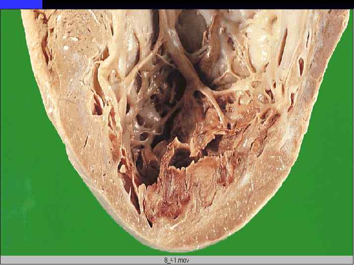
Клинико-анатомические формы артериальной гипертензии: n. Сердечная n. Мозговая n. Почечная
 in addition to")
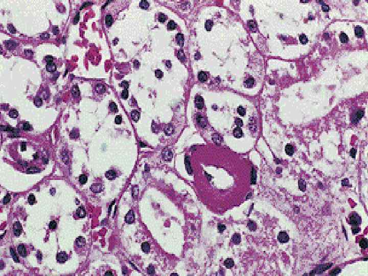
There are two other forms of arteriosclerosis (hardening of the arteries) in addition to atherosclerosis: arteriolosclerosis and medial calcific sclerosis. Arteriolosclerosis is typically seen in the kidneys. One form, called hyaline arteriolosclerosis, is demonstrated by the markedly thickened arteriole to the lower right of this glomerulus with PAS stain. Hyaline arteriolosclerosis is seen in the elderly, but more advanced lesions are seen in persons with diabetes mellitus and/or with hypertension.
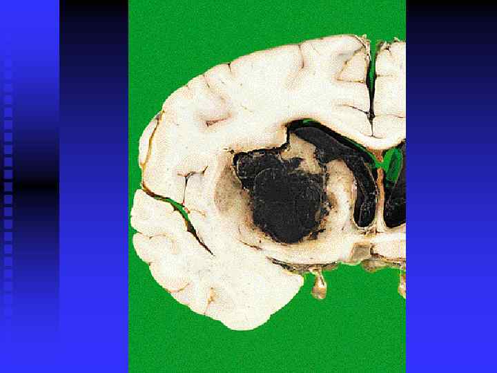
подкорковые (гематома или диядра")
Мозговая форма АГ Локализация Виды инсульта кровоизлияний n Геморрагический инсульт а)подкорковые (гематома или диядра апедезные геморрагии б)мозжечок n Ишемический инсульт

This intermediate infarct of the frontal lobe shows liquefactive necrosis with formation of cystic spaces as resolution begins.
 located 7 cm above the aortic valve and proximal")
There is a tear (arrow) located 7 cm above the aortic valve and proximal to the great vessels in this aorta with marked atherosclerosis. This is an aortic dissection.
Атеросклероз+ГБ.ppt