98924690603b278e0f9249fab3e70d54.ppt
- Количество слайдов: 65


GI bleeding Mackay Memorial Hospital Department of Internal Medicine Division of Gastroenterology R 4 陳泓達 97/6/22

n GI Bleeding n UGI bleeding n Peptic ulcer disease n Variceal bleeding n LGI bleeding

UGI bleeding: 5 times more common than LGI bleeding. Men > Women Elderly persons. n Despite ongoing advances, fundamental principles are the same !!!! immediate assessment and stabilization of hemodynamic status n

Determine the source of bleeding Stop active bleeding Treat underlying abnormality Prevent recurrent bleeding
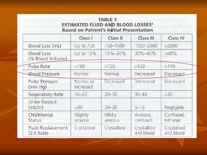
 Severity of bleeding normal < 10 minor Orthostatic")
hemodynamics Blood loss(% of intravascular volume) Severity of bleeding normal < 10 minor Orthostatic hypotension or tachycardia 10 -20 moderate shock 20 -25 massive

Resuscitation In hemodynamically unstable… Set up two large-bore IV catheter Colloid solution (NS or lactated Ringer’s) To restore vital sign !! ICU monitor is indicated Central venous monitoring F/U vital sign and urine output

History taking and physical examination UGI or LGI ? UGI peptic ulcer disease or portal hypertension related (EV or GV)? n

Differentiate LGI and UGI Melena – upper GI cause in 90% n. Hematochezia – upper GI cause in 10% n

The intermediate patient Take more time…. Re-examine, Monitor vital signs, Re-check CBC, BUN

Transfusion ? In hemodynamic unstable, any sign of poor tissue oxygenation, continued bleeding, persistent low Ht level(20 -25%) Maintain adequate perfusion Target ? n

Other Blood tests on the bleeding patient… INR, PTT – coagulopathy anyone?

“There is no single value of hemoglobin concentration that justifies or requires transfusion; an evaluation of the patient’s clinical situation should also be a factor in the decision. ” Capital Health Guide to Blood Transfusion

You’ve decided to give blood… Options?

O neg – immediately available Type Specific – 10 – 15 min. – 30 – 60 min. Full Cross Match

What is in a unit of packed cells? 250 m. L volume n. Contains citrate (anticoagulant), and preservative. n 1 unit packed cells will increase the Hb concentration by approx. --? 0. 5 mg/d. L n
 transfused within 24")
Massive Transfusion Greater than 1 blood volume( or 10 units ) transfused within 24 hours n. May dilute platelets and clotting factors n
")
Dilution coagulopathy Monitor the patient for coagulopathy n. Follow the resuscitation (CBC, INR, PTT) n

Treatment of dilution coagulopathy Plasma /FFP 10 – 15 m. L / kg Usual adult dose 2 units. 5 – 8 m. L / kg dose for warfarin reversal

Treatment of dilution coagulopathy Platelets Keep the count greater than 50 , 000 in the bleeding patient 1 unit should increase platelet count by 5 , 000– 10, 000 / L Dose: 6 pack
")
Massive Transfusion What else can go wrong? Hypothermia Potassium Citrate toxicity (hypocalcemia)

Vomiting Blood Hematemesis Upper GI Bleeding

Etiology Peptic Ulcer 50 % n. Gastritis 20% n. Esophageal varices 10% n. The rest: Tears, AVM, CA, etc 20% n

More about bleeds…. 80 % of Non – variceal upper GI bleeds will stop spontaneously n 60 % of variceal bleeds will stop spontaneously n

What else can I do for GI bleeding, before endoscopy NG lavage Drug ABC Patient and family Agree ( Sign permit first)

Urgent Endoscopy ? Initial evaluation: 初始出血量是否大量 ? 出血量大者 , rebleeding 機會也大 觀察重點 : vital sign (tachycardia, orthostatic hypotension resting hypotension, shock), 吐血或 血便黑便的頻次與量 , NG lavage的結果 n

NG lavage 15 – 20 % of upper GI bleeds have a negative aspirate Sensitivity 79%, Specificity 55% Cuellar et al, Arch of Int Med Jul 1990 • For endoscopic preparation ( not contraindicated in patients with varices)

Endoscopy Diagnostic n. Therapeutic n. Prognostic n

Endoscopic features and risk of re-bleeding Active bleeding 55 – 90%

Endoscopic features and risk of rebleeding Non bleeding visible vessel 40 – 50 %

Endoscopic features and risk of rebleeding Adherent clot 10 – 33%

Endoscopic features and risk of rebleeding Flat spot 7 – 10 %

Endoscopic features and risk of rebleeding Clean base 3 – 5%

Variceal bleeding Non-variceal bleeding

Drugs: Peptic ulcer bleeding Manipulation of gastric p. H

Use of PPI’s Theory : raise gastric p. H n. Better platelet activity n. Pepsinogen requires acid to become activated to pepsin n. Clots will form, clots not digested

High Risk Patients Elderly n. Co – Morbidity n. More severe bleeding (hemo-dynamically unstable, ongoing bleeding n

Other helpful medication somatostatin / octreotide associated with a reduced risk of continued bleeding and rebleeding in PUD

When endoscopic / pharmacological treatment fail… ◎ angiography to localize bleeder and hemostasis generally reserved for patient: poor surgical candidates control of bleeding in an unstable patient awaiting surgery
")
Surgery n n Hemodynamic instability despite vigorous resuscitation (more than a three unit transfusion) Recurrent hemorrhage after initial stabilization (attempts at obtaining endoscopic hemostasis) Shock associated with recurrent hemorrhage Continued slow bleeding with a transfusion requirement exceeding three units per day.

Variceal Bleeding EGD finding: F 1 -4 Ls-m-i Cb / Cw Red color sign
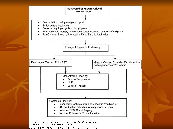

After endoscopic treatment… Fail to achieve hemostasis or rebleeding n Balloon tamponade Transjugular Intrahepatic Portosystemic Shunt (TIPS) Surgery for shunt

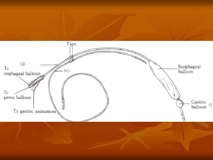
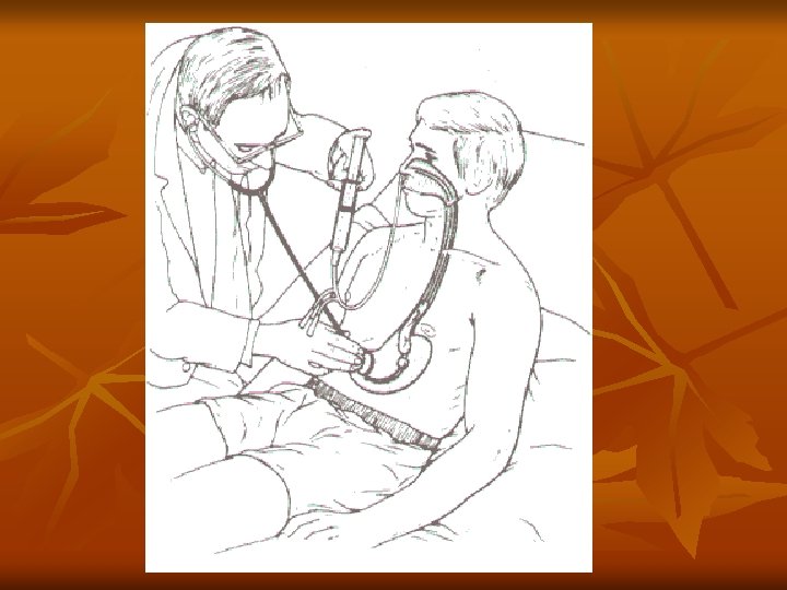
Balloon Tamponade -Buy time Available in MMH S-B tube

Esophageal ballon SB tube Mc. Cormick. British Journal of Hospital Medicine. 43, Apr. 1990 Gastric ballon
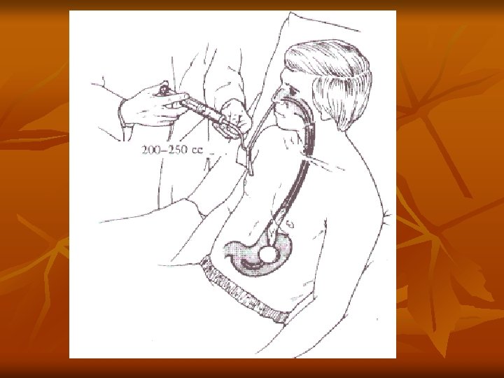
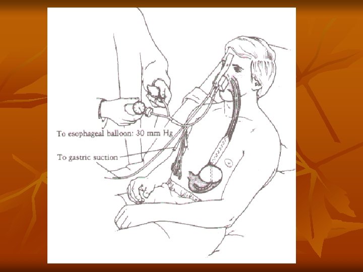

never exceed 45 mm. Hg. Volume 200 ml Mc. Cormick. British Journal of Hospital Medicine. 43, Apr 1990
 tube Radiographic confirmation of the gastric balloon’s position -- 30")
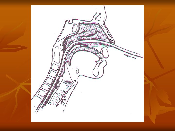
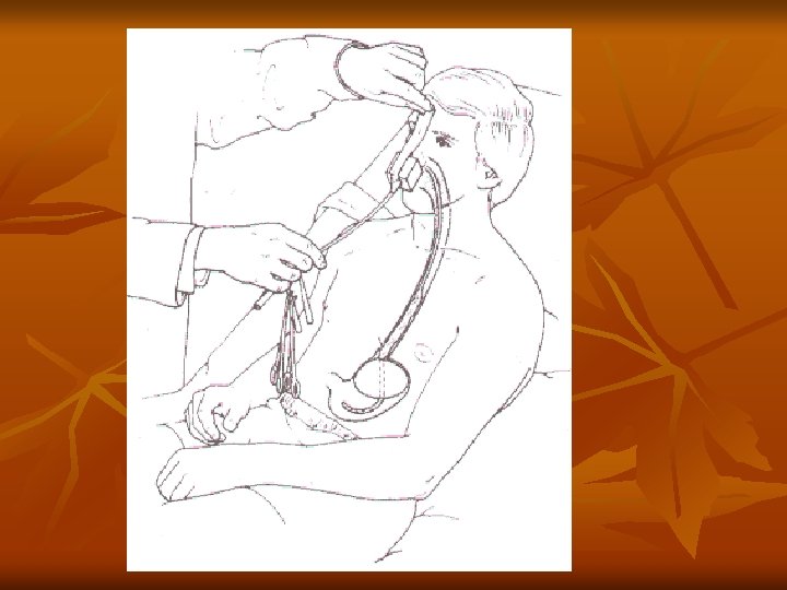
Tamponade Tube Sengstaken-Blakemore (S-B) tube Radiographic confirmation of the gastric balloon’s position -- 30 cc air inflate the gastric balloon Insufflation of the esophageal balloon to 35 mm. Hg

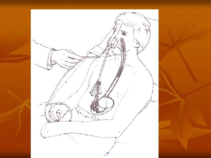
n n Compression of varices for not excess 48 hours Deflate the esophageal balloon for about 30 mins every 12 hours Major complications -- aspiration and esophageal perforation Control hemorrhage >90%, but it is temporary

n Bridging procedure buy time Definite therapeutic management must be performed.

Lower GI Bleeding Hematochezia 90% Melena 10%

Etiology Most blood passed per rectum is from the upper GI tract. Lower GI Bleeds Diverticulosis, angiodysplasia, CA, colitis, ischemia, hemorrhoids

More about Lower GI Bleeds 80% resolve spontaneously n 25 % will re–bleed n. Usually painless n. If painful, r/o mesenteric ischemia n

Investigation of the lower GI bleed The usual suspects: CBC, BUN, Creatinine, INR, PTT, T/S

Investigation of the lower GI bleed Plain X-rays and abd. CT – not much help unless you clinically suspect perforation, obstruction, ischemia (PAIN)

Diagnostic procedure Endoscopy : 80% accuracy Poor visibility with heavy bleeding n • Angiography : 40– 80% accuracy Requires heavy bleeding Able to perform embolization or vasopressin infusion

Diagnostic procedure RBC scans n 25– 90% accurate n. Able to do with lower bleeding rates n

What if the patient is really bleeding? Involve your consultants early. Radiologist for angiography Procto. If tumor or ischemic bowel
98924690603b278e0f9249fab3e70d54.ppt