GENITAL ORGANS THE MALE INTERNAL GENITAL


- Размер: 27.4 Mегабайта
- Количество слайдов: 39
Описание презентации GENITAL ORGANS THE MALE INTERNAL GENITAL по слайдам
 GENITAL ORGANS
 THE MALE INTERNAL GENITAL ORGANS The testis The epididymis The ductus (vas) deferens The seminal vesicles The ejaculatory ducts The prostate gland The bulbourethral gland
 THE MALE EXTERNAL GENITAL ORGANS the penis the scrotum the spermatic cord
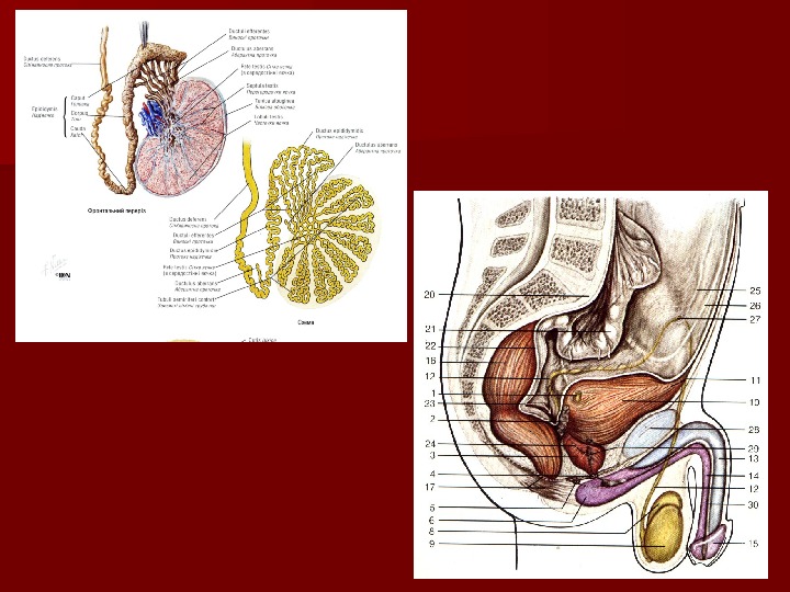
 The testis The exocrine function means producing male genital cells – spermatozoa ТТ he endocrine function means producing testosterone (male sexual hormone)
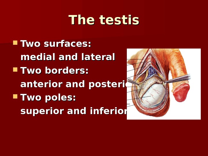
 The testis Two surfaces: medial and lateral ТТ wo borders: anterior and posterior ТТ wo poles: superior and inferior
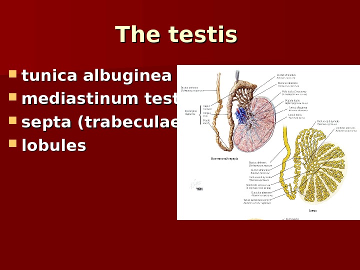
 The testis tunica albuginea mediastinum testis septa (trabeculae) lobule ss
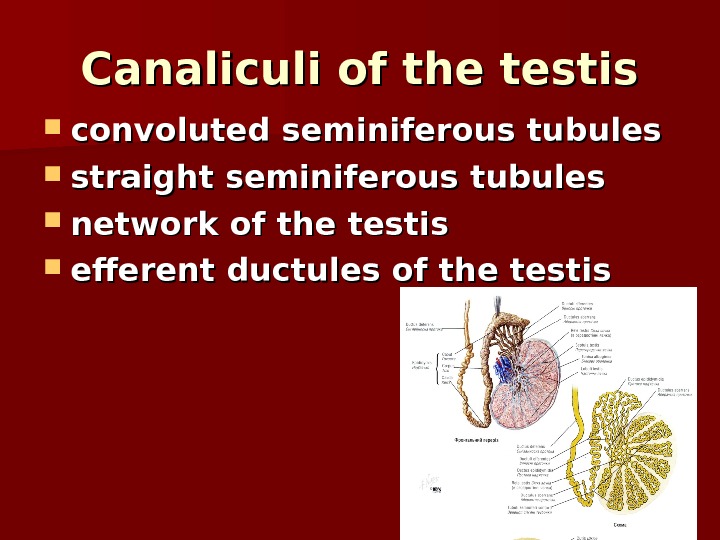
 Canaliculi of the testis convoluted seminiferous tubules straight seminiferous tubules network of the testis efferent ductules of the testis
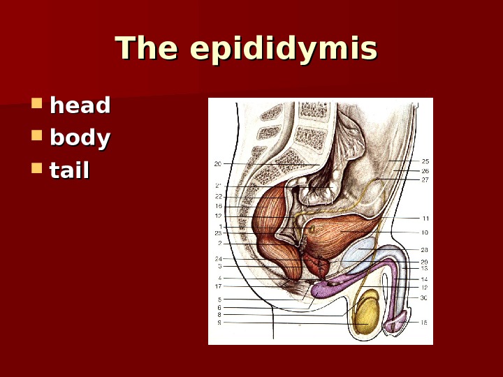
 The epididymis head bb odyody tail
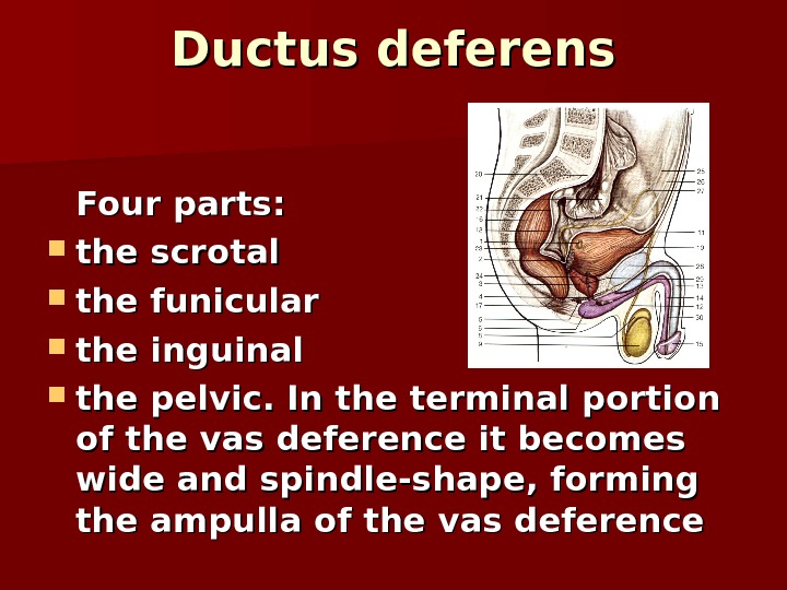
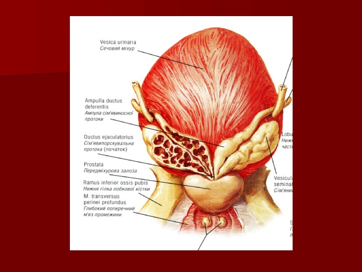
 Ductus deferens Four parts: the scrotal the funicular the inguinal the pelvic. In the terminal portion of the vas deference it becomes wide and spindle-shape, forming the ampulla of the vas deference

 The wall of the ductus deference is composed of three layers: an outer adventitious coat a middle muscular coat an inner mucous coat
 The seminal vesicles It is a tubular gland The seminal vesicles contribute 60 percent of the volume of semen The junction of the duct of the seminal vesicles with ampulla of the ductus deferens marks the ejaculatory duct Opens into prostate part of the urethra

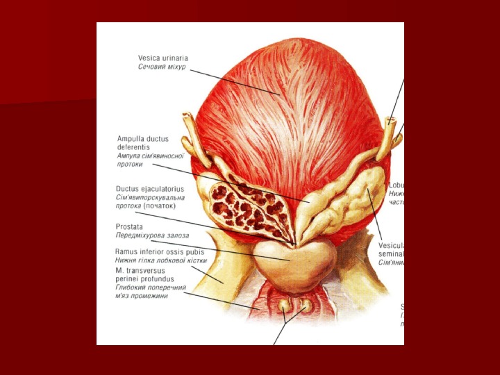
 The prostate gland is partly muscular and partly glandular organ It secretes a component of semen The prostate has: base and apex anterior, posterior and inferolateral surfaces left, right and middle lobes

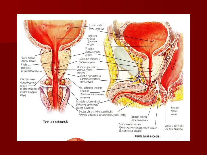
 The bulbourethral glands or Cowper’s glands They are situated at the base of the penis, covered by the fascia of the urogenital diaphragm The excretory duct opens into the spongy portion of the urethra The secretion of the glands protects the urethral walls from irritation by the urine

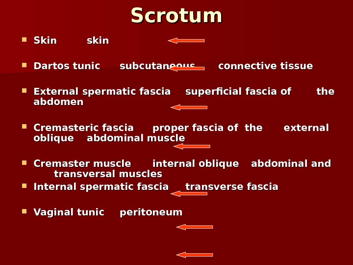
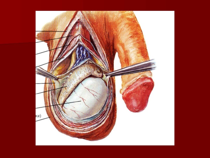
 Scrotum Skin skin Dartos tunic subcutaneous connective tissue External spermatic fascia superficial fascia of of the abdomen Cremasteric fascia proper fascia of the external oblique abdominal muscle Cremaster muscle internal oblique abdominal and transversal muscles Internal spermatic fascia transverse fascia Vaginal tunic peritoneum

 Spermatic cord the vas deferens; testicular artery; convoluted (venous) pampiniform plexus; the artery and veins of the vas deferens; the lymphatic vessels of the testis and the epididymis; the nerves; the vestige of the processus vaginalis
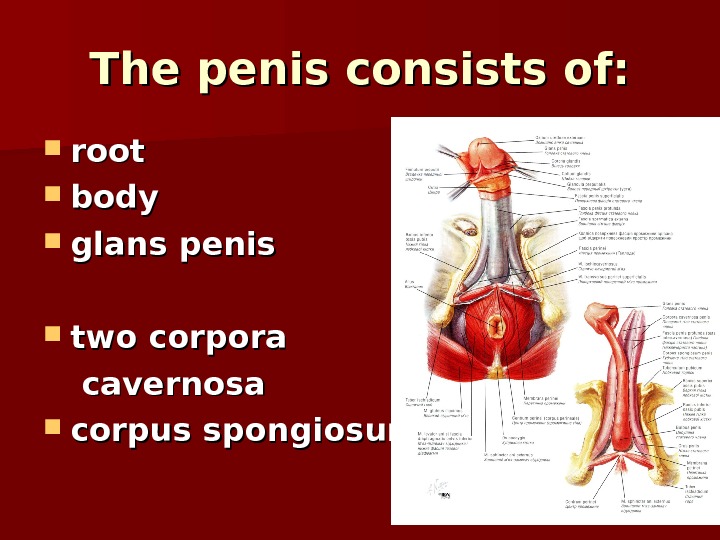
 The penis consists of: root body glans penis two corpora cavernosa corpus spongiosum
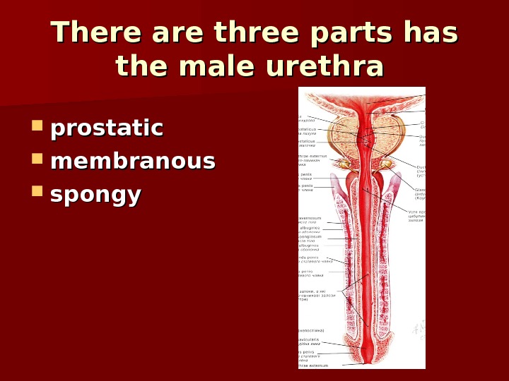
 There are three parts has the male urethra prostatic membranous spongy
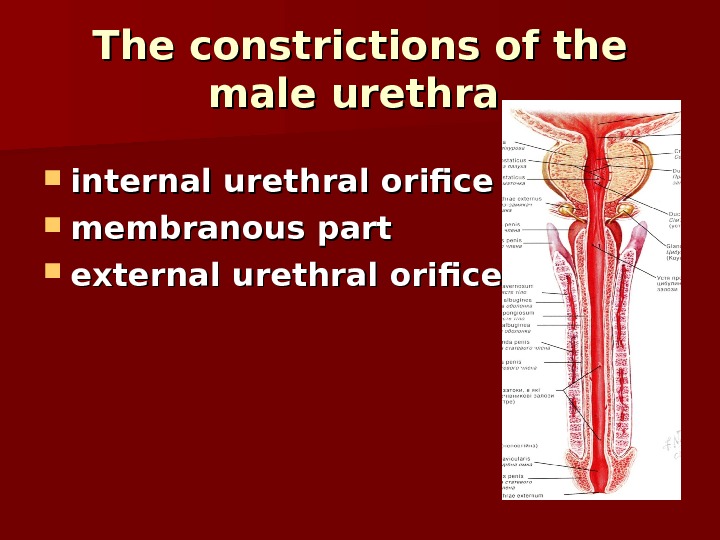
 The constrictions of the male urethra internal urethral orifice membranous part external urethral orifice
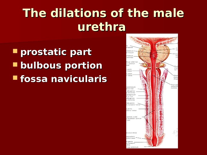
 The dilations of the male urethra prostatic part bulbous portion fossa navicularis
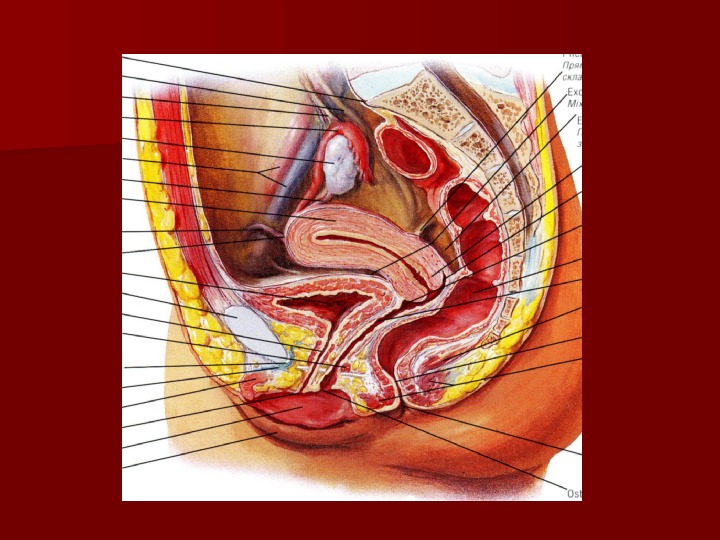
 Internal female organs ovaries uterine tubes uterus vagina
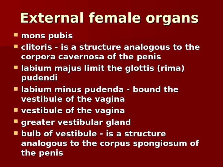
 External female organs mons pubis clitoris — is a structure analogous to the corpora cavernosa of the penis labium majus limit the glottis (rima) pudendi labium minus pudenda — bound the vestibule of the vaginavestibule of the vagina greater vestibular gland bulb of vestibule — is a structure analogous to the corpus spongiosum of the penis
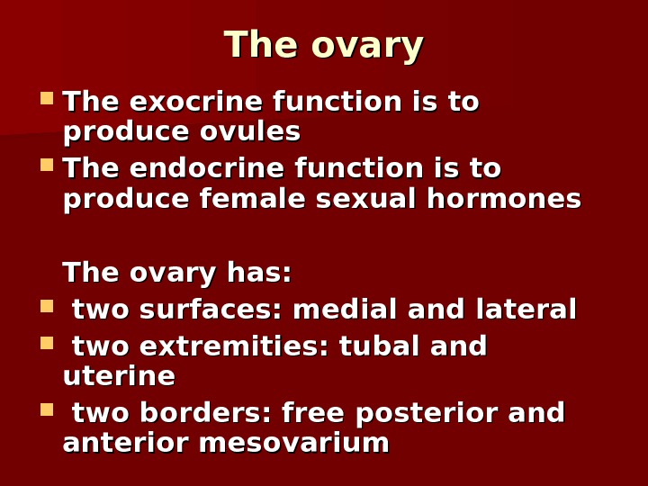
 The ovary The exocrine function is to produce ovules The endocrine function is to produce female sexual hormones The ovary has: two surfaces: medial and lateral two extremities: tubal and uterine two borders: free posterior and anterior mesovarium


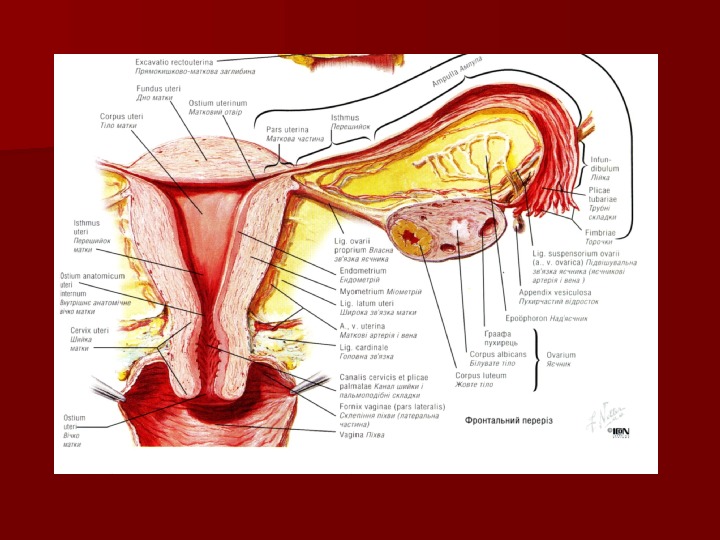
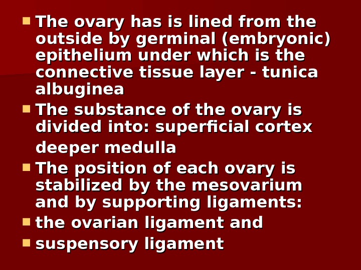
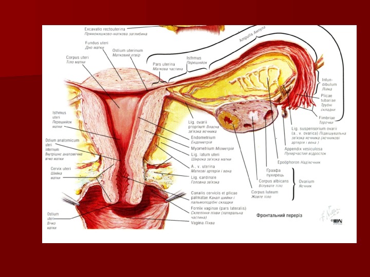
 The ovary has is lined from the outside by germinal (embryonic) epithelium under which is the connective tissue layer — tunica albuginea The substance of the ovary is divided into: superficial cortex deeper medulla The position of each ovary is stabilized by the mesovarium and by supporting ligaments: the ovarian ligament and suspensory ligament

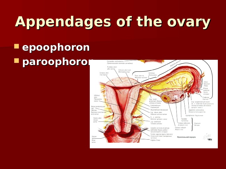
 Appendages of the ovary epoophoron paroophoron
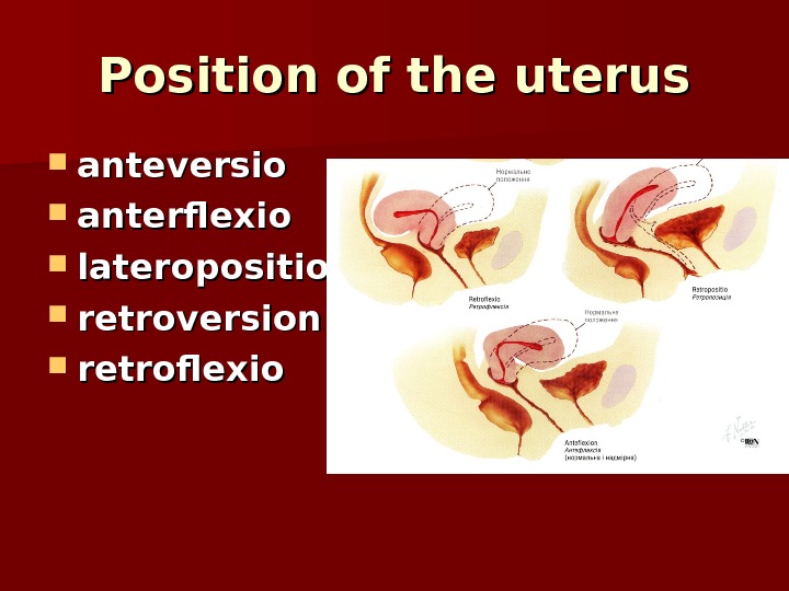
 Position of the uterus anteversio anterflexio lateroposition retroversion retroflexio
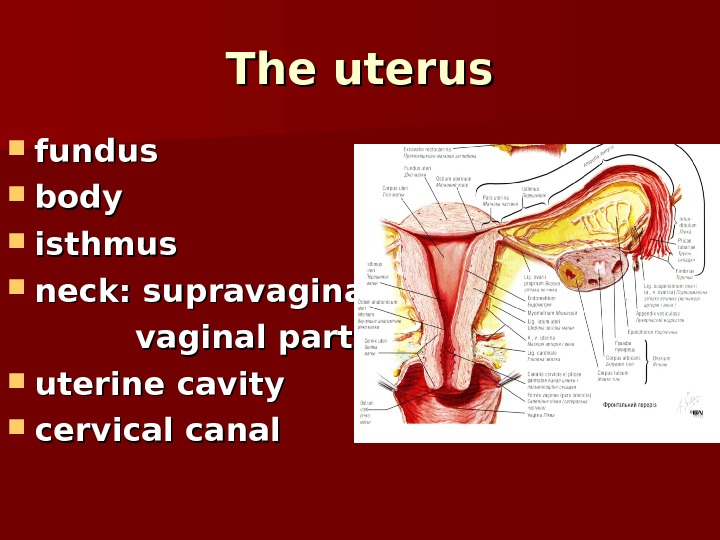
 The uterus fundus body isthmus neck: supravaginal vaginal parts uterine cavity cervical canal
 The wall of the uterus serous coat — the perimetrium muscular coat – myometrium: internal and external longitudinal and middle circular layers mucosa — endometrium
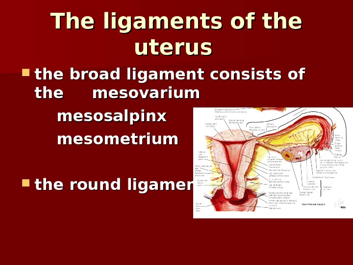
 The ligaments of the uterus the broad ligament consists of the mesovarium mesosalpinx mesometrium the round ligament
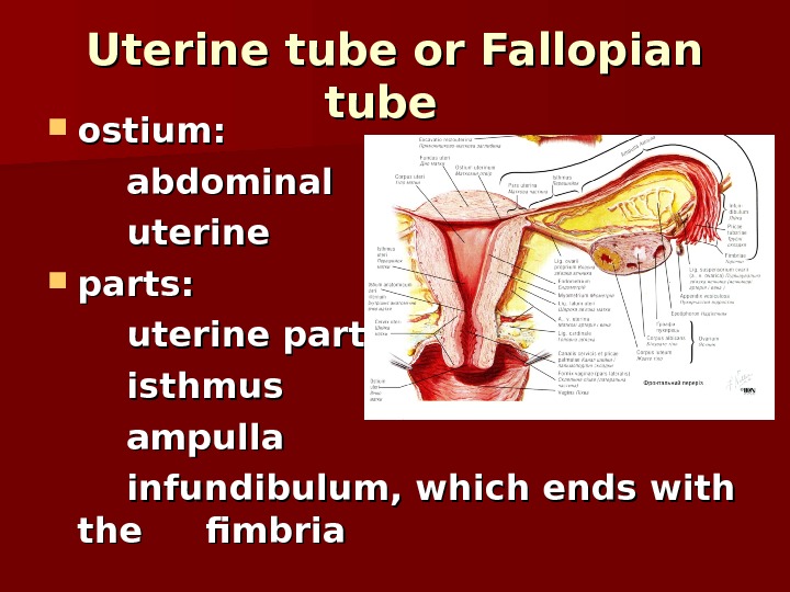
 Uterine tube or Fallopian tube ostium: abdominal uterine parts: uterine part isthmus ampulla infundibulum, which ends with the fimbria
 The wall of the uterine tube the serous coat the muscular coat: internal circular and external longitudinal the mucosa
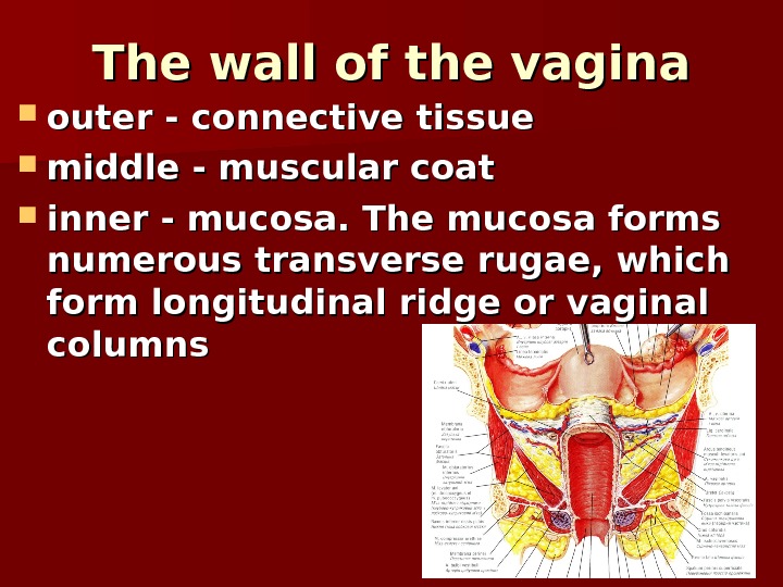
 The wall of the vagina outer — connective tissue middle — muscular coat inner — mucosa. The mucosa forms numerous transverse rugae, which form longitudinal ridge or vaginal columns

