

Лекция 7 Общее учение о воспалении 2011.ppt
- Количество слайдов: 87
 General doctrine of inflammation Lecture
 Process essence: o Complex local vascularmesenchymal reaction of an organism aimed at the destruction of causative agent and tissue structure recovery
 Etiological factors of inflammation: o Biological o Chemical o Physical
 Biological factors: o o o viruses; bacteria; fungi; animals-parasites; circulating in blood antibodies and activated immune complexes.
 Chemical factors: o o acids; alkali; mineral and organic substances; Endogenous toxins (bile acids, nitrogen metabolism products).
; ionizing,") Physical factors: o o trauma (cuts, injections, bites, contusions, vibration, noise exposure, compression); ionizing, ultraviolet radiation; electric energy; high (fire) and low (cold) temperatures.
Physical factors: o o trauma (cuts, injections, bites, contusions, vibration, noise exposure, compression); ionizing, ultraviolet radiation; electric energy; high (fire) and low (cold) temperatures.
 Clinical signs o o o Reddening Edema Temperature rise Pain Function derangement
: Alteration o Exudation o Proliferation o") Morphologic signs (stages): Alteration o Exudation o Proliferation o
Morphologic signs (stages): Alteration o Exudation o Proliferation o
 Alteration o o Parenchyma and stroma dystrophy up to necrosis Release of bioactive substances
 Important !!!!!! o o Any inflammation begins with alteration!!! Inflammation triggering mechanism – release of bioactive substances !!!
: -kallikrein-kinin system -complement system -blood coagulation") The sources of bioactive substances o Plasma (circulating): -kallikrein-kinin system -complement system -blood coagulation system o Cellular (local): labrocytes, thrombocytes, basophiles, macrophages, lymphocytes, fibroblasts, neutrophils etc.
The sources of bioactive substances o Plasma (circulating): -kallikrein-kinin system -complement system -blood coagulation system o Cellular (local): labrocytes, thrombocytes, basophiles, macrophages, lymphocytes, fibroblasts, neutrophils etc.
 Histamine and serotonin are released from tissue basophiles and thrombocytes. Histamine and serotonin lead to vasodilation and rapid increase of vascular wall permeability.
 Bradykinin, the end product of kinin system, is formed as a result of kallikrein influence on the protein precursor in plasma. It is leading to vasodilation, minor increase of vascular wall permeability and causes pain, irritating the pain receptors.
 Blood coagulation system Coagulation system, leading to fibrin formation, is activated by the Hageman`s factor (activated factor XII). Fibrinopeptides, which are formed in fibrin catabolism (fibrinolysis), also stimulate the increase of vascular permeability and are chemoattractants for neutrophils.
 Complement system C 5 a and C 3 a, which are formed during the complement activation, cause the increase of vascular permeability, stimulating the histamine release by tissue basophiles. C 5 a – is a powerful hemotaxic agent for neutrophils and macrophages.
 Neutrophils factors protease and toxic oxygenous free radicals, which are formed in neutrophils, cause endothelial injury, leading to increase of vascular permeability.
 Exudation It is a complicated process of effusion formation. Exudate consists of 2 parts: o Liquid part (water, proteins, mineral salts) o Cells: plasma and tissue cells.
 2 main components of the exudation phase: o o Microcirculatory changes; Cellular reactions.
 Microcirculatory changes: o o Short-term vasoconstriction Vasodilation and stasis Increase of vascular wall permeability Increase of fluid transition from microcirculatory system into tissues
 standing of leukocytes Active migration of inflammatory cells") Cellular reactions: o o Marginal (edge) standing of leukocytes Active migration of inflammatory cells from the blood into lesion area (1 - neutrophils and erythrocytes, 2 - macrophages) Phagocytosis (1 -recognition, 2 absorption, 3 -destruction of microorganisms with the help of Н 2 О 2, myeloperoxydase, lysozyme) Exudate formation
Cellular reactions: o o Marginal (edge) standing of leukocytes Active migration of inflammatory cells from the blood into lesion area (1 - neutrophils and erythrocytes, 2 - macrophages) Phagocytosis (1 -recognition, 2 absorption, 3 -destruction of microorganisms with the help of Н 2 О 2, myeloperoxydase, lysozyme) Exudate formation
 Differential diagnostics of exudate and transudate Features Transudate Exudate Vascular permeability normal increased Proteins content 0 -1, 5 gl 1, 5 -6 gl Proteins types albumins all Fibrin no yes Cells no inflammation


") Proliferation - increase of cells quantity in the inflammation zone of the local (histiogenous) and hematogenous origin
Proliferation - increase of cells quantity in the inflammation zone of the local (histiogenous) and hematogenous origin
 Histiogenous cells o Epithelial cells o Fibroblasts - fibrocytes
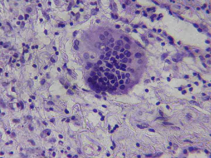
 Hematogenous cells o o Monocytes - macrophages - epithelioid cells - giant cells В-lymphocytes - labrocytes - plasma cells Neutrophils – are dying Т-lymphocytes – are dying


 Inflammation regulation o Hormonal factors o Neural factors o Immune factors
 mineralocorticoids, somatotropin (growth hormone), hypophysial") Hormonal factors: o o Proinflammatory (increase the inflammatory reaction) mineralocorticoids, somatotropin (growth hormone), hypophysial thyrotropin, aldosterone Anti-inflammatory (depress the inflammation) – glucocorticoids and adrenocorticotropic hormone (ACTH)
Hormonal factors: o o Proinflammatory (increase the inflammatory reaction) mineralocorticoids, somatotropin (growth hormone), hypophysial thyrotropin, aldosterone Anti-inflammatory (depress the inflammation) – glucocorticoids and adrenocorticotropic hormone (ACTH)
 Neural factors: o o Proinflammatory – cholinergic substances stimulate the output of inflammation mediators Anti-inflammatory – adrenergic substances depress the activity of mediators like the anti-inflammatory hormones
 Immune factors: Immunity state has an influence on the intensity of inflammatory reaction, its development rates and character. Especially rapid the inflammation develops in the conditions of antigenous stimulation (sensitization). In that case we say about the immune or allergic inflammation.
 Inflammation course: o Acute o Subacute o Chronic
 Inflammation forms according to morphology: o Exudative o Proliferative
 Exudative inflammation
 Process essence: o Exudation prevalence over alteration and proliferation with the reaction of microcirculatory bed and exudate formation
 Exudate composition: o o o Fluid Proteins Blood uniform elements Cells of local tissue Lysis (decay) products Microbes
 Types of exudative inflammation: o o o o Serous Fibrinous Suppurative Catarrhal Hemorrhagic Putrid Mixed
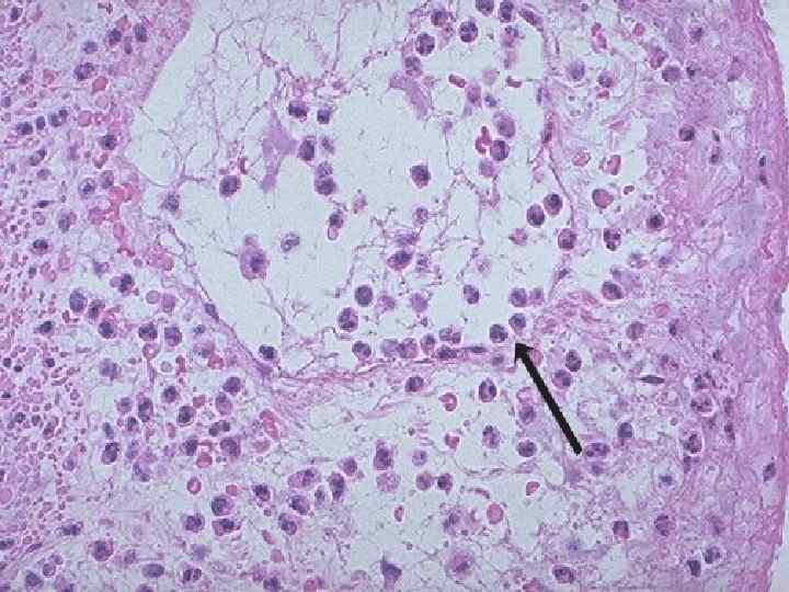
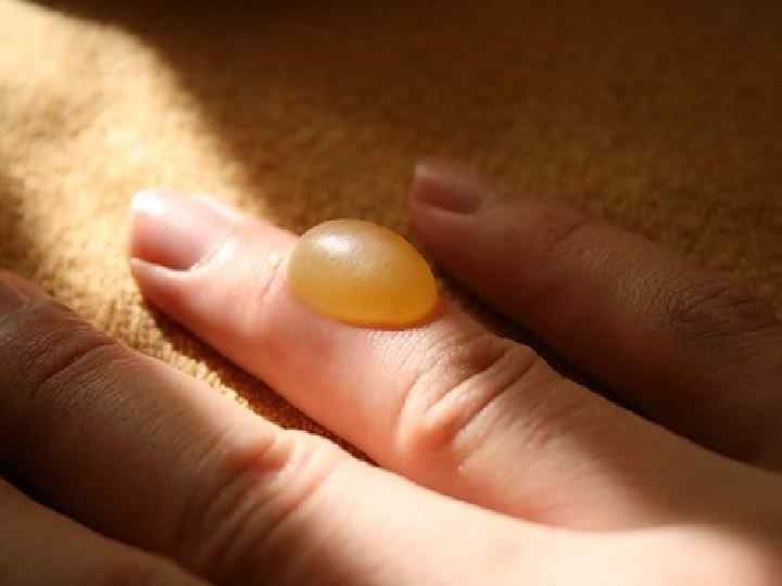
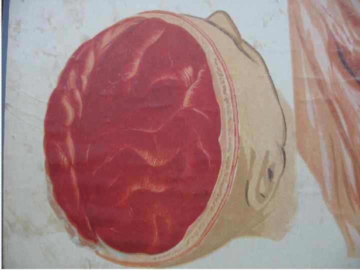
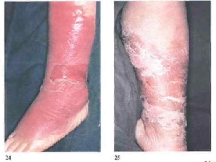
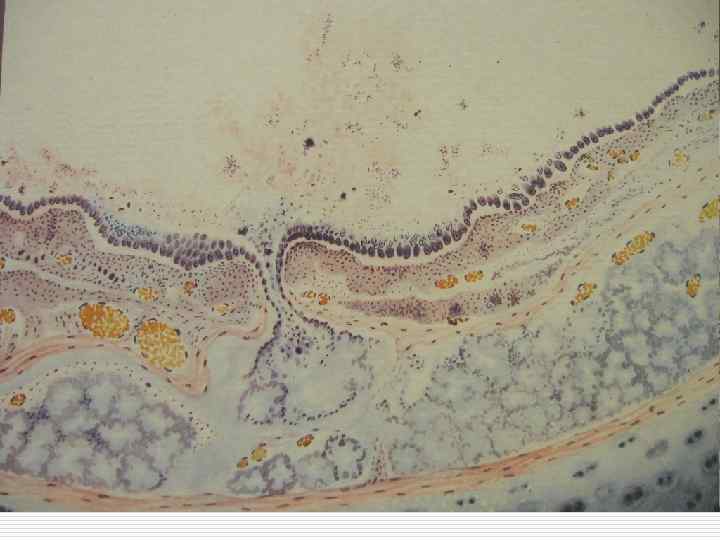
 Serous inflammation o o o Localization: mucous and serous membranes, lungs, meninx vasculosa, skin Exudate composition – fluid+plasma proteins, single cells Outcome – exudate resorption


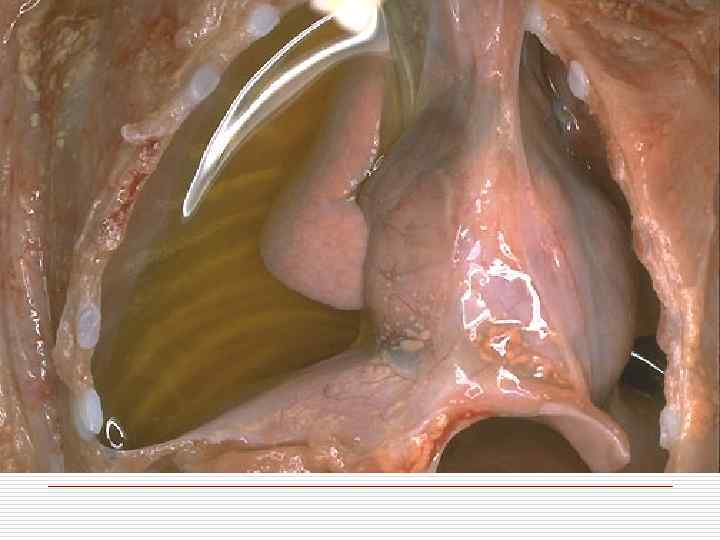
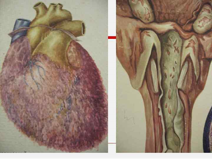
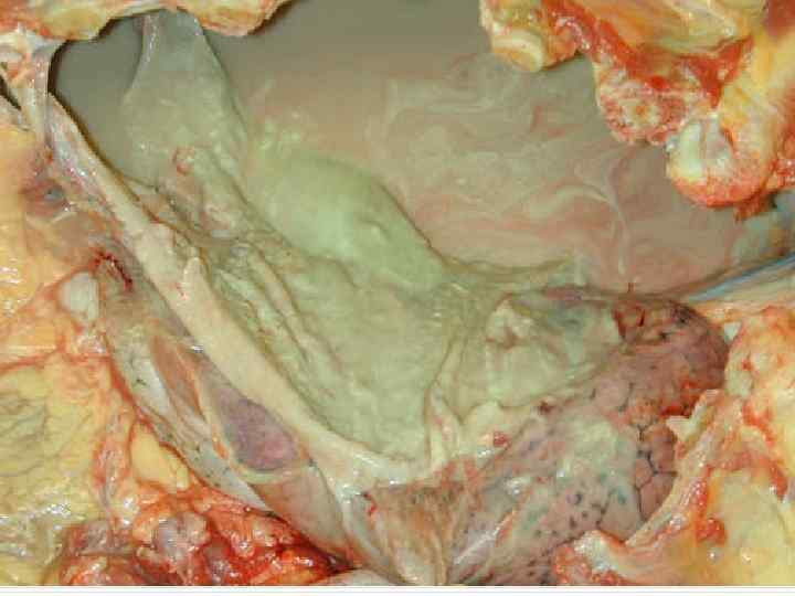
 Fibrinous inflammation o o - Localization: mucous and serous membranes, lungs Varieties depending on epithelium: croupous diphtheritic
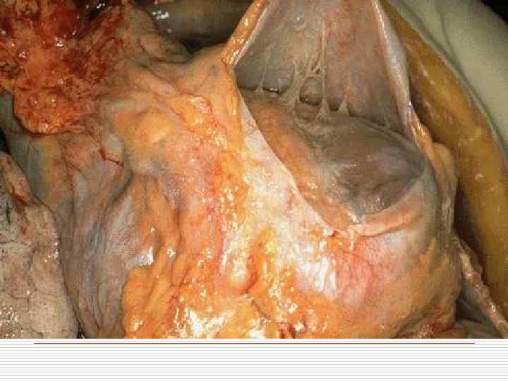
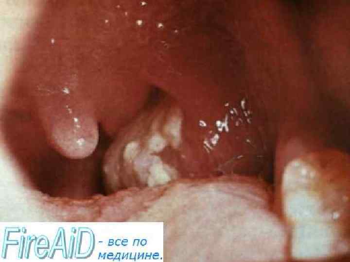
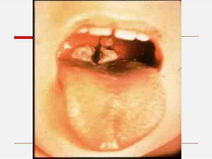

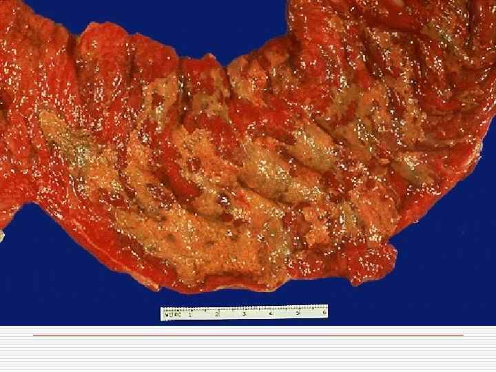

 Outcomes of fibrinous inflammation: o o o exudate resorption exudate organization commissures formation epithelization ulcer formation
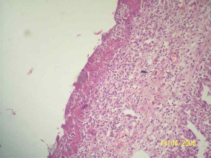
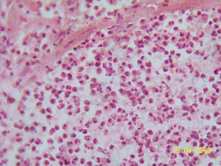
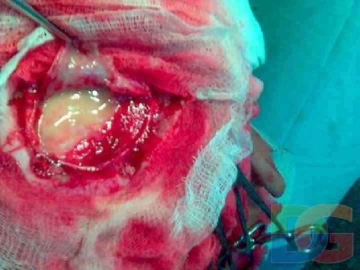
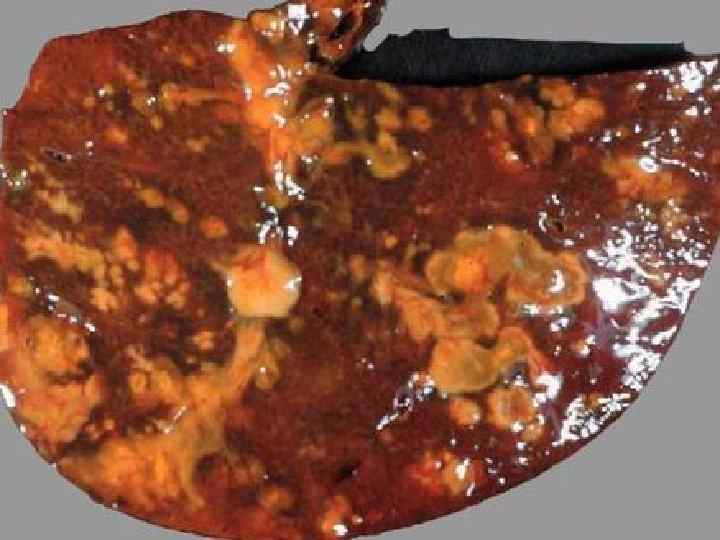
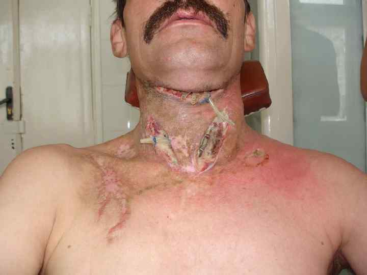
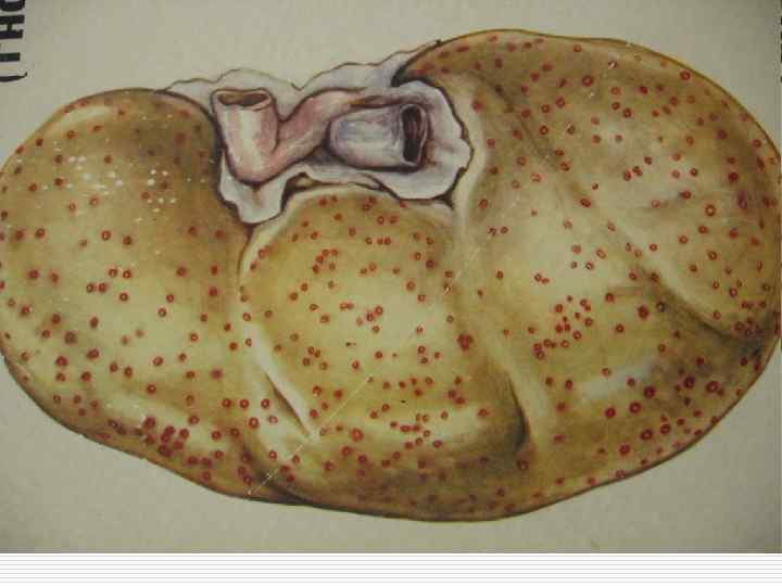
 Localization –") Suppurative inflammation o o o Exudate basis – neutrophilic leukocytes (purulent bodies) Localization – any of the organs and tissues Forms: - abscess - phlegmon - empyema - apostema abscess
Suppurative inflammation o o o Exudate basis – neutrophilic leukocytes (purulent bodies) Localization – any of the organs and tissues Forms: - abscess - phlegmon - empyema - apostema abscess


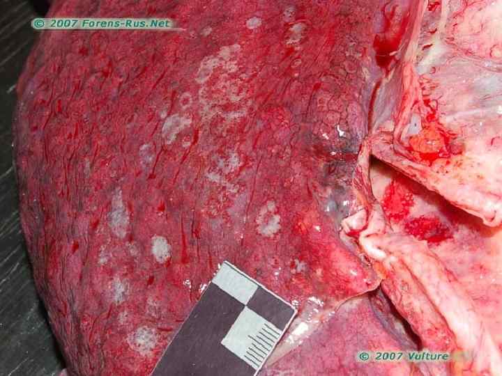


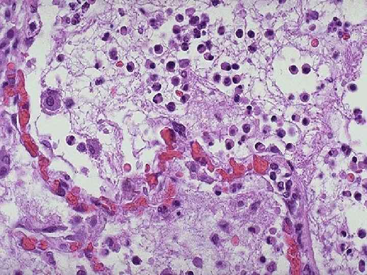
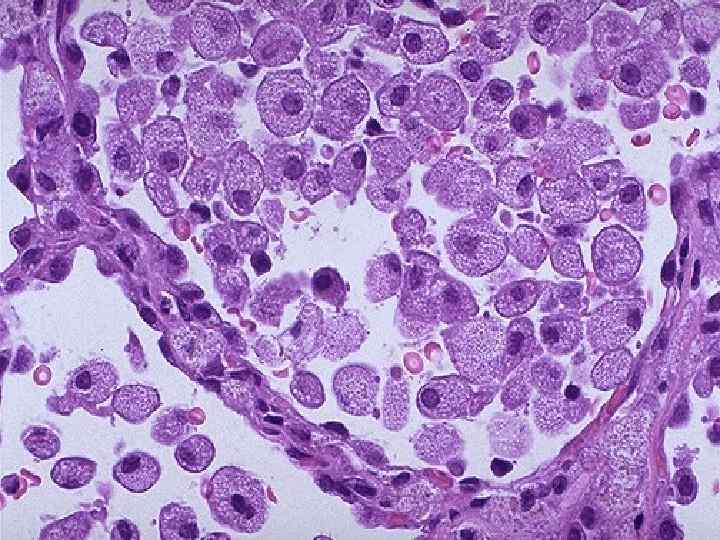
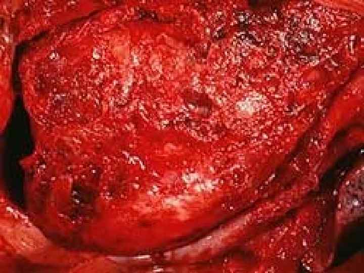
 Hemorrhagic inflammation o o Exudate basis – erythrocytes Occurs during the especially perilous infections: plague, anthrax, influenza


 Localization: postpartum uterus,") Putrid inflammation o o Develops by joining of putrefactive microorganisms (clostridium) Localization: postpartum uterus, wounds, large intestines of the newborn
Putrid inflammation o o Develops by joining of putrefactive microorganisms (clostridium) Localization: postpartum uterus, wounds, large intestines of the newborn
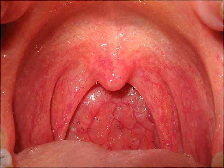
. Serous catarrh") Catarrhal inflammation o o Localization – mucous membranes. Exudate contains slime (mucus). Serous catarrh Mucous catarrh Purulent catarrh
Catarrhal inflammation o o Localization – mucous membranes. Exudate contains slime (mucus). Serous catarrh Mucous catarrh Purulent catarrh

 Mixed inflammation