Gene Expression Regulation Fundamentals of Biochemistry of Hormones

Gene Expression Regulation Fundamentals of Biochemistry of Hormones THE MINISTRY OF PUBLIC HEALTH OF UKRAINE ZAPOROZHYE STATE MEDICAL UNIVERSITY Produced by Ass.professor Krisanova N.V., 2015
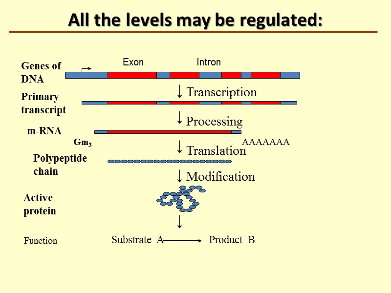
All the levels may be regulated: Transcription Processing Translation AAAAAAA Genes of DNA Primary transcript m-RNA Polypeptide chain Modification Substrate A Product B Function Active protein Gm3 Intron Exon
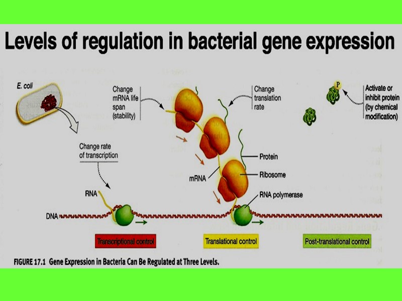
All the genes of DNA in prokaryotic cell are divided in types: House keeping genes (constitutive) Inducible (structural) Gene-regulators Gene-operators
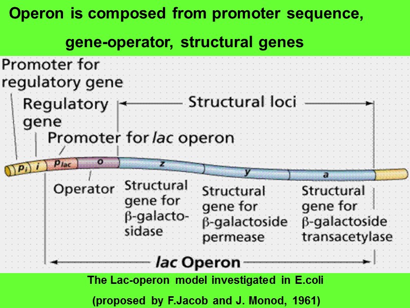
Operon is composed from promoter sequence, gene-operator, structural genes The Lac-operon model investigated in E.coli (proposed by F.Jacob and J. Monod, 1961)
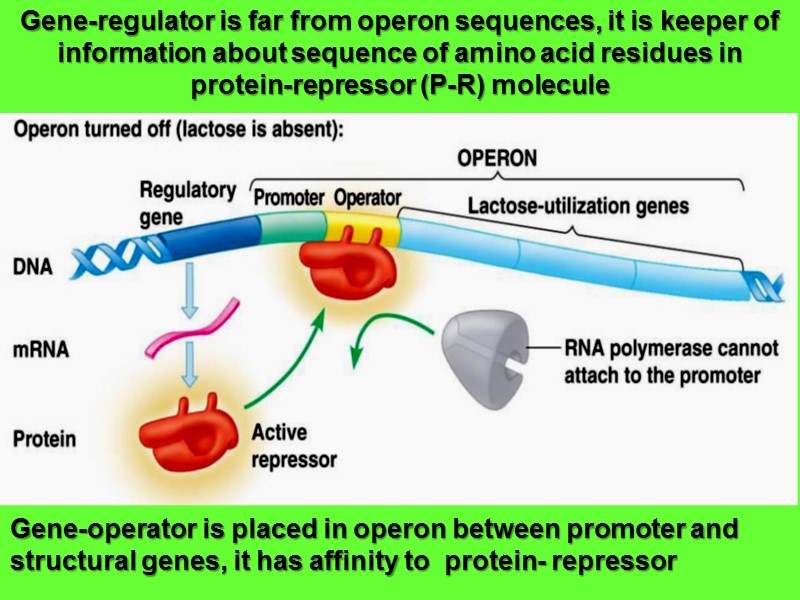
Gene-regulator is far from operon sequences, it is keeper of information about sequence of amino acid residues in protein-repressor (P-R) molecule Gene-operator is placed in operon between promoter and structural genes, it has affinity to protein- repressor
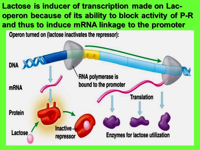
Lactose is inducer of transcription made on Lac-operon because of its ability to block activity of P-R and thus to induce mRNA linkage to the promoter
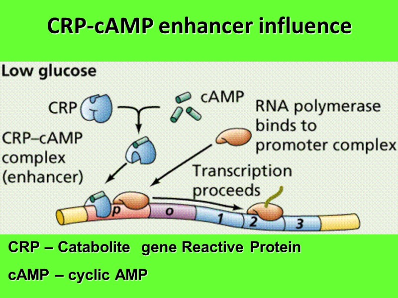
CRP-cAMP enhancer influence CRP – Catabolite gene Reactive Protein cAMP – cyclic AMP
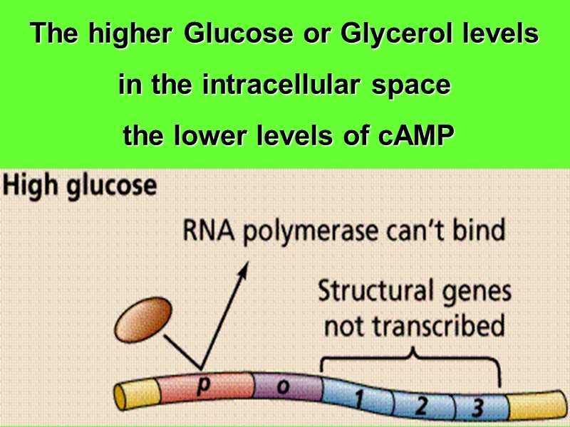
The higher Glucose or Glycerol levels in the intracellular space the lower levels of cAMP
Different Genes are found in eukaryotic DNA House keeping genes Genes required during cellular differentiation Genes which get triggered as a response to some external factors Genes which get triggered during apoptosis
Points for Gene Expression in Eukaryotes Synthesis of proteins is controlled right from the chromatin stage. Expression of gene is controlled at many steps during the process of transcription and translation.
Two forms of chromatin : Euchromatin – A lesser coiled transcriptionally active region which can be easily accessed by the RNA polymerases. Heterochromatin – A highly condensed transcriptionally inactive region. The genes in this region cannot be accessed by the RNA polymerases for active transcription .
Mechanisms which affect the chromatin structure and hence the expression of gene are: Acetylation of Histones : ↑ Acetylation ----↓ Condensation of DNA ----- ↑ Transcription of genes in that region Methylation of histone H4 on R4 (arginine residue at the 4th position) ->-> opens the chromatin structure ->-> leading to transcriptional activation
Mechanisms which affect the chromatin structure and hence the expression of gene are: Methylation of histone H3 on K4 and K79 (lysine residues at the 4th and 79th position) ->-> opens the chromatin structure ->-> leading to transcriptional activation Methylation of histone H3 on K9 and K27 (lysine residues at the 9th and 27th position) ->-> condenses the chromatin structure ->-> leading to transcriptional inactivation
Ubiquitination Ubiquitination of H2A – Transcriptional inactivation Ubiquitination of H2B - Transcriptional activation Methylation of DNA Target sites of methylation are - The cytidine residues which exist as a dinucleotide, CG (written as CpG) ↑ methylated cytidine -- ↓Transcriptional activity
3` 5`
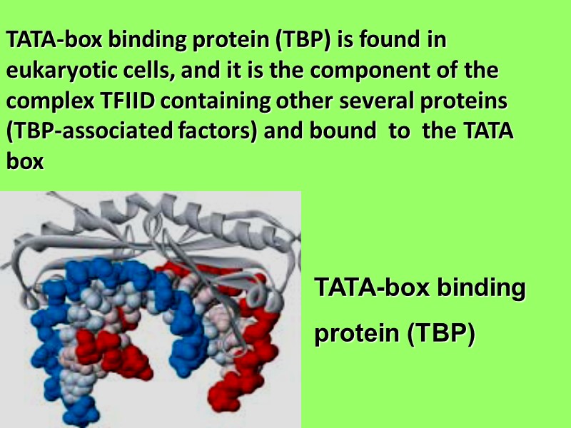
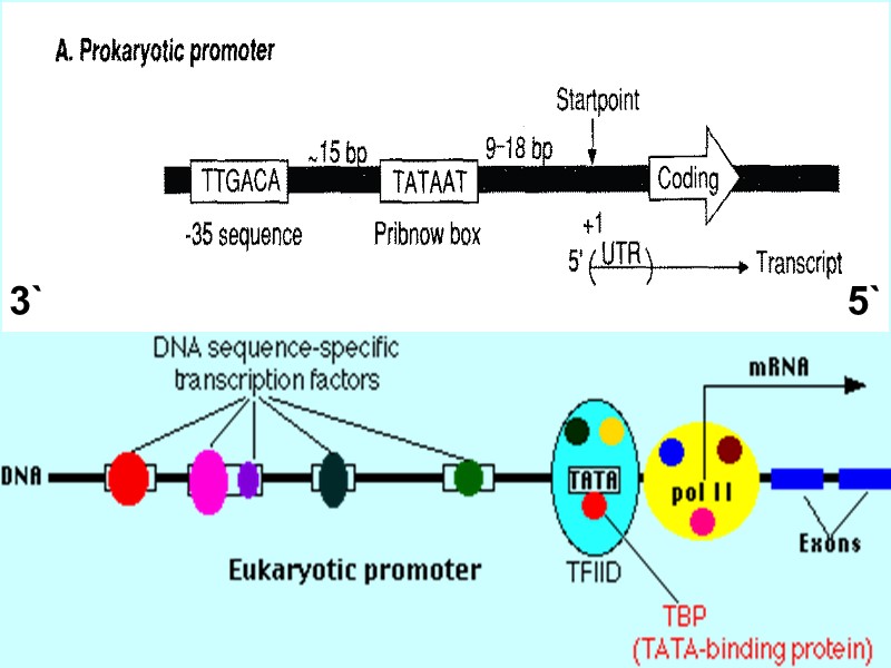
TATA-box binding protein (TBP) is found in eukaryotic cells, and it is the component of the complex TFIID containing other several proteins (TBP-associated factors) and bound to the TATA box TATA-box binding protein (TBP)
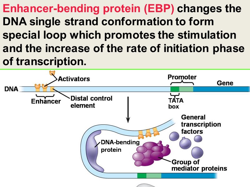
Enhancer-bending protein (EBP) changes the DNA single strand conformation to form special loop which promotes the stimulation and the increase of the rate of initiation phase of transcription.
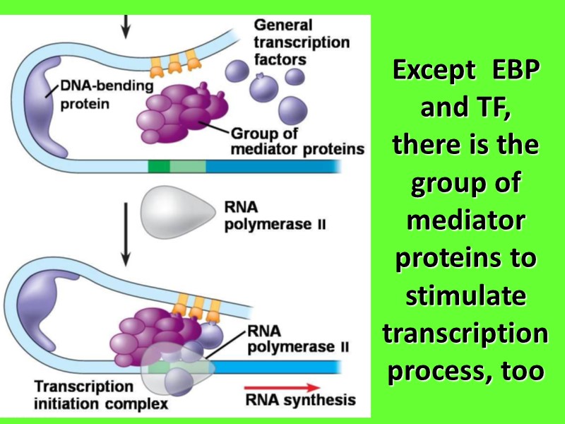
Except EBP and TF, there is the group of mediator proteins to stimulate transcription process, too
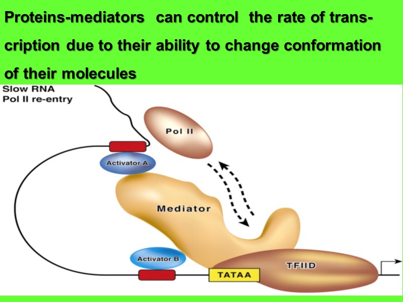
Proteins-mediators can control the rate of trans- cription due to their ability to change conformation of their molecules
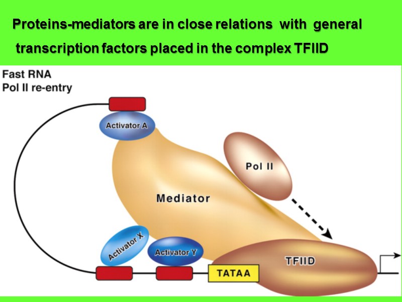
Proteins-mediators are in close relations with general transcription factors placed in the complex TFIID
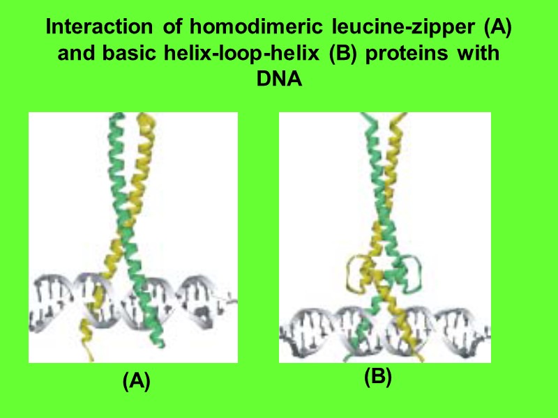
Interaction of homodimeric leucine-zipper (A) and basic helix-loop-helix (B) proteins with DNA (A) (B)
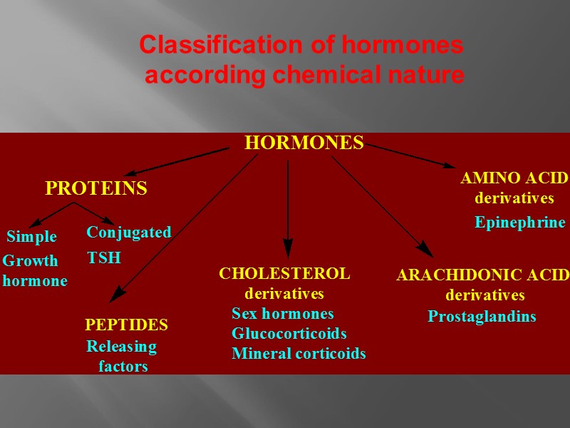
Classification of hormones according chemical nature
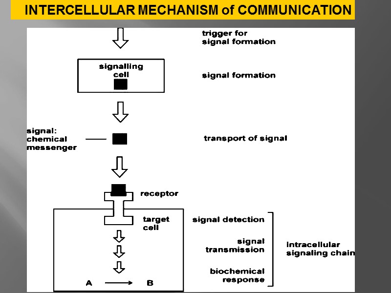
INTERCELLULAR MECHANISM of COMMUNICATION
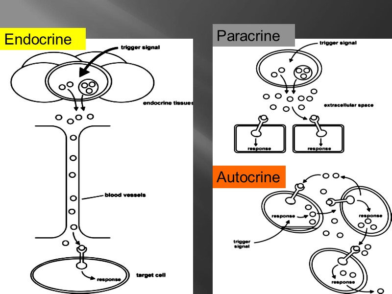
Endocrine Paracrine Autocrine
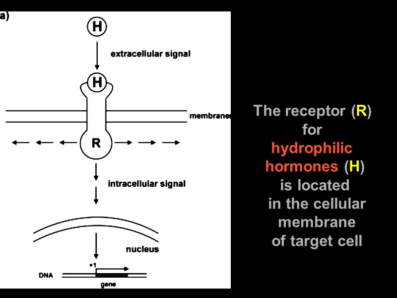
The receptor (R) for hydrophilic hormones (H) is located in the cellular membrane of target cell
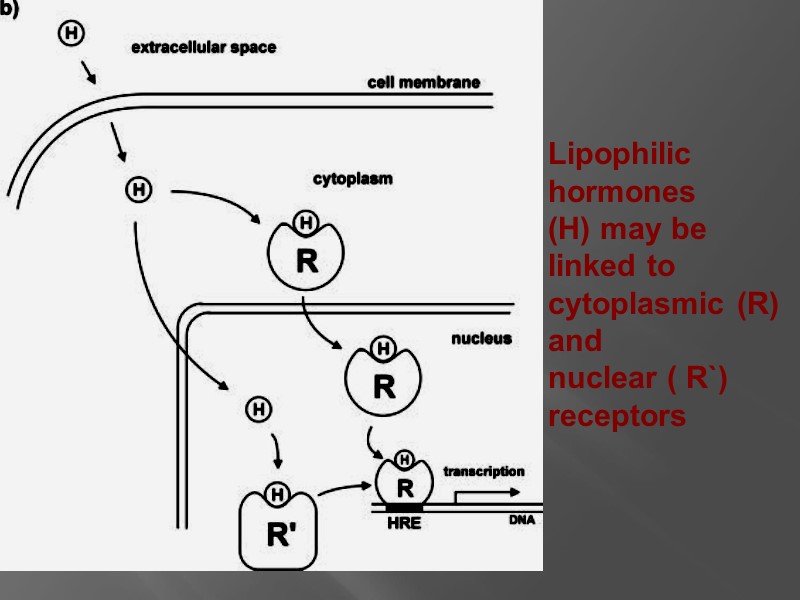
Lipophilic hormones (H) may be linked to cytoplasmic (R) and nuclear ( R`) receptors
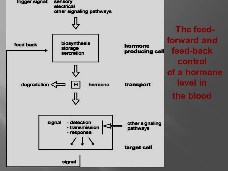
The feed-forward and feed-back control of a hormone level in the blood
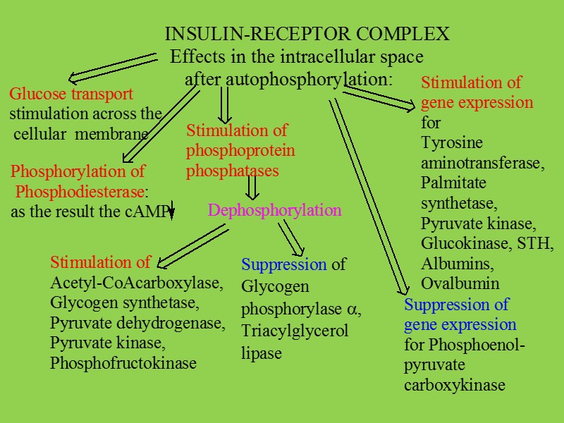
All of the steps below are subject to regulation: biosynthesis of the hormone storage, secretion of the hormone transport of the hormone to the target cell reception of the signal by the hormone receptor transmission and amplification of the signal, biochemical reaction in the target cell degradation and excretion of the hormone.
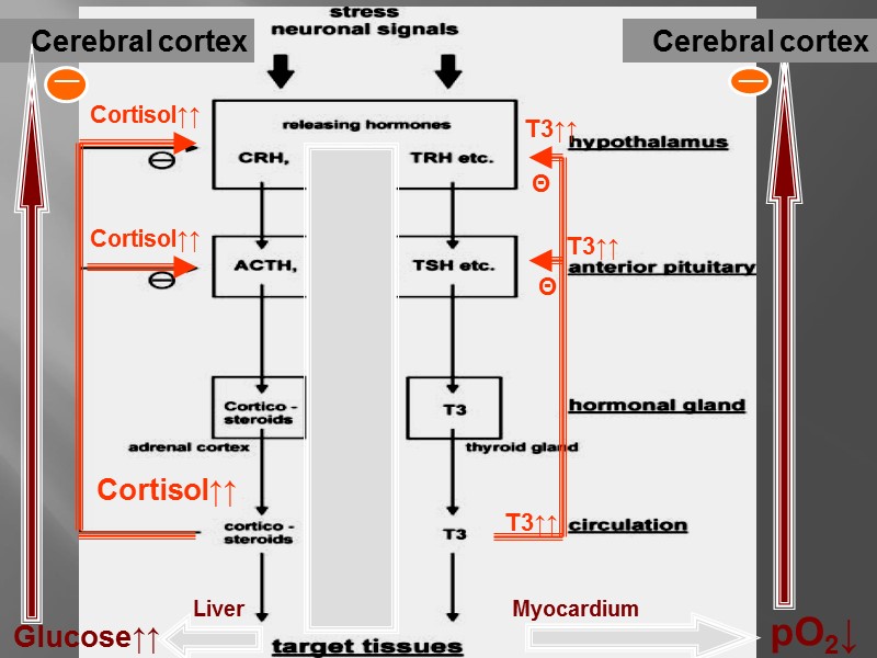
Cerebral cortex T3↑↑ Cortisol↑↑ Θ Θ Cortisol↑↑ Cortisol↑↑ T3↑↑ T3↑↑ Glucose↑↑ pO2↓ Cerebral cortex Liver Myocardium __ __
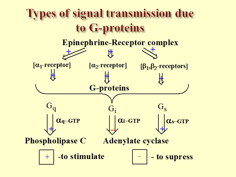
Types of signal transmission due to G-proteins
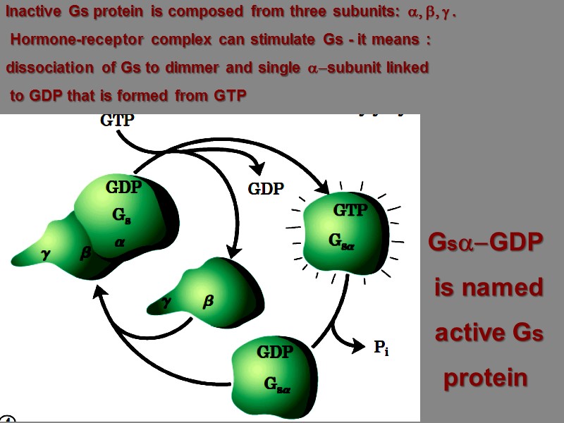
Inactive Gs protein is composed from three subunits: a, b, g . Hormone-receptor complex can stimulate Gs - it means : dissociation of Gs to dimmer and single a-subunit linked to GDP that is formed from GTP Gsa-GDP is named active Gs protein
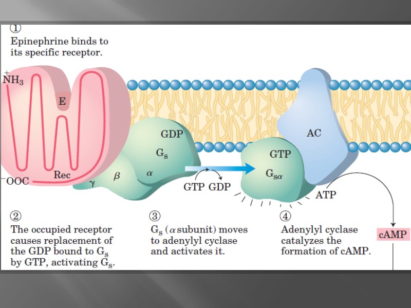
Some factors influenced G-proteins Cholera toxin modifies a-subunit of Gs as the result – the block of hydrolysis of GTP to GDP and superstimulation of Adenylate cyclase Pertussis toxin (produced at whooping cough) modifies a-subunit of Gi to allow Adenylate cyclase to produce cAMP in excess levels
GAP function: GTPase-Activating Proteins, or GAPs can bind to activated G-proteins and stimulate their GTPase activity, with the result of terminating the signaling event. GAPs are also known as regulator of G protein signaling proteins, or RGS proteins, and these proteins are crucial in controlling the activity of G proteins. GAP role is to turn the G protein activity off .
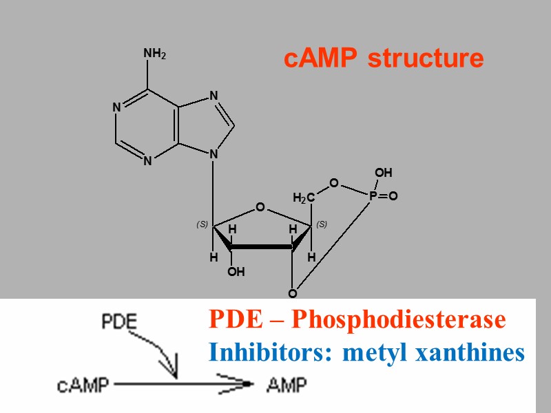
cAMP structure PDE – Phosphodiesterase Inhibitors: metyl xanthines
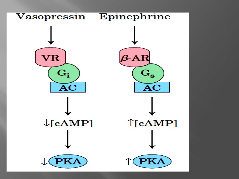
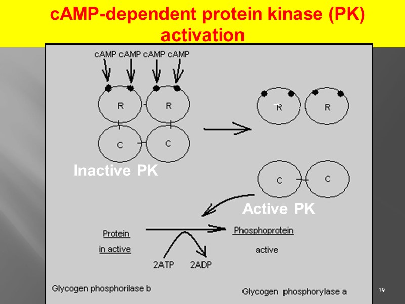
39 cAMP-dependent protein kinase (PK) activation Inactive PK Active PK _
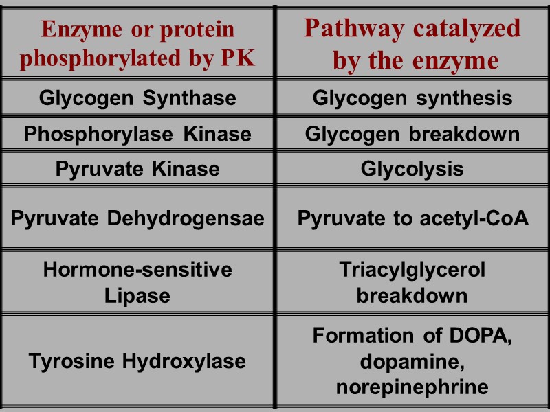
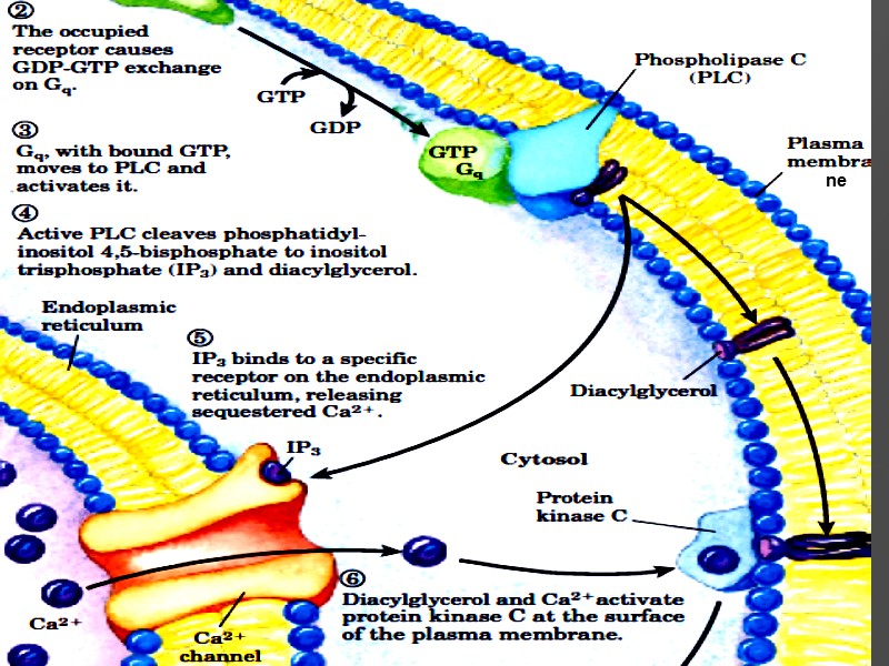
ne
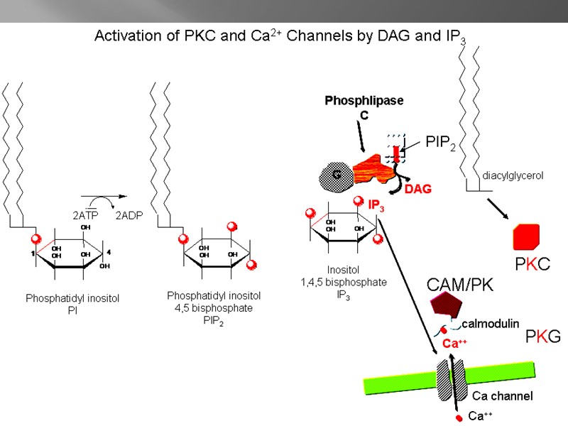
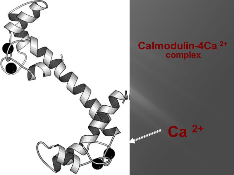
Calmodulin-4Ca 2+ complex Ca 2+
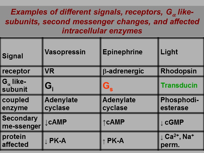
Examples of different signals, receptors, Ga like-subunits, second messenger changes, and affected intracellular enzymes
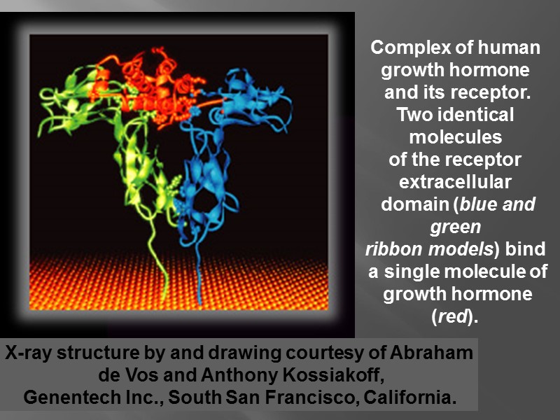
X-ray structure by and drawing courtesy of Abraham de Vos and Anthony Kossiakoff, Genentech Inc., South San Francisco, California. Complex of human growth hormone and its receptor. Two identical molecules of the receptor extracellular domain (blue and green ribbon models) bind a single molecule of growth hormone (red).
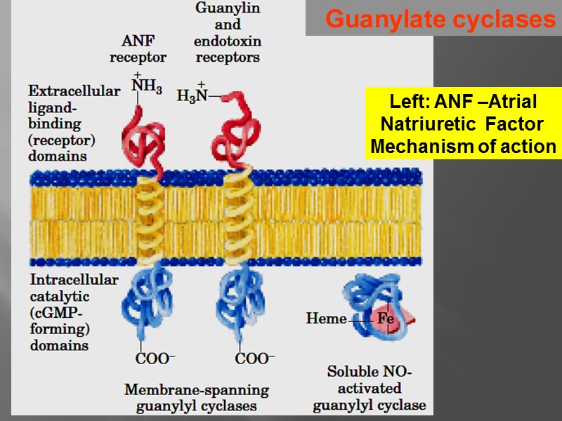
Guanylate cyclases Left: ANF –Atrial Natriuretic Factor Mechanism of action
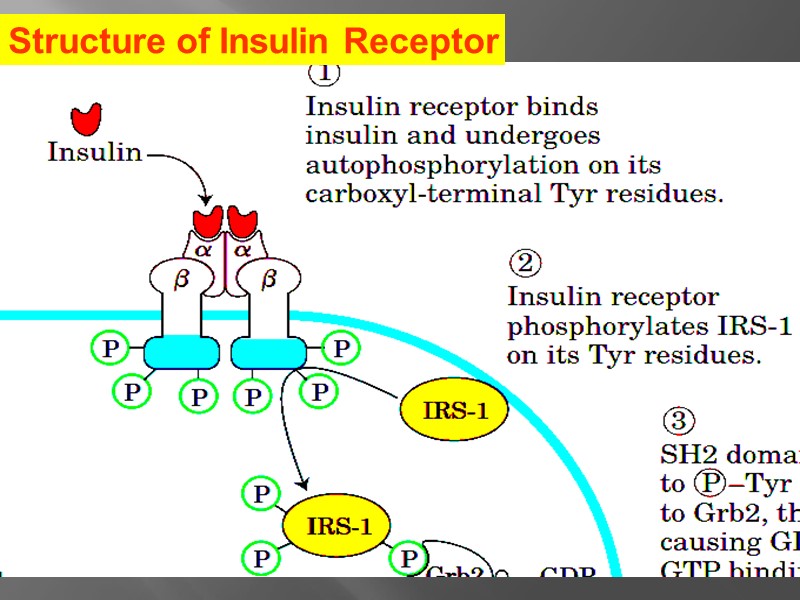
Structure of Insulin Receptor
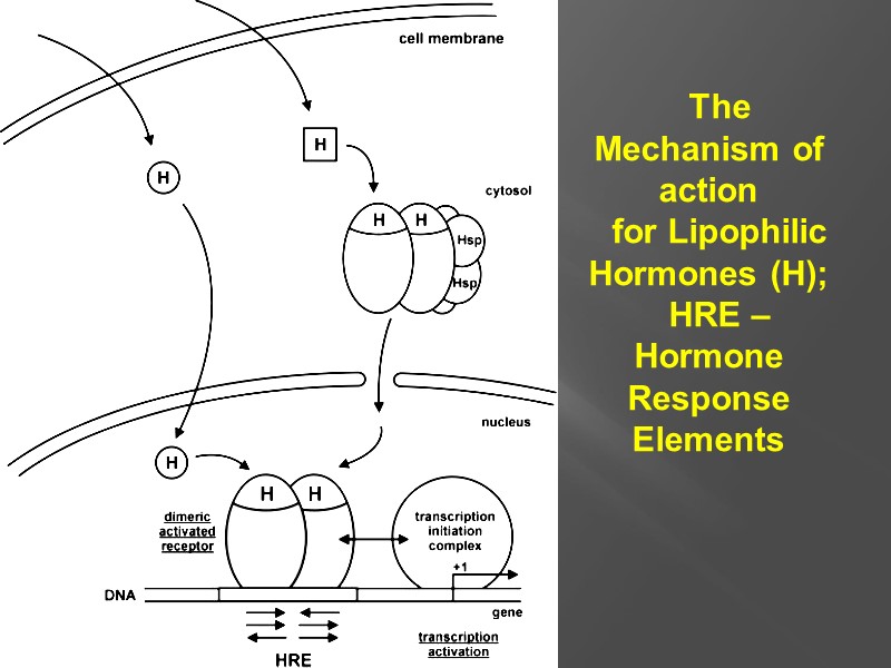
The Mechanism of action for Lipophilic Hormones (H); HRE –Hormone Response Elements
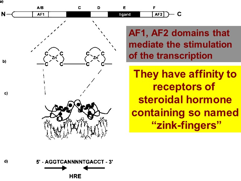
They have affinity to receptors of steroidal hormone containing so named “zink-fingers” AF1, AF2 domains that mediate the stimulation of the transcription
51 THANK YOU For ATTENTION!
25558-gene_exp_and_hormones_part_1.ppt
- Количество слайдов: 51

