
Eye Emergencies BARAKZAY 21 th February 2017


 • Blow out fracture")


 All penetrating trauma require urgent referral to ophthalmologist – immediate consult")

 scan of the orbits and brain (axial and coronal")


eye_emergencies_11.pptx
- Размер: 6.0 Мб
- Автор:
- Количество слайдов: 20
Описание презентации Eye Emergencies BARAKZAY 21 th February 2017 по слайдам
Eye Emergencies BARAKZAY 21 th February
Eye Emergencies Trauma • Lid laceration • Ocular trauma (blunt/penetrating) • Blow out fracture Non trauma • Corneal foreign body • Chemical burns • Acute red eye • Acute visual disturbances
ENT Emergencies Trauma • Fracture of Nasal bone • Traumatic perforation of tympanic membrane Non-trauma • Epistaxis • Foreign body in ear/nose/throat • Otitis media • Peritonsillar abscess • Epiglotitis • Angioedema
1. Lid laceration Any laceration other than superficial skin that involves the lid margin will need ophthalmological referral. An eyelid laceration is a potential penetrating eye injury until proven otherwise. History Four basic questions are: -which eye is injured? -how did it happen? -when did it happen? -what are the symptoms? Nature of injury – was there any possibility of penetration into the lid/orbit?
Examination -Wound examination – size and depth. All wounds should be explored fully for extent of damage. -Visual acuity. -Superficial ocular examination with magnification to assess for any corneal/ conjunctival laceration or penetration. -Further ocular examination including dilated fundus examination as determined by history and examination findings. Treatment/Investigation Orbital X-Ray or CT if indicated foreign bodies or orbital fracture. If superficial laceration: • Clean the area and surrounding skin with antiseptic such as Betadine. Subcutaneous anaesthetic with vasoconstrictor (2% Lignocaine with Adrenaline). Irrigate and debride the wound thoroughly with saline. Identify foreign bodies if applicable. Suture with a 6/0 non-absorbable suture. When to refer? • If the eyelid laceration is associated with ocular trauma requiring surgery such as ruptured globe or intraorbital foreign body. • If the laceration position is nasal to either the upper or the lower eyelid punctum, for the possibility of damage to the nasolacrimal drainage system. • If there is extensive tissue loss or distortion of the anatomy. • If there is full thickness laceration or the laceration involves the lid margin.
2. Ocular trauma History Mechanism of trauma – any history suggestive of a penetrating trauma. The type of projectile and the likely velocity (e. g. low or high) should be documented. Small projectiles at high velocities increase the likelihood of penetrating trauma. Symptoms include loss of vision, pain on movement and diplopia. Was the patient wearing eye protection? Any previous history of ocular trauma or previous surgery is to be documented and may suggest reduced integrity of the wall of the globe. Blunt Closed globe injury Blunt trauma to the eye may result in considerable damage to the intraocular contents. Fracture of the orbital wall may occur due to the transfer of mechanical energy to relatively thin orbital bone. Ruptured globe Trauma of sufficient force may result in globe rupture and typically occurs in the areas where the scleral wall is thinnest: at the limbus (which would be visible via the slit lamp) or behind the insertion of the rectus muscle (which would result in reduced ocular motility, loss of red reflex and vitreous bleeding).
Examination Visual acuity. Ocular movements – if there is considerable eyelid oedema, carefully lift up the lid while viewing the eye to ensure there is no obvious rupture. Reduced movement may suggest ruptured globe or orbital wall fracture. Slit lamp – looking for evidence of rupture (often at the limbus). Examine for blood in the anterior chamber Examine the eyelid for lacerations Ophthalmoscopy – Red reflex (missing in intraocular haemorrhage or retinal detachment). Look for any retinal pathology (after dilating the pupil). Investigations CT scan (axial and coronal) for orbital wall fracture if indicated (see. p 40). Follow up — When to refer? Non-urgent referral within 3 days if the above findings are negative. Urgent referral to ophthalmologist — immediate consult by phone if findings are suggestive of intraocular haemorrhage, ruptured globe or orbital wall fracture Treatment Topical antibiotic drops for superficial trauma. Suture any eyelid lacerations as indicated in eyelid trauma section
Sharp (penetrating) All penetrating trauma require urgent referral to ophthalmologist – immediate consult by phone following appropriate pre-op workup. Examination may only need to be cursory if the trauma is obvious otherwise: – -Visual acuity. -Direct ophthalmoscopy – loss of red reflex may suggest retinal trauma or detachment. -Slit lamp – looking for distorted anterior chamber structures or corneal/scleral breaks. Treatment -Ensure nil by mouth status. Strict bed rest. Injectable analgesia/antiemetic if required. -CT scan of the orbit to exclude retained ocular/orbital foreign body after discussion with ophthalmologist. -Shield (not pad) the eye making sure not to increase the intraocular pressure with further loss of ocular contents. -No ointment for penetrating eye injury. -Check for tetanus immunisation status as per current protocol. -Commence broad spectrum IV antibiotics.
3. Blow out fracture History Mechanism of injury — for example squash ball, punch with a fist. Symptoms -Pain (especially on vertical movement), local tenderness, diplopia, eyelid swelling and crepitus after nose blowing. -A “white” blow-out fracture occurs with orbital injury with the findings of minimal periorbital haemorrhage, sunken (enophthalmic) globe, restricted eye movement in an unwell child. — Exclude head injury and refer urgently. Signs Nose bleed, ptosis and localised tenderness. Examination -Complete ophthalmological examination. -Sensation of affected cheek compared with that of the contralateral side. -Infraorbital nerve involvement is demonstrated by anaesthetised upper teeth and gums on the affected side. Inferior rectus entrapment (diplopia on upward gaze) Palpate eyelid for crepitus.
Investigation Computed tomography (CT) scan of the orbits and brain (axial and coronal views). Treatment -Nasal decongestants for 7 -10 days. -Broad spectrum oral antibiotics. -Instruct patient not to blow his or her nose. (Avoid Valsalva manoeuvre). -Ice pack to the orbit for 24 -48 hours. Follow up — When to refer? -Non-urgent referral – contact ophthalmogist for time frame for all cases with: -Suspected or documented orbital floor fractures. -Patients should be seen within 1 -2 weeks post trauma and evaluated for persistent diplopia or enophthalmos. -Surgical repair if necessary is usually performed 7 to 14 days after trauma.
4. Corneal foreign bodies Any foreign body penetration of the cornea or retained foreign body will require urgent referral to ophthalmologist — immediate consult by phone. History -What is the likely foreign body? -Examples include dirt, glass, metal and inorganic material. — Retained organic material may lead to infection; retained metallic foreign bodies may lead to the formation of rust rings that produce persistent inflammation and corneal epithelial defect. -Velocity of impact? High speed motor drilling without eye protection may lead to a penetrating corneal/scleral injury. Examination -Visual acuity -Slit lamp — assess for the size, site/s and nature of foreign body and the depth of penetration. — Examine the cornea, anterior chamber, iris, pupil and lens for any distortion that may indicate ocular penetration and require urgent referral to an ophthalmologist. -Evert the eyelids to exclude retained foreign bodies – remove if appropriate.
5. Chemical burns History -When did it occur? -What is the chemical? (e. g. acid/alkali; alkalis are more harmful to the eye). Examples of acids include: toilet cleaner, car battery fluid, pool cleaner. Examples of alkalis include: lime, mortar & plaster, drain cleaner, oven cleaner, ammonia. -Any first aid administered and how soon after the incident? Examination -Use topical anaesthesia. -The degree of vascular blanching, particularly at the limbus, is proportional to severity of chemical burn Immediate treatment as indicated ** Chemical Burns -Measure p. H using universal indicator paper to sample the forniceal space -Test and record visual acuity. -Contact poisons information or the chemical manufacturer for further information if required.
1. Instil local anaesthetic drops to affected eye/eyes. 2. Commence irrigation with 1 litre of a neutral solution, eg N/Saline (0. 9%), Hartmann’s. 3. Evert the eyelid and clear the eye of any debris / foreign body that may be present by sweeping the conjunctival fornices with a moistened cotton bud. 4. a. Continue to irrigate, aiming for a continuous irrigation with giving set regulator fully open. b. If using a Morgan Lens, carefully insert the device now. 5. Review the patient’s pain level every 10 minutes and instil another drop of local anaesthetic as required. 6. a. After one litre of irrigation, review. b. If using a Morgan Lens, remove the device prior to review. 7. Wait 5 minutes after ceasing the irrigation luid then check p. H. Acceptable p. H range 6. 5 -8. 5. 8. Consult with the senior medical oficer and recommence irrigation if necessary. 9. Severe burns will usually require continuous irrigation for at least 30 minutes.
6. Acute red eye
Acute Angle Closure Glaucoma Eye pain, headache, cloudy vision, colored halos around lights, conjunctival injection Fixed, mid-dilated pupil Increased IOP (40 -70 mm Hg) Normal range is 10 – 20 mm Hg Nausea, vomiting
Scleritis -Vision may be impaired. Sclera is thickened and discoloured. The globe is tender to palpation. -Associated history of life-threatening vascular or connective tissue disease – may require appropriate physician consult (NB look for medications involving systemic steroids, NSAIDs, antimetabolites). Urgent referral – see ophthalmologist within 24 hours. Anterior chamber involvement Acute Anterior Uveitis (Iritis) -Pain, photophobia, and red eye. -Anterior chamber appears cloudy from cells and flare. Urgent referral – see ophthalmologist within 24 hours. Hypopyon -Visible accumulation of white cells inferiorly seen in severe uveitis. Urgent referral – immediate consult by phone — for investigation of infection, inflammation or ocular malignancy. Hyphaema -Usually trauma related but consider non-accidental injury in children and blood dyscrasias. Bed rest. Urgent referral – see ophthalmologist within 24 hours.
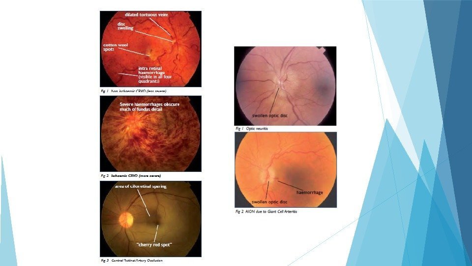
7. Acute visual loss
Amaurosis Fugax Symptoms -Monocular visual loss that usually lasts seconds to minutes, but may last 1 -2 hours. Vision returns to normal. Signs -Essentially normal fundus exam (an embolus within a retinal arteriole is only occasionally seen Other neurological signs associated with ischemia of cerebral hemispheres. Investigation -As per protocol but usually includes assessment of cardiovascular risk factors: -Blood count/electrolytes/lipids/fasting blood sugar. -Thrombophilia screen. -Echocardiogram. -Carotid doppler studies. Management -Commence aspirin. Referral to neurology/cardiology or vascular surgery as appropriate. Patients with recurrent episodes of amaurosis fugax require immediate diagnostic and therapeutic intervention.
TAKE HOME NOTE : WHEN TO REFER IMMEDIATELY? ? Decreased visual acuity Deep, rather than superficial, pain Pain unrelieved by topical anaesthetics Corneal edema Flare or cells in the anterior chamber Ciliary flush Pain in the contralateral eye on direct exposure of the unaffected eye Corneal or conjunctival foreign body which cannot be removed after one attempt in the office In the case of corrosive burn, implement saline irrigation immediately prior to the patient being transported to hospital. Irrigate until the effluent is p. H neutral or mildly acidic as shown by the use of blue and pink litmus paper