
External Anatomy of the Eye Lacrimal Apparatus

- Размер: 3.3 Mегабайта
- Количество слайдов: 20
Описание презентации External Anatomy of the Eye Lacrimal Apparatus по слайдам
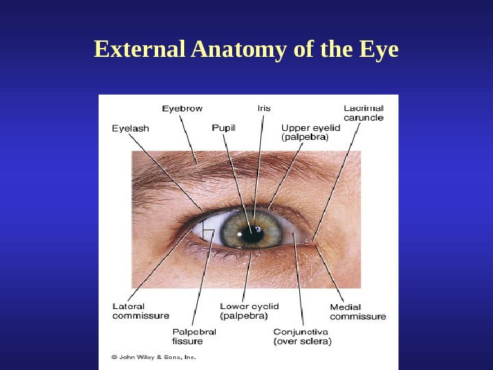
External Anatomy of the Eye
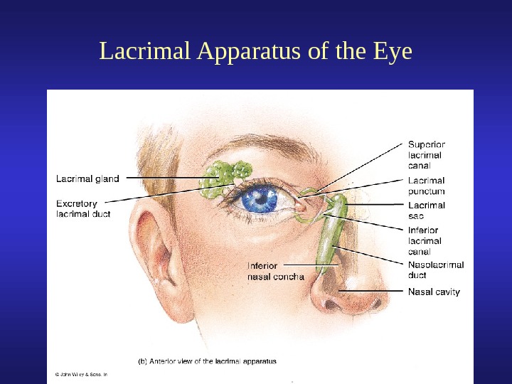
Lacrimal Apparatus of the Eye
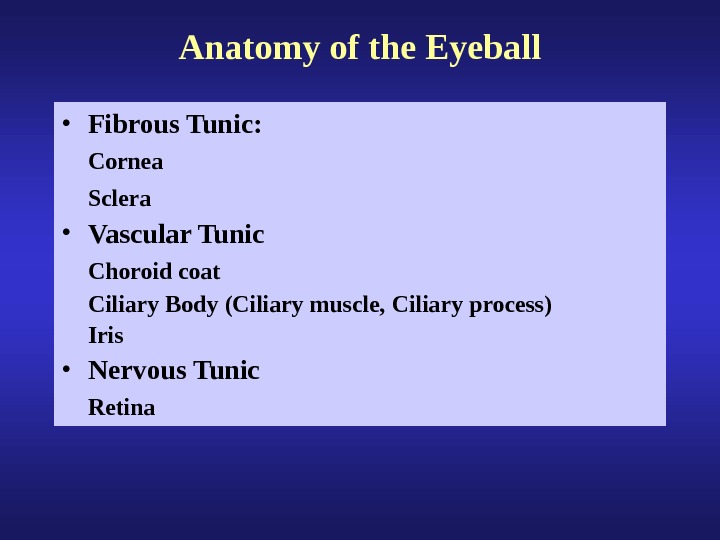
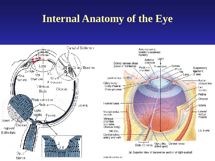
Anatomy of the Eyeball • Fibrous Tunic: Cornea Sclera • Vascular Tunic Choroid coat Ciliary Body (Ciliary muscle, Ciliary process) Iris • Nervous Tunic Retina
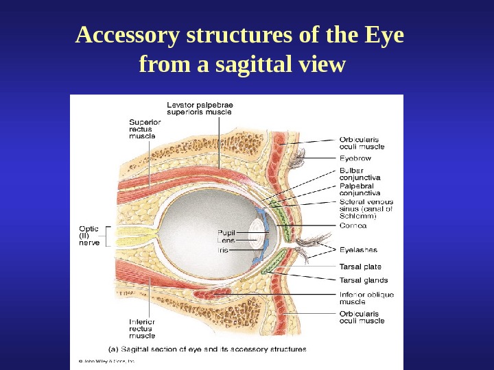
Accessory structures of the Eye from a sagittal view
Internal Anatomy of the Eye
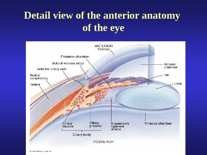
Detail view of the anterior anatomy of the eye
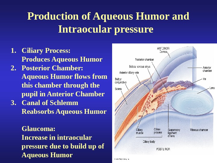
Production of Aqueous Humor and Intraocular pressure 1. Ciliary Process: Produces Aqueous Humor 2. Posterior Chamber: Aqueous Humor flows from this chamber through the pupil in Anterior Chamber 3. Canal of Schlemm Reabsorbs Aqueous Humor Glaucoma: Increase in intraocular pressure due to build up of Aqueous Humor
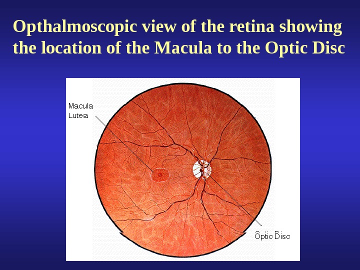
Opthalmoscopic view of the retina showing the location of the Macula to the Optic Disc
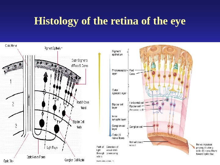
Histology of the retina of the eye
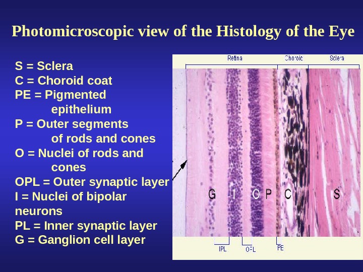
Photomicroscopic view of the Histology of the Eye S = Sclera C = Choroid coat PE = Pigmented epithelium P = Outer segments of rods and cones O = Nuclei of rods and cones OPL = Outer synaptic layer I = Nuclei of bipolar neurons PL = Inner synaptic layer G = Ganglion cell layer
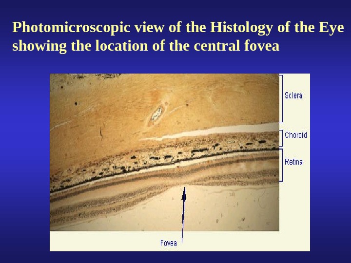
Photomicroscopic view of the Histology of the Eye showing the location of the central fovea
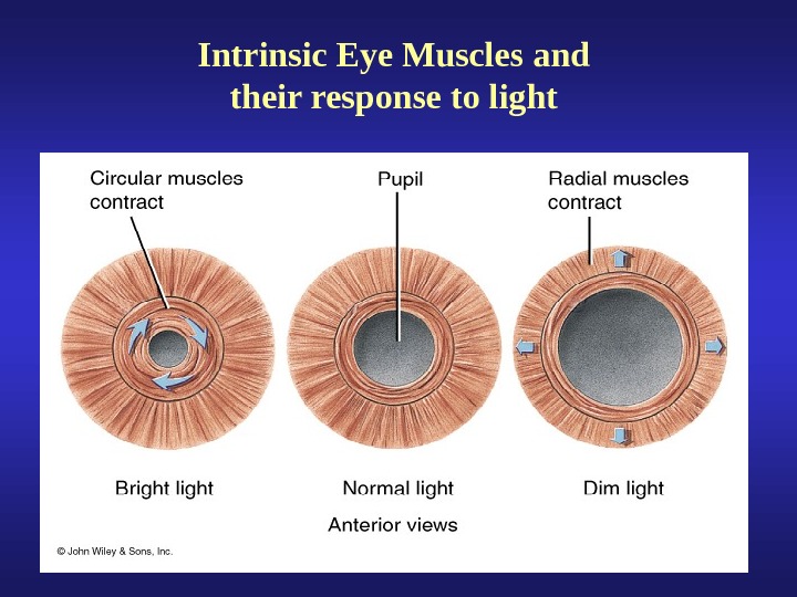
Intrinsic Eye Muscles and their response to light
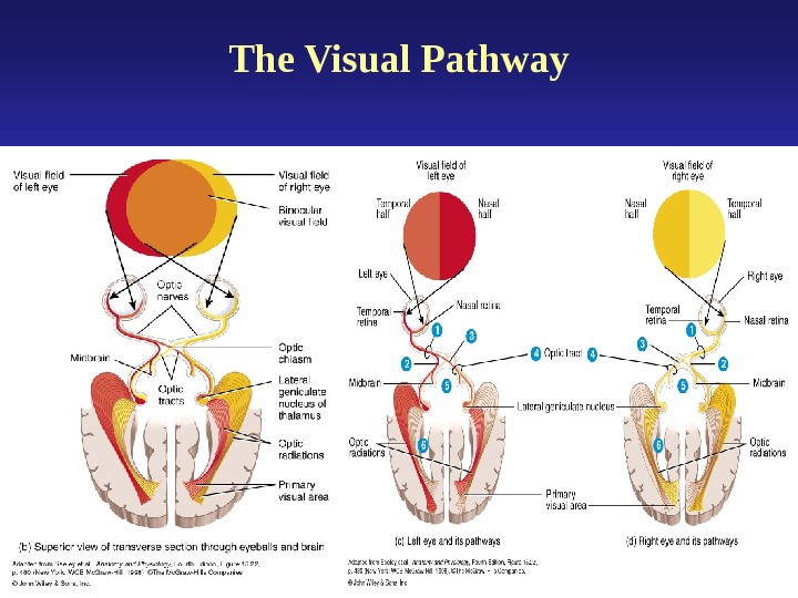
The Visual Pathway
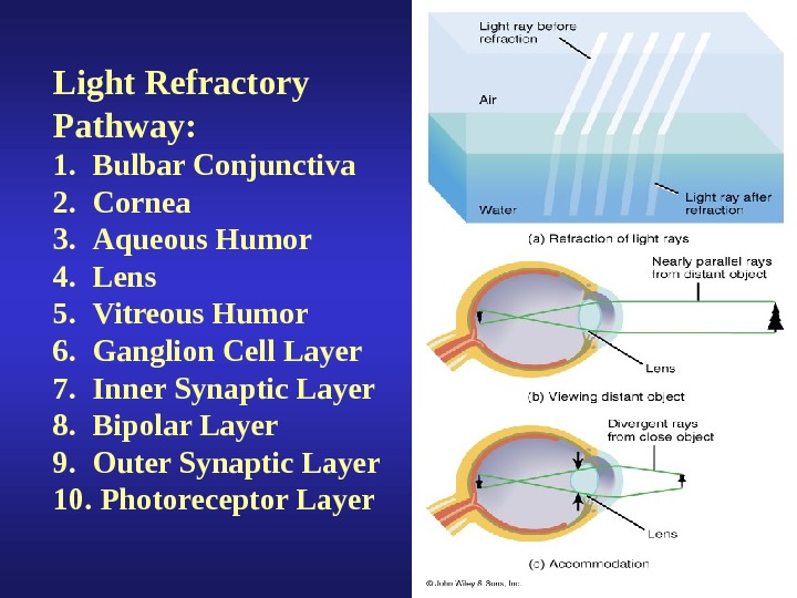
Light Refractory Pathway: 1. Bulbar Conjunctiva 2. Cornea 3. Aqueous Humor 4. Lens 5. Vitreous Humor 6. Ganglion Cell Layer 7. Inner Synaptic Layer 8. Bipolar Layer 9. Outer Synaptic Layer 10. Photoreceptor Layer
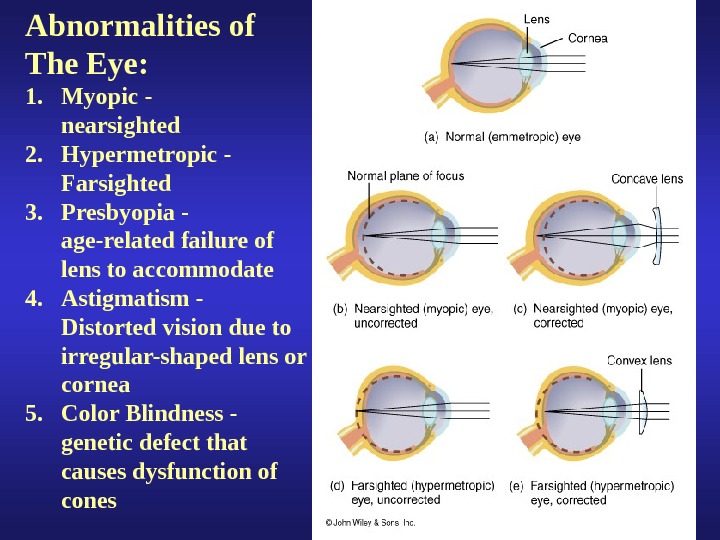
Abnormalities of The Eye: 1. Myopic — nearsighted 2. Hypermetropic — Farsighted 3. Presbyopia — age-related failure of lens to accommodate 4. Astigmatism — Distorted vision due to irregular-shaped lens or cornea 5. Color Blindness — genetic defect that causes dysfunction of cones
Accommodation of the Lens for near vision • Ciliary muscles contract • Ciliary body pulls forward and inward • Tension on suspensory ligaments of lens is decreased • Lens becomes thicker (rounder) due to its elasticity • Pupils constricts
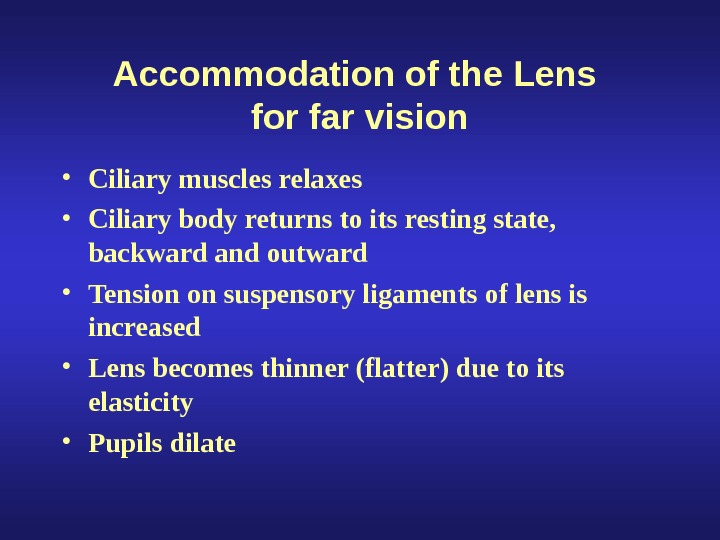
Accommodation of the Lens for far vision • Ciliary muscles relaxes • Ciliary body returns to its resting state, backward and outward • Tension on suspensory ligaments of lens is increased • Lens becomes thinner (flatter) due to its elasticity • Pupils dilate
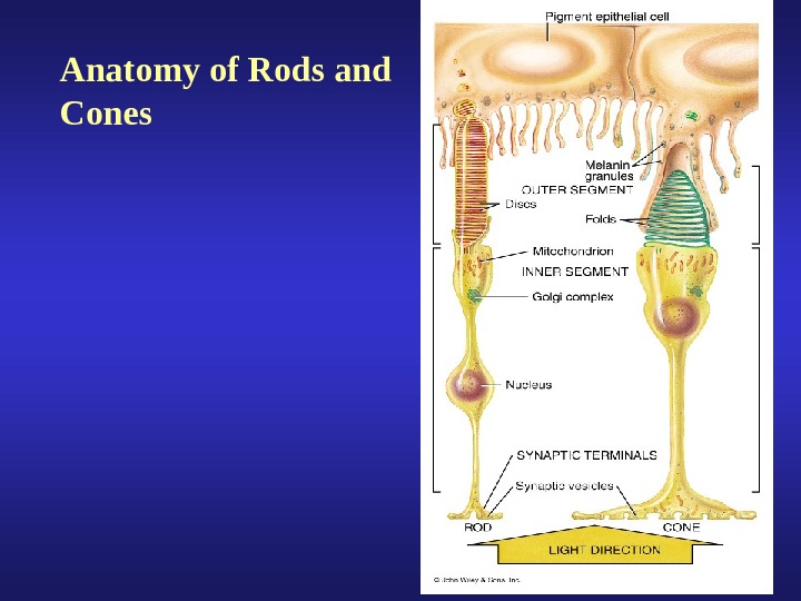
Anatomy of Rods and Cones
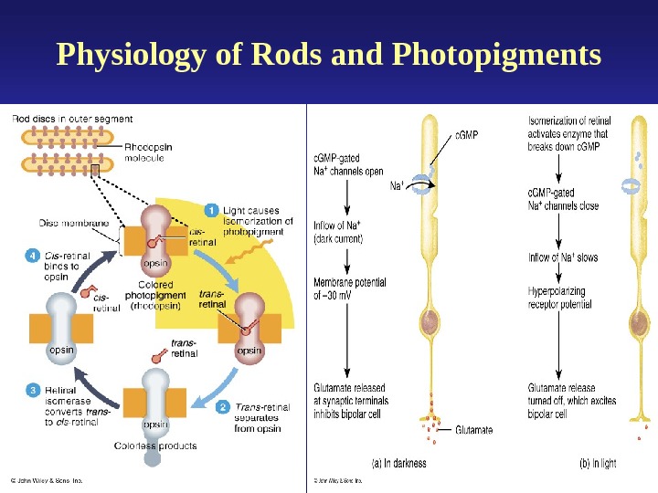
Physiology of Rods and Photopigments
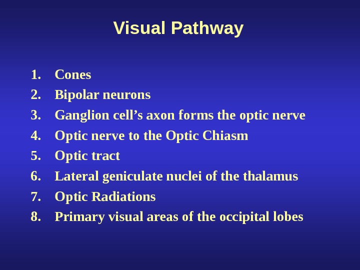
Visual Pathway 1. Cones 2. Bipolar neurons 3. Ganglion cell’s axon forms the optic nerve 4. Optic nerve to the Optic Chiasm 5. Optic tract 6. Lateral geniculate nuclei of the thalamus 7. Optic Radiations 8. Primary visual areas of the occipital lobes