

480718b4279859468402fa88b7447ed1.ppt
- Количество слайдов: 27
 Eugene Yevstratov, MD Institute of Cardiology and Cardiovascular Surgery, Favaloro Foundation Buenos Aires, Argentina October/2002
 Goals of Myocardial protection 1. Protect against ischemic injury 2. Provide a motionless, bloodless field 3. Allow effective post-ischemic myocardial resuscitation
 Spectrum of myocardial ischemic injury Acute ischemic disfunction Preconditioning Stunning Hibernation Necrosis vs. Apoptosis
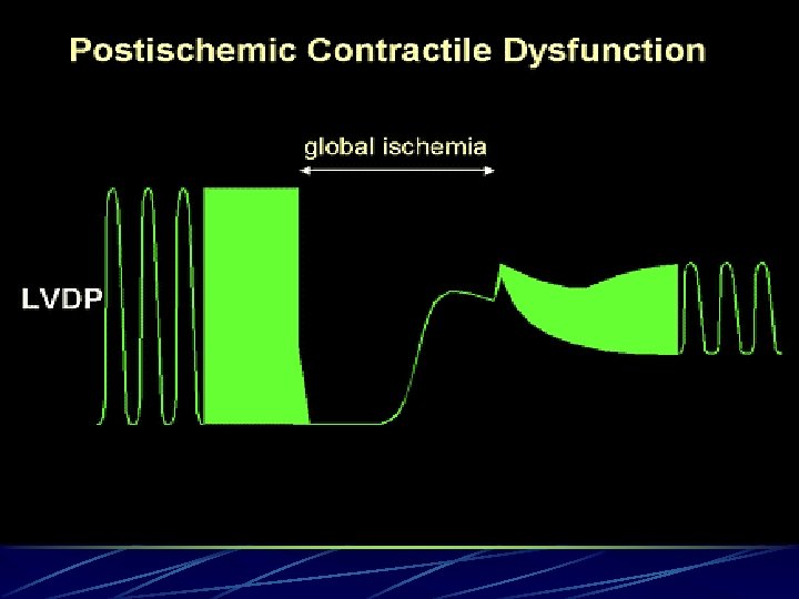
 Acute ischemic disfunction Reversible contractile failure Perfusion pressure O 2 supply Inmediate recovery
 Preconditioning Reversible Slowed energy utilization Reduction in myocardial necrosis Increase protective abilities of myocardium Presented as a normal proper protective reaction of the ischemic myocardium Recovery Hs, Ds
 causing reduced coronary blood") Stunning Parcialy Reversible May be accompained by endothelial disfunction (NO) causing reduced coronary blood flow Result of ischemia-reperfusion insult Mediated by increased intracellular Ca accumulation Recovery in Hs, Wks
Stunning Parcialy Reversible May be accompained by endothelial disfunction (NO) causing reduced coronary blood flow Result of ischemia-reperfusion insult Mediated by increased intracellular Ca accumulation Recovery in Hs, Wks
 Hibernation Parcialy Reversible Related to poor myocardial blood flow Chronic Recovery Wks, Mo
 Necrosis Irreversible Hyper contracture - “contracture band necrosis”, “stone heart” Osmotic/ionic dysregulation, membrane injury Cell swelling&disruption Lysis
 Apoptosis Irreversible Death signal Cell shrinkage Cytoplasmic and nuclear condensation Phagocytosis
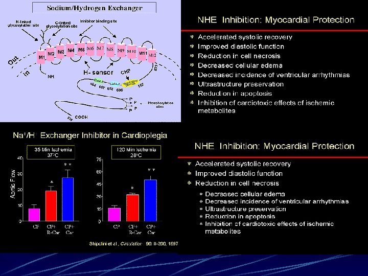
 Adenosine dependent receptors K+ATP dependent") Systems involved into membrane injury MAC( membrane attack complex) Adenosine dependent receptors K+ATP dependent chanels NHE(sodium hydrogen exchanger)
Systems involved into membrane injury MAC( membrane attack complex) Adenosine dependent receptors K+ATP dependent chanels NHE(sodium hydrogen exchanger)
 Cellular swelling") Cellular effects of ischemia Altered membrane potential Altered ion distribution(increase intracellular. Ca++/Na++) Cellular swelling Cytoskeletal Disorganisation Increased hypoxantine Decreased ATP Decreased phosphocreatine Decreased Glutatione Cellular Acidosis
Cellular effects of ischemia Altered membrane potential Altered ion distribution(increase intracellular. Ca++/Na++) Cellular swelling Cytoskeletal Disorganisation Increased hypoxantine Decreased ATP Decreased phosphocreatine Decreased Glutatione Cellular Acidosis
 Straqtegies for Heart protection Increase the O 2 offer Decreae oxygen demand Methabolical intervention Prevention of demand increased Substrate disponsability
 10 ml/100 gr/min Beating") Myocardial O 2 consumptions at 37 C Beating (full, perfused) 10 ml/100 gr/min Beating (empty, perfused) 5, 5 ml/100 gr/min Fibrilating(empty, perfused) 6, 5 ml/100 gr/min K+ Cardioplegia(empty, crossclamp) 1, 0 ml/100 gr/min
Myocardial O 2 consumptions at 37 C Beating (full, perfused) 10 ml/100 gr/min Beating (empty, perfused) 5, 5 ml/100 gr/min Fibrilating(empty, perfused) 6, 5 ml/100 gr/min K+ Cardioplegia(empty, crossclamp) 1, 0 ml/100 gr/min
") Myocardial O 2 consumption ml/100 gr/min Temperatura C 37 32 28 22 Beating (empty) 5, 5 5, 0 4, 0 2, 9 Fibrilating (empty) 6, 5 3, 8 3, 0 2, 0 K+ cardioplegia 1, 0 0, 8 0, 6 0, 3
Myocardial O 2 consumption ml/100 gr/min Temperatura C 37 32 28 22 Beating (empty) 5, 5 5, 0 4, 0 2, 9 Fibrilating (empty) 6, 5 3, 8 3, 0 2, 0 K+ cardioplegia 1, 0 0, 8 0, 6 0, 3
 Cardioplegia - Options No cardioplegia Cardioplegia Type ( blood vs crystalloid, cont vs intermittent ) Route ( antegrade vs retrograde ) Temperature ( warm vs cold ) Additives Special consideration ( Acute infarction, Neonate)
 Rivero Cardioplegia solutions 1 2
 Mechanism of Cardioplegic Protection Mechanical arrest ( K – induced, 80% reduction in O 2 consumption) Hypotermia (10 -15% furter reduction in O 2 consumption) Aerobic metabolism – oxygenated cardioplegia Maintain hypotermic arrest with readministration every 15 -20 min Retrograde delivery LV RV protection
 Acute") Other consideration Protect from rewarming Systemic hypotermia Aortic/ventricular vents Total bypass (caval oclusion) Acute Ischemia Waqrm induction Substrate enhancement Controlled reperfusion Warm, hypocalcemic, alkaline cardioplegia Retrograde or low flowpressure antegrade perfusion Energy replacement while arrested Uniform warming
Other consideration Protect from rewarming Systemic hypotermia Aortic/ventricular vents Total bypass (caval oclusion) Acute Ischemia Waqrm induction Substrate enhancement Controlled reperfusion Warm, hypocalcemic, alkaline cardioplegia Retrograde or low flowpressure antegrade perfusion Energy replacement while arrested Uniform warming
 Cardioplegic Composition Blood vs Crystalloid Buffers Calcium Potassium Free radical scavengers Others
 Buffers") Blood vs Crystalloid O 2 carrying capacity ( Hematocrit 15 – 20 %) Buffers –histidine Free radical scavengers in RBCs Improved rheologic / oncotic properties Metabolic substrate
Blood vs Crystalloid O 2 carrying capacity ( Hematocrit 15 – 20 %) Buffers –histidine Free radical scavengers in RBCs Improved rheologic / oncotic properties Metabolic substrate
 Buffers THAM Histidine Na. HCO 3 Slightly alkaline reperfusion
 Calcium, Potassium Small amounts of calcium ( 0. 1 – 0. 5 m. M/L ) Ca chelated in blood with citrate 10 – m. M/L of potassium ( first dose highest ) > 30 m. M/L – endothelial dysfunction
 Free radical scavengers. Others Allopurinol Propofol Deferoxamine Metabolic substrates ( adenosine, nucleotid transport inhibitors. . . ) K- channel openers ( Nicorandil )
 The ways of pharmacological therapy Addition of metabolites or cofactors Activation of enzymes or complexes involving in generation of reduced equivalents, and their utilisation Control of synthesis of mitochondrial factors, or genesis of mitochondria, and protection of mitochondria Improving Ph balance in the ischemic heart
 Institute of Cardiology and Cardiovascular Surgery, Favaloro Foundation Buenos Aires, Argentina The End Eugene Yevstratov E-mail Fax 001775 679 2870