
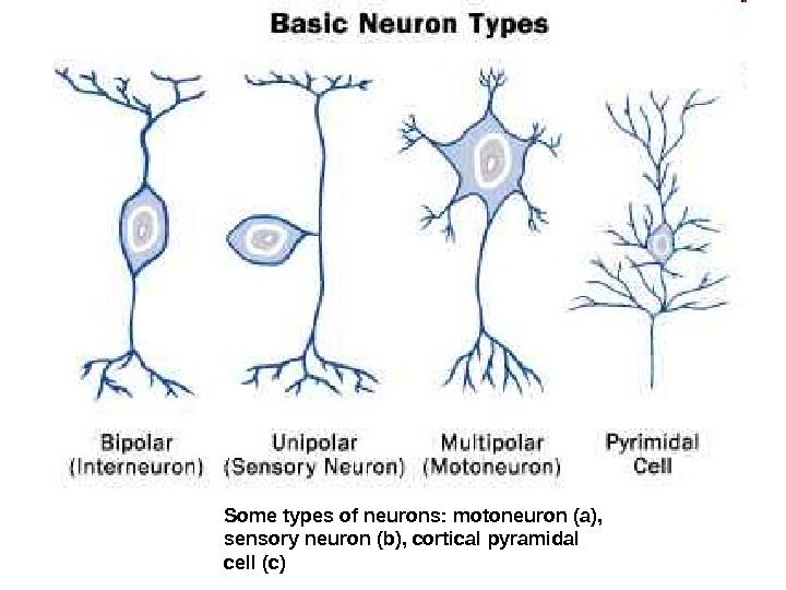
Endbrain Interbrain Midbrain Hindbrain Some types of


- Размер: 37.9 Mегабайта
- Количество слайдов: 134
Описание презентации Endbrain Interbrain Midbrain Hindbrain Some types of по слайдам
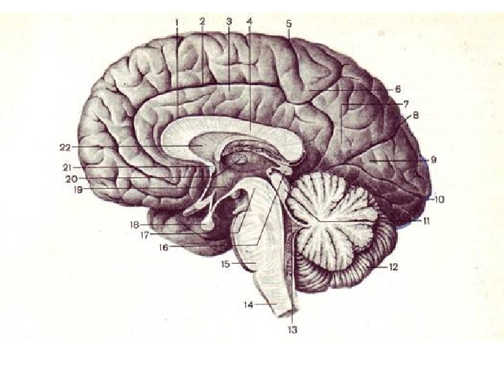
Endbrain Interbrain Midbrain Hindbrain
Some types of neurons: motoneuron (a), sensory neuron (b), cortical pyramidal cell (c)
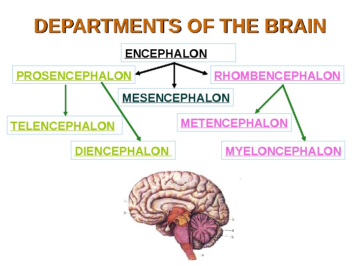
DEPARTMENTS OF THE BRAIN ENCEPHALON PROSENCEPHALON MESENCEPHALON RHOMBENCEPHALON TELENCEPHALON DIENCEPHALON METENCEPHALON MYELONCEPHALON
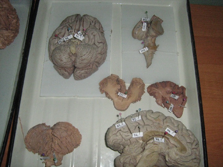
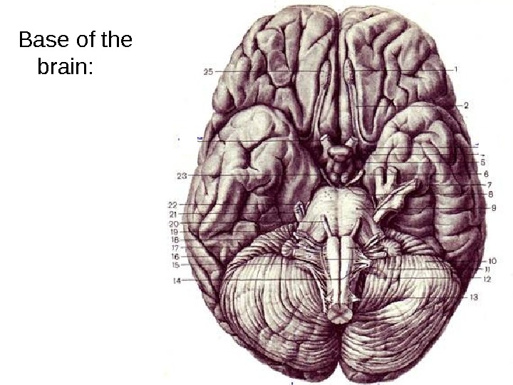
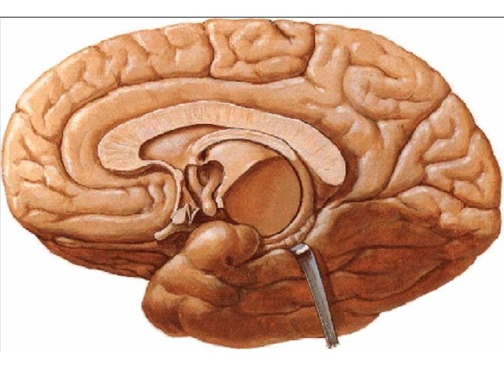
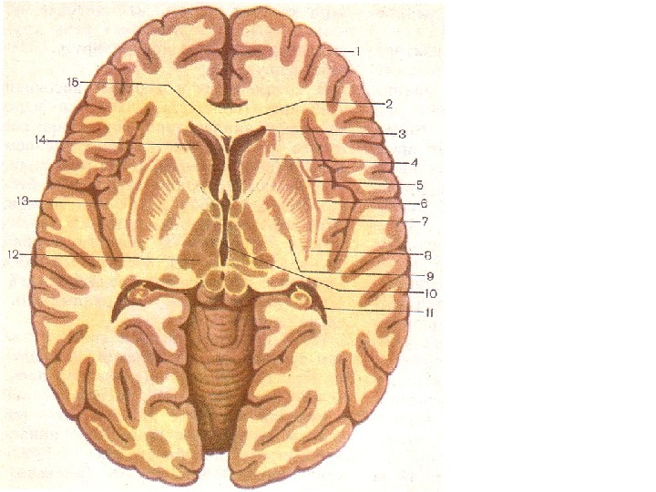
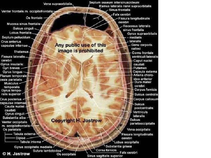
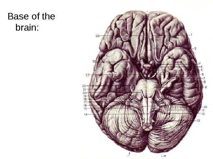
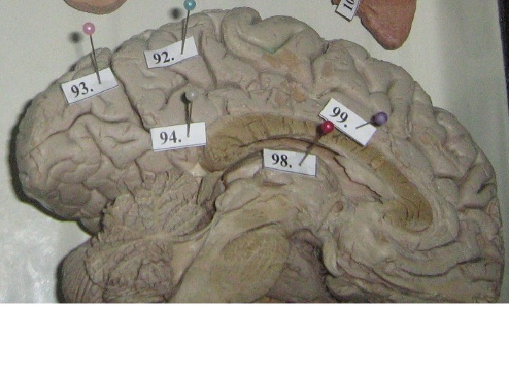
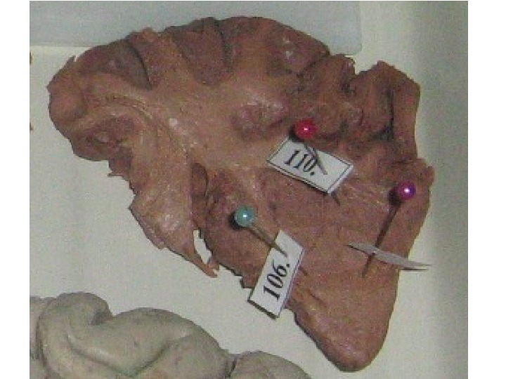
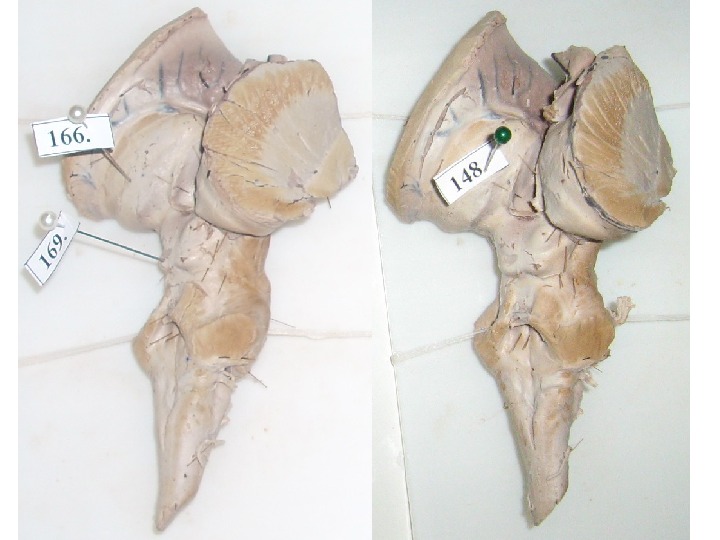
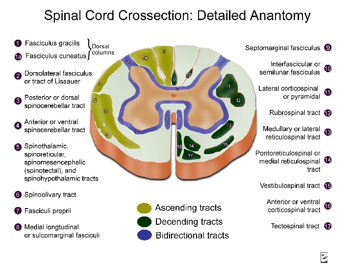
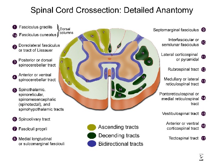
Base of the brain:
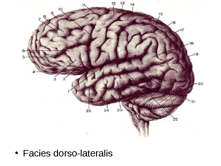
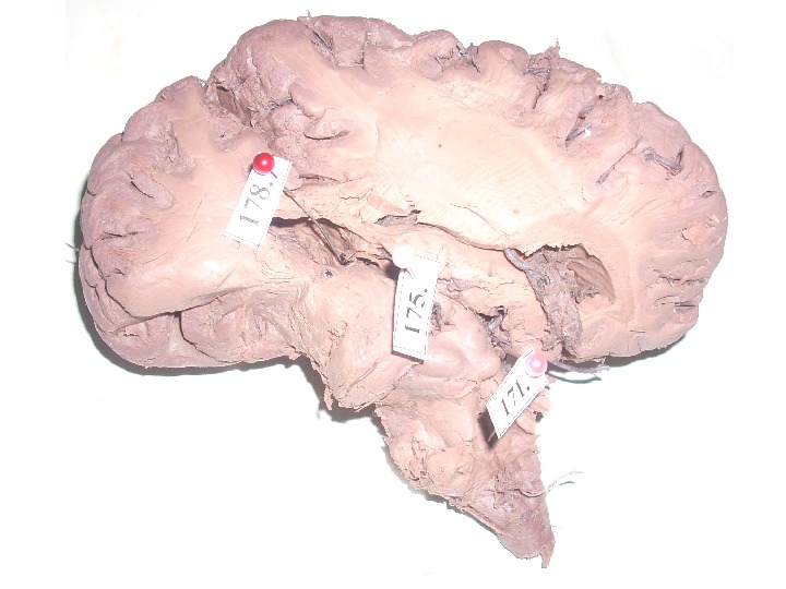
• Facies dorso-lateralis
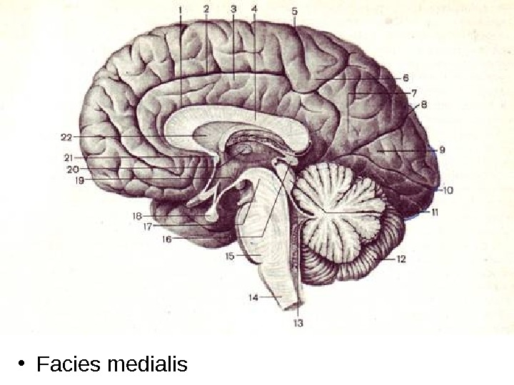
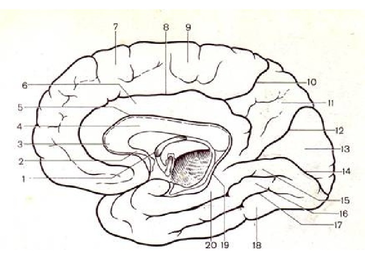
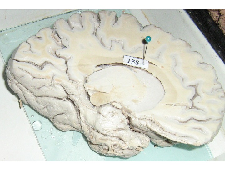
• Facies medialis
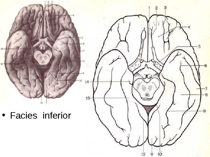
• Facies inferior
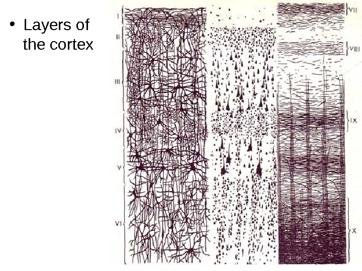
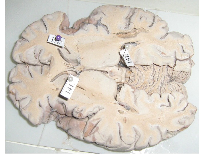
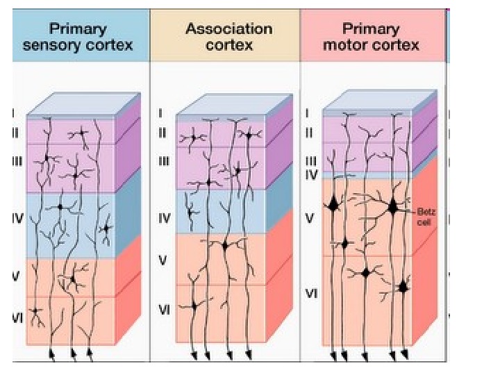
• Layers of the cortex
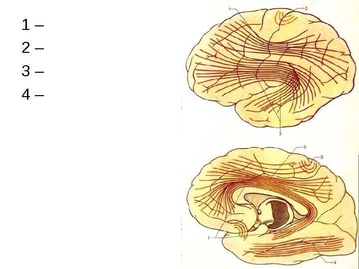
1 – 2 – 3 – 4 –
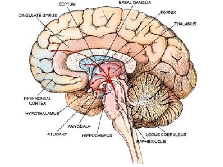
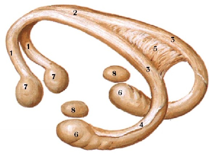
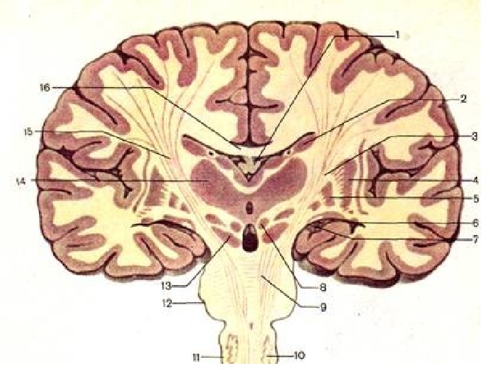
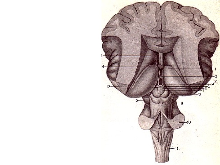
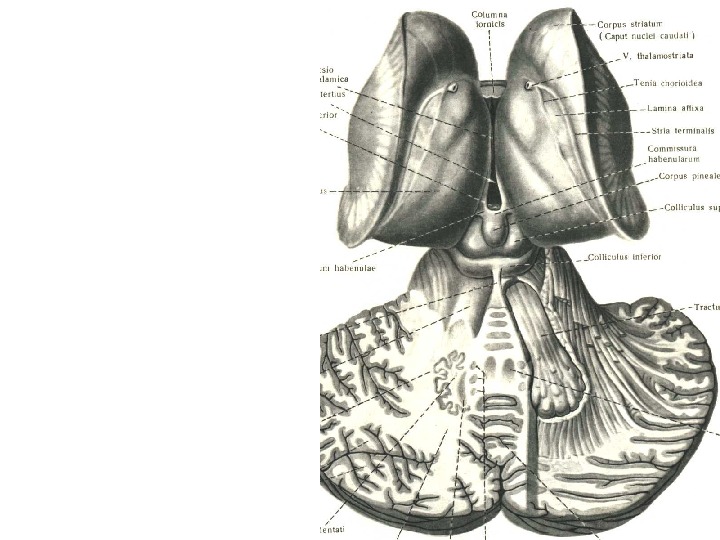
Fornix
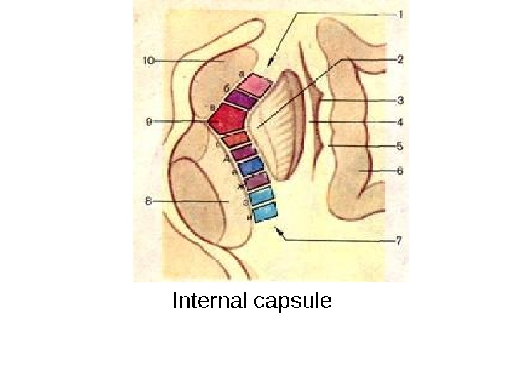
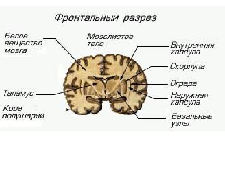
Internal capsule
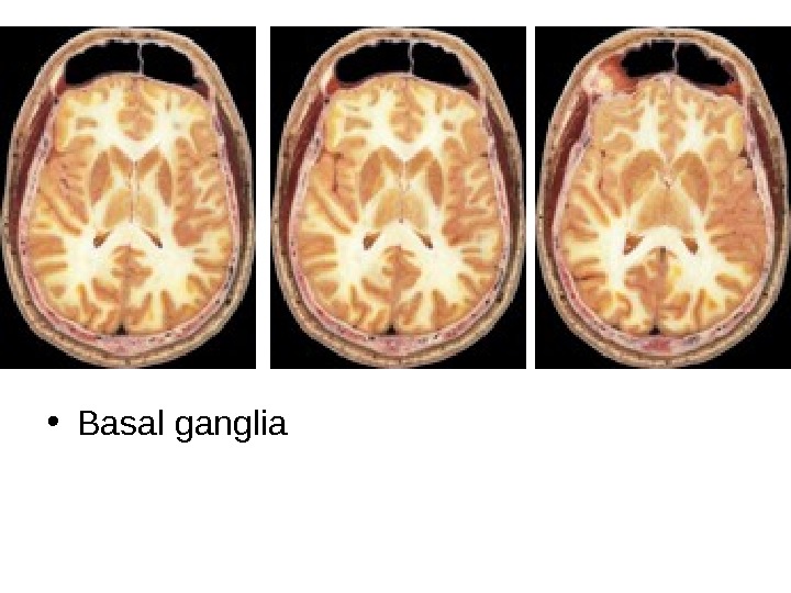
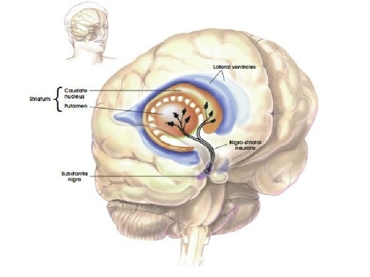
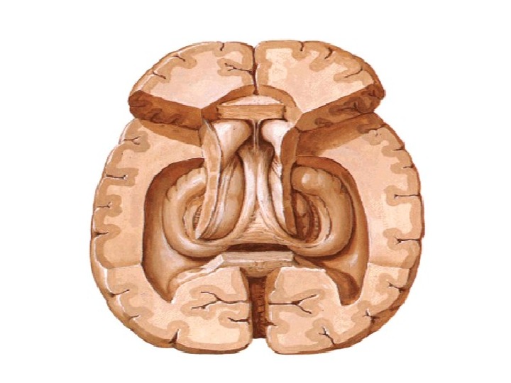
• Basal ganglia
Base of the brain:
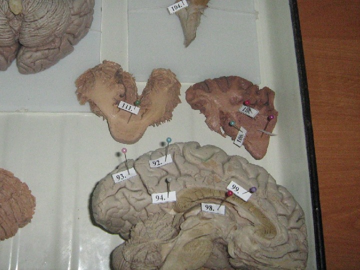
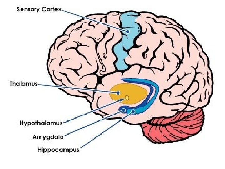
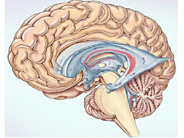
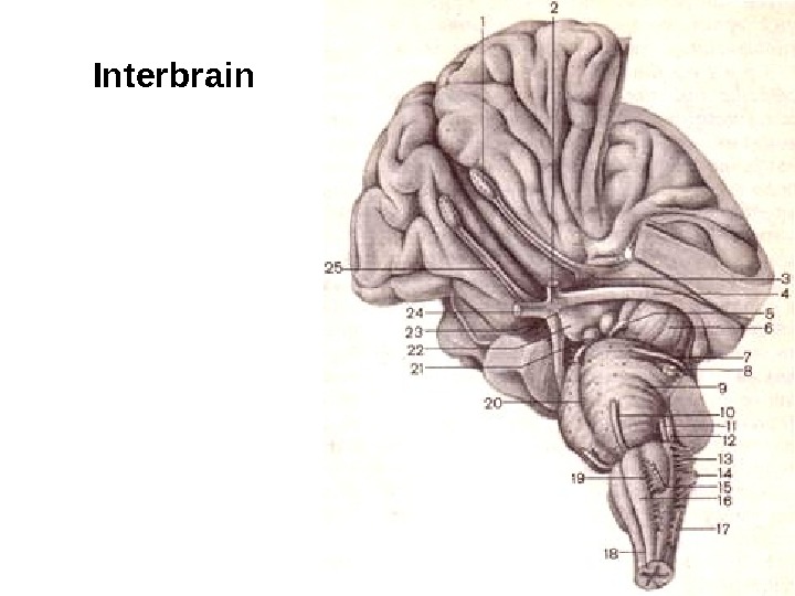
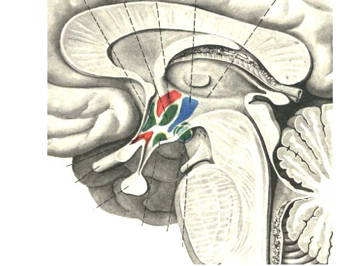
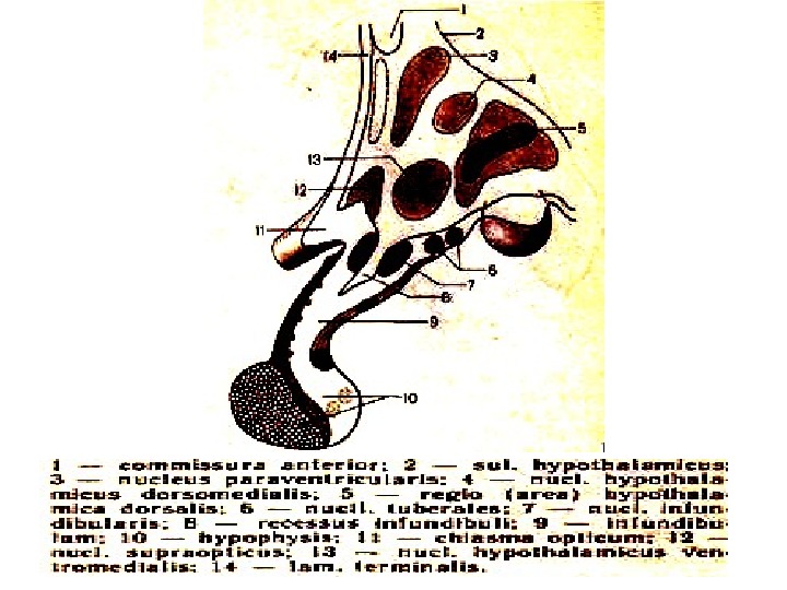
Interbrain
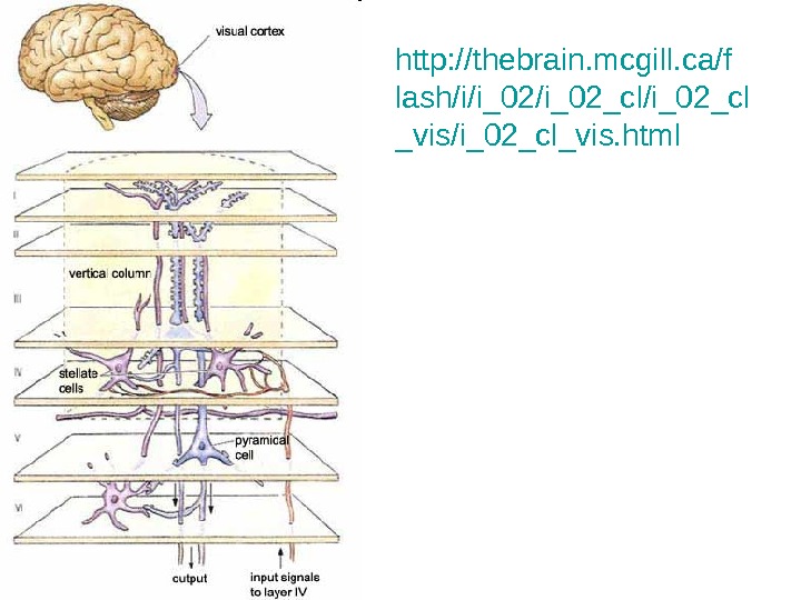
http: //thebrain. mcgill. ca/f lash/i/i_02_cl/i_02_cl _vis/i_02_cl_vis. html
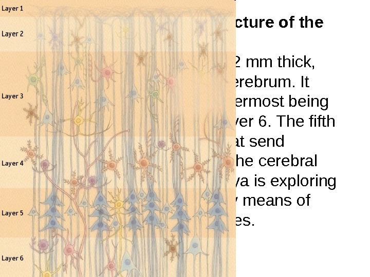
• Figure 1: The six-layer structure of the cerebral cortex. The cerebral cortex is about 2 mm thick, covering the surface of the cerebrum. It consists of six layers, the outermost being layer 1, and the innermost layer 6. The fifth layer contains output cells that send information to the outside of the cerebral cortex, and other cells. Hosoya is exploring important circuit structures by means of genetic engineering techniques.
http: //www. users. globalnet. co. uk/~ lka/conz 3 a. htm
http: //www. google. kz/imgres? imgurl=http: //www. nature. com/nrn/journal/v 5/n 3/images/nrn 1345 -i 1. jpg &imgrefurl=http: //www. nature. com/nrn/journal/v 5/n 3/box/nrn 1345_BX 1. html&usg=__okf. Ev. U 27 F 7 w. XEx 2 zjd. C 1 l. XGIf. PQ=&h=635&w=600&sz=5 9&hl=ru&start=10&zoom=1&tbnid=Jc. QPk-lk_r 08 j. M: &tbnh=137&tbnw=1 29&ei=2 D-j. Tc. OPI 8 qa. Oqi. Pn. DU&prev=/search%3 Fq%3 Dreticular %2 Bformation%26 hl%3 Dru%26 client%3 Dfirefox-a%26 sa%3 DG%26 rls %3 Dorg. mozilla: ru: official%26 biw%3 D 1280%26 bih%3 D 613%26 gbv %3 D 2%26 tbm%3 Disch 0%2 C 342&itbs=1&biw=1280&bih=
• http: //www. ncbi. nlm. nih. gov/books/NBK 110 81/
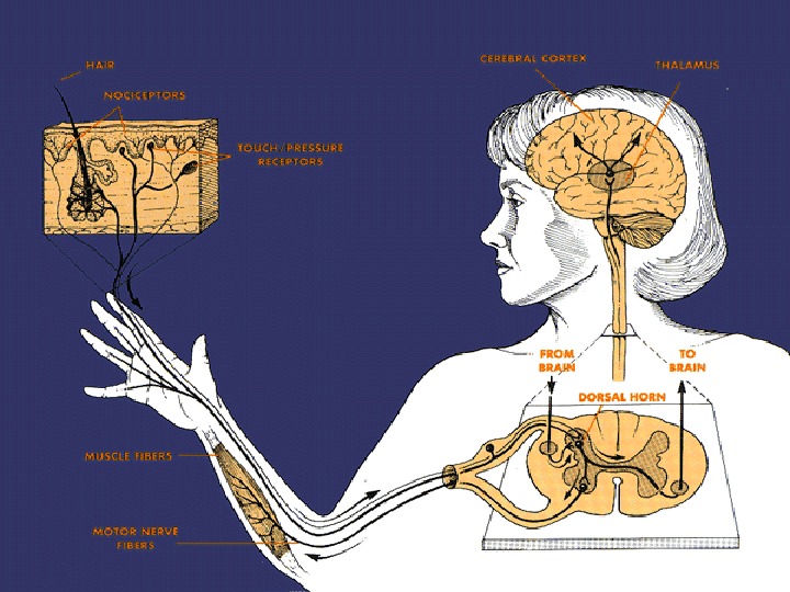
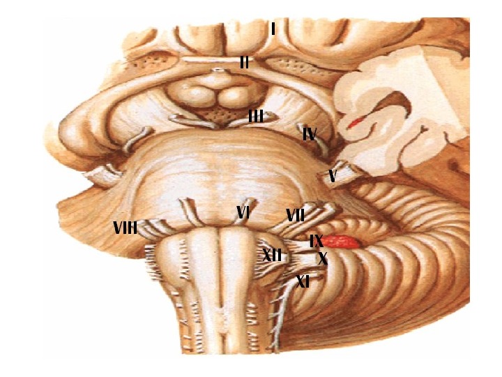
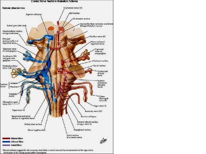
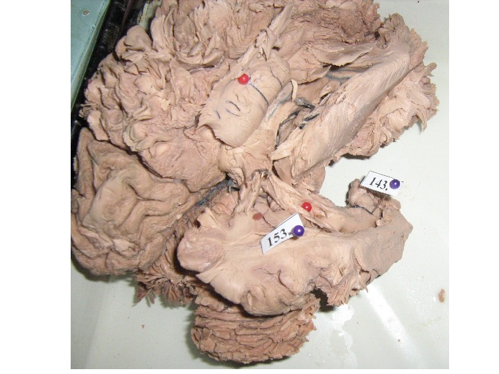
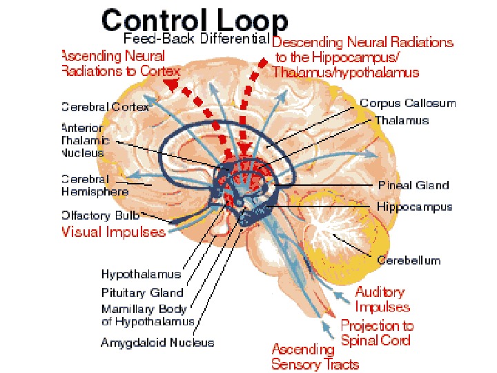
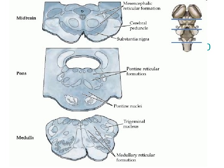
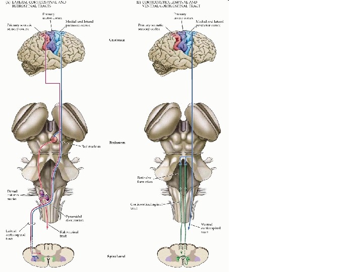
• The reticular formation is a complicated network of circuits located in the core of the brainstem and extending from the rostral midbrain to the caudal medulla ( Figure 17. 3 ). Unlike the well-defined sensory and motor nuclei of the cranial nerves , the reticular formation comprises clusters of neurons scattered among a welter of interdigitating axon bundles; it is therefore difficult to subdivide anatomically. The neurons within the reticular formation have a variety of functions, including cardiovascular and respiratory control (see Chapter 21), governance of myriad sensory motor reflexes (see Chapter 16), the organization of eye movements (see Chapter 20), regulation of sleep and wakefulness (see Chapter 28), and, most important for present purposes, motor control. The descending motor control pathways from the reticular formation to the spinal cord are similar to those of the vestibular nuclei: They terminate primarily in the medial parts of the gray matter where they influence the local circuit neurons that coordinate axial and proximal limb muscles
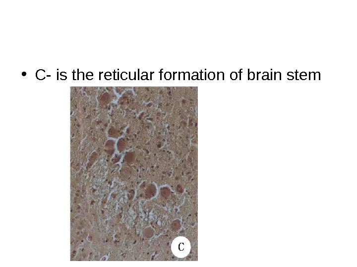
• C- is the reticular formation of brain stem
• The importance of the reticular formation for feedforward mechanisms of postural control has been explored in more detail in cats trained to use a forepaw to strike an object. As expected, the forepaw movement is accompanied by feedforward postural adjustments in the other legs to maintain the animal upright. These adjustments shift the animal’s weight from an even distribution over all four feet to a diagonal pattern, in which the weight is carried mostly by the contralateral , nonreaching forelimb and the ipsilateral hindlimb. Lifting of the forepaw and postural adjustments in the other limbs can also be induced in an alert cat by electrical stimulation of the motor cortex. After pharmacological inactivation of the reticular formation , however, electrical stimulation of the motor cortex evokes only the forepaw movement, without the feedforward postural adjustments that normally accompany them. • The results of this experiment can be understood in terms of the fact that the upper motor neurons in the motor cortex influence the spinal cord circuits by two routes: direct projections to the spinal cord and indirect projections to brainstem centers under consideration here that in turn project to the spinal cord (Figure 17. 6). The reticular formation is one of the major destinations of these latter projections from the motor cortex; thus, cortical upper motor neurons initiate both the reaching movement of the forepaw and also the postural adjustments in the other limbs necessary to maintain body stability. The forepaw movement is initiated by the direct pathway from the cortex to the spinal cord (and possibly by the red nucleus as well), whereas the postural adjustments are mediated via pathways from the motor cortex that reach the spinal cord indirectly, after an intervening relay in the reticular formation (the corticoreticulospinal pathway). • http: //www. ncbi. nlm. nih. gov/books/NBK 11081/
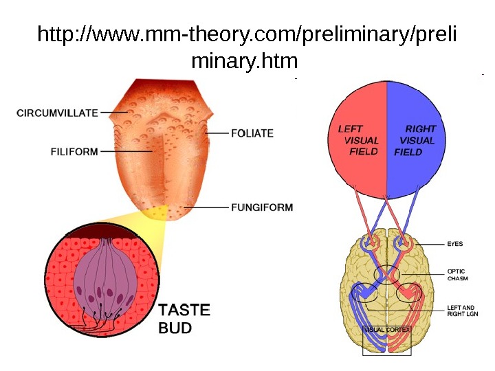
http: //www. mm-theory. com/preliminary/preli minary. htm
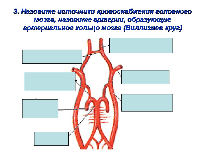
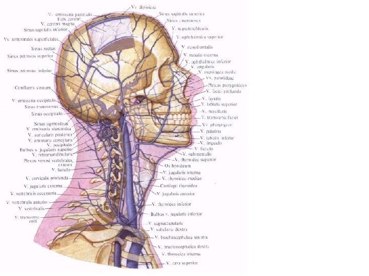
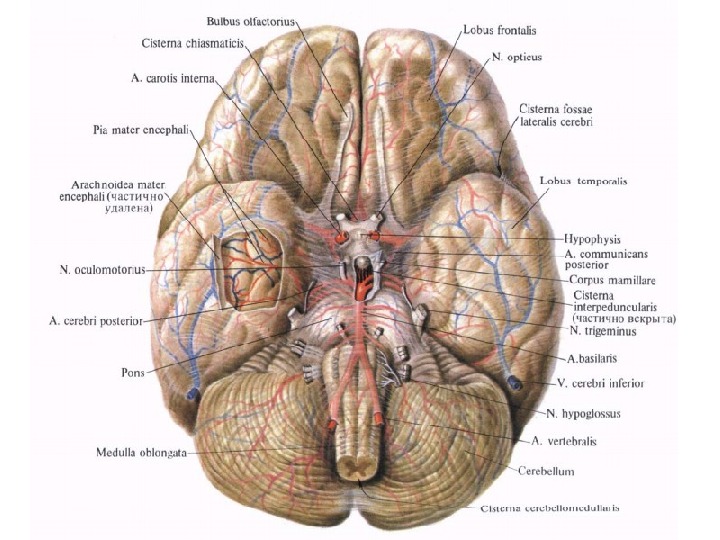
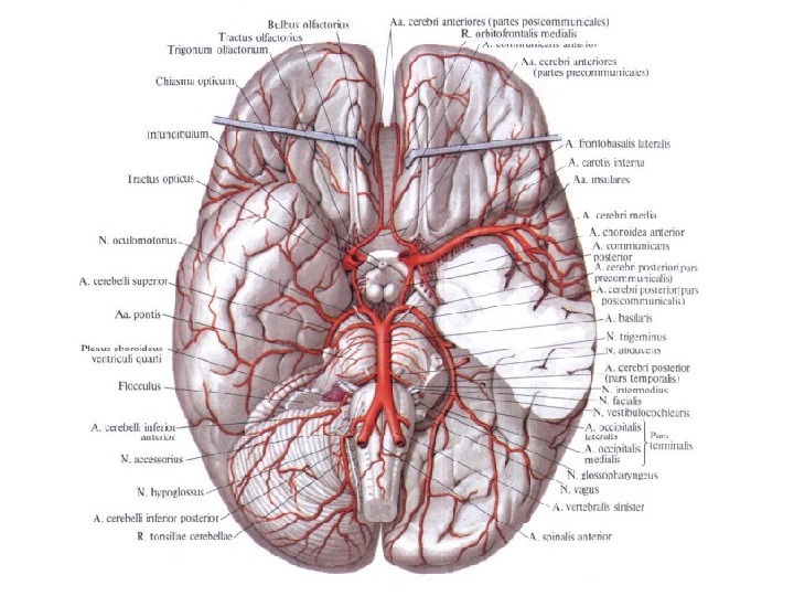
3. Назовите источники кровоснабжения головного мозга, назовите артерии, образующие артериальное кольцо мозга (Виллизиев круг)
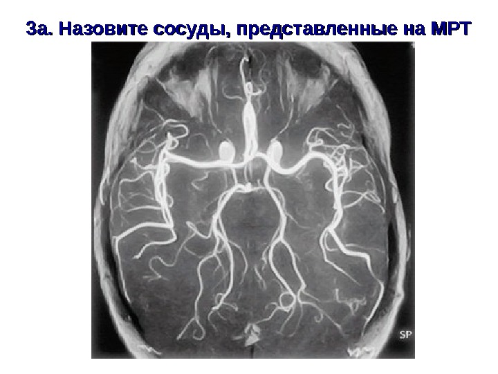
3 а. Назовите сосуды, представленные на МРТ
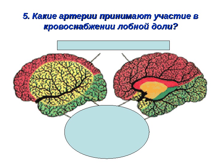
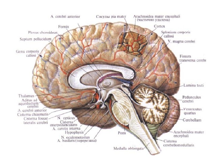
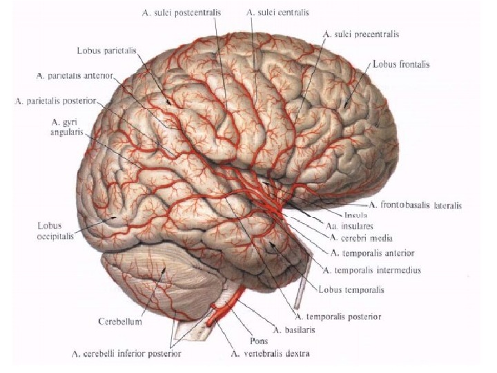
5. Какие артерии принимают участие в кровоснабжении лобной доли?
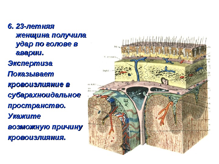
6. 23 -летняя женщина получила удар по голове в аварии. Экспертиза Показывает кровоизлияние в субарахноидальное пространство. Укажите возможную причину кровоизлияния.
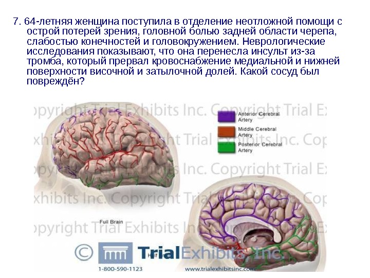
7. 64 -летняя женщина поступила в отделение неотложной помощи с острой потерей зрения, головной болью задней области черепа, слабостью конечностей и головокружением. Неврологические исследования показывают, что она перенесла инсульт из-за тромба, который прервал кровоснабжение медиальной и нижней поверхности височной и затылочной долей. Какой сосуд был повреждён?
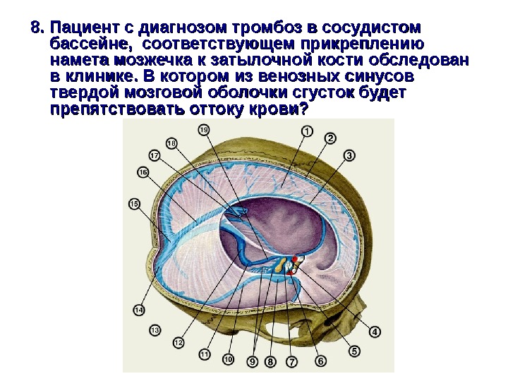
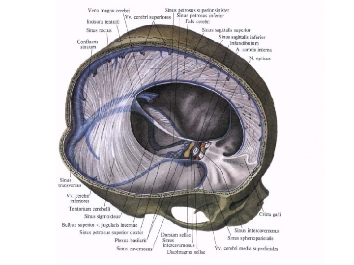
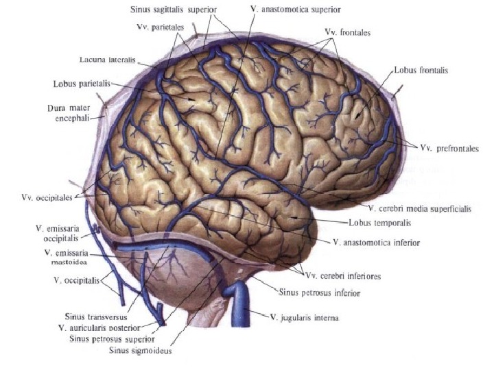
8. Пациент с диагнозом тромбоз в сосудистом бассейне, соответствующем прикреплению намета мозжечка к затылочной кости обследован в клинике. В котором из венозных синусов твердой мозговой оболочки сгусток будет препятствовать оттоку крови?
МРТ нормального головного мозга. • Пациент Б, 8 лет. Комплексная форма гидроцефалии, обусловленная внутриутробной инфекцией и стенозом водопровода мозга. Обследован в связи с прогрессирующими расстройствами статики, походки и координации, прогрессирующей макрокранией. На момент постановки диагноза имелись выраженные признаки внутричерепной гипертензии на глазном дне. Окружность головы 62, 5 см (значительно больше возрастной нормы). • А – Данные МРТ исследования головного мозга
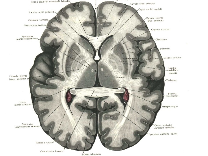
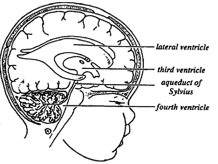
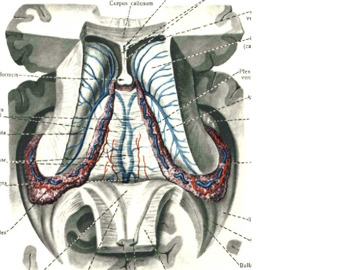
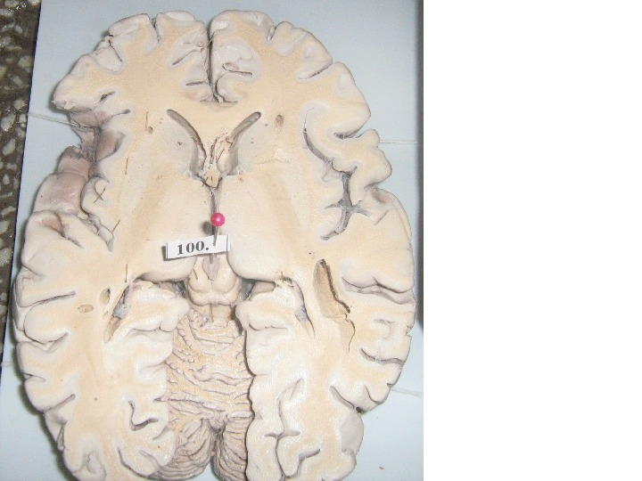
9. Назовите и покажите структурные компоненты вентрикулярной системы мозга
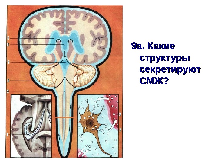
9 а. Какие структуры секретируют СМЖ?
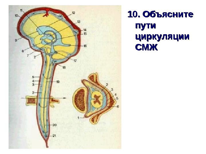
10. Объясните пути циркуляции СМЖСМЖ
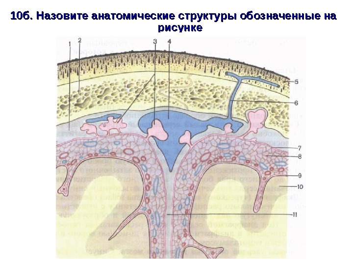
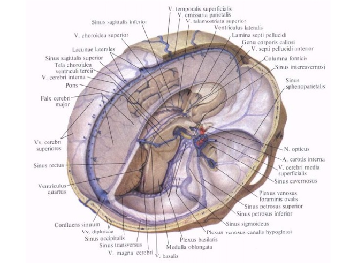
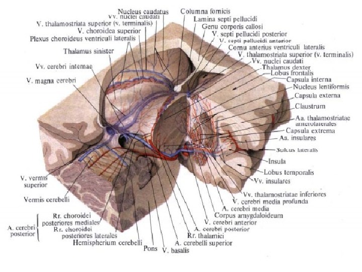
10 б. Назовите анатомические структуры обозначенные на рисунке