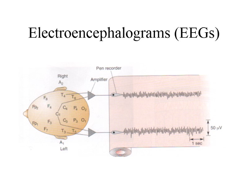
 Electroencephalograms (EEGs)
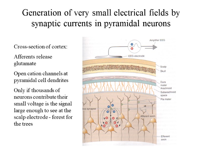 Generation of very small electrical fields by synaptic currents in pyramidal neurons Cross-section of cortex: Afferents release glutamate Open cation channels at pyramidal cell dendrites Only if thousands of neurons contribute their small voltage is the signal large enough to see at the scalp electrode - forest for the trees
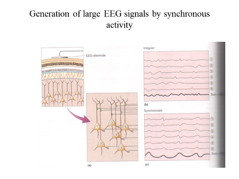 Generation of large EEG signals by synchronous activity
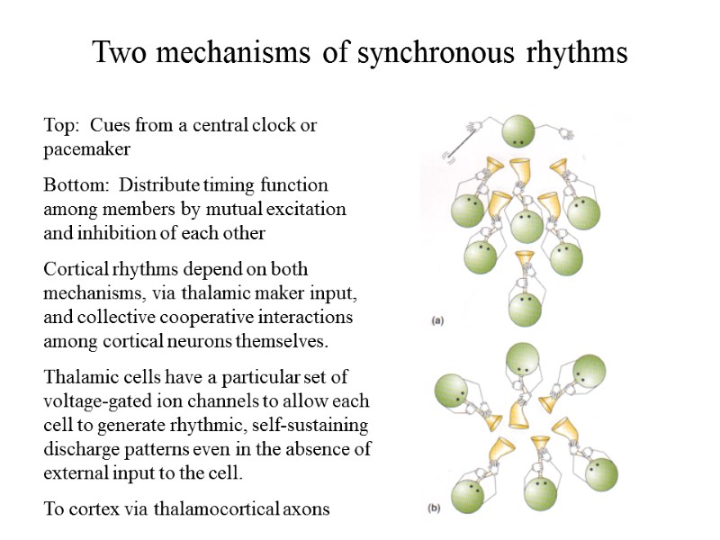 Two mechanisms of synchronous rhythms Top: Cues from a central clock or pacemaker Bottom: Distribute timing function among members by mutual excitation and inhibition of each other Cortical rhythms depend on both mechanisms, via thalamic maker input, and collective cooperative interactions among cortical neurons themselves. Thalamic cells have a particular set of voltage-gated ion channels to allow each cell to generate rhythmic, self-sustaining discharge patterns even in the absence of external input to the cell. To cortex via thalamocortical axons
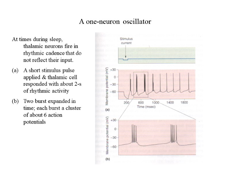 A one-neuron oscillator At times during sleep, thalamic neurons fire in rhythmic cadence that do not reflect their input. A short stimulus pulse applied & thalamic cell responded with about 2-s of rhythmic activity (b) Two burst expanded in time; each burst a cluster of about 6 action potentials
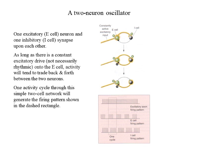 A two-neuron oscillator One excitatory (E cell) neuron and one inhibitory (I cell) synapse upon each other. As long as there is a constant excitatory drive (not necessarily rhythmic) onto the E cell, activity will tend to trade back & forth between the two neurons. One activity cycle through this simple two-cell network will generate the firing pattern shown in the dashed rectangle.
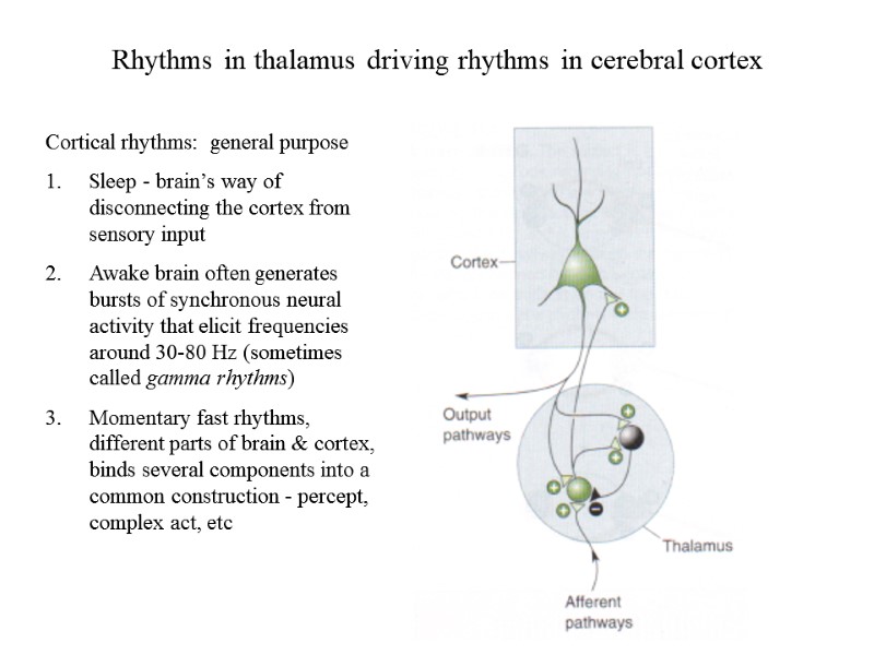 Rhythms in thalamus driving rhythms in cerebral cortex Cortical rhythms: general purpose Sleep - brain’s way of disconnecting the cortex from sensory input Awake brain often generates bursts of synchronous neural activity that elicit frequencies around 30-80 Hz (sometimes called gamma rhythms) Momentary fast rhythms, different parts of brain & cortex, binds several components into a common construction - percept, complex act, etc
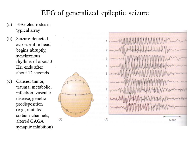 EEG of generalized epileptic seizure EEG electrodes in typical array Seizure detected across entire head, begins abruptly, synchronous rhythms of about 3 Hz, ends after about 12 seconds Causes: tumor, trauma, metabolic, infection, vascular disease, genetic predisposition (e.g., mutated sodium channels, altered GAGA synaptic inhibition)
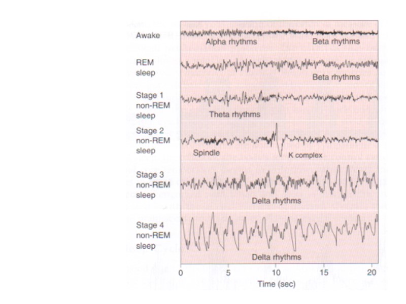
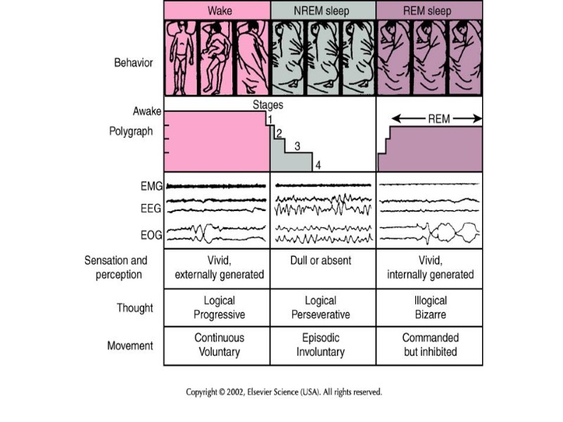
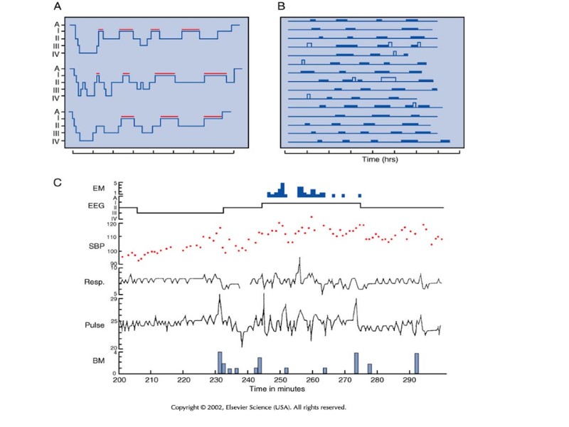
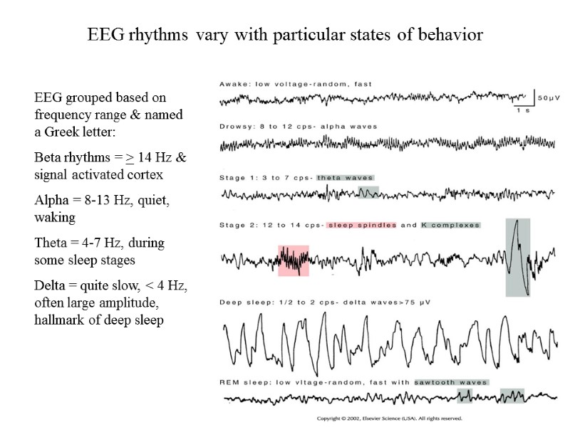 EEG rhythms vary with particular states of behavior EEG grouped based on frequency range & named a Greek letter: Beta rhythms = > 14 Hz & signal activated cortex Alpha = 8-13 Hz, quiet, waking Theta = 4-7 Hz, during some sleep stages Delta = quite slow, < 4 Hz, often large amplitude, hallmark of deep sleep
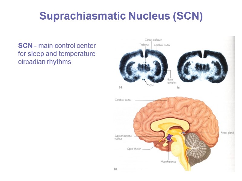 Suprachiasmatic Nucleus (SCN) SCN - main control center for sleep and temperature circadian rhythms
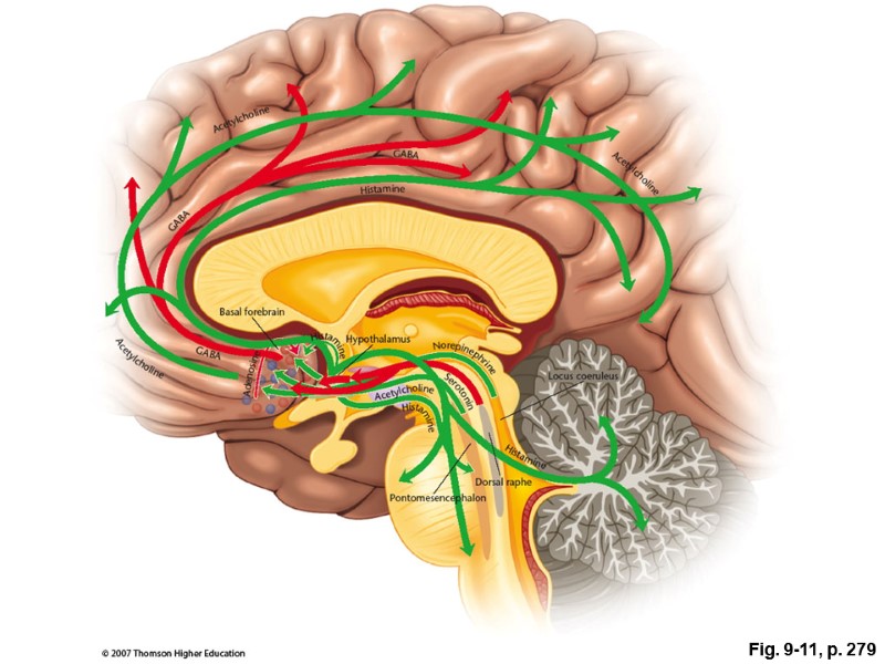 Fig. 9-11, p. 279
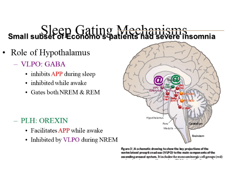 Sleep Gating Mechanisms Role of Hypothalamus VLPO: GABA inhibits APP during sleep inhibited while awake Gates both NREM & REM PLH: OREXIN Facilitates APP while awake Inhibited by VLPO during NREM Small subset of Economo’s patients had severe insomnia @ @
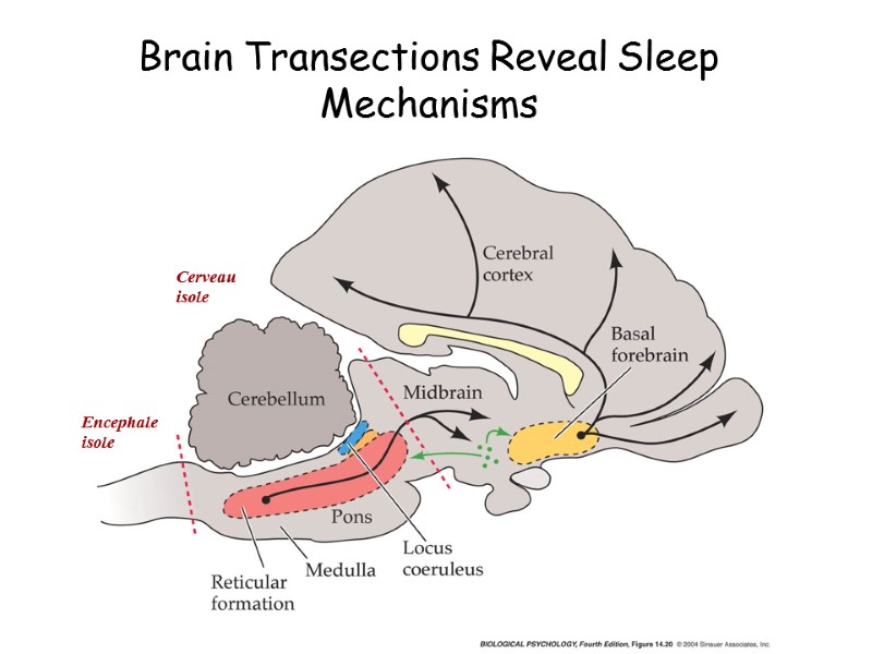 Brain Transections Reveal Sleep Mechanisms Encephale isole Cerveau isole
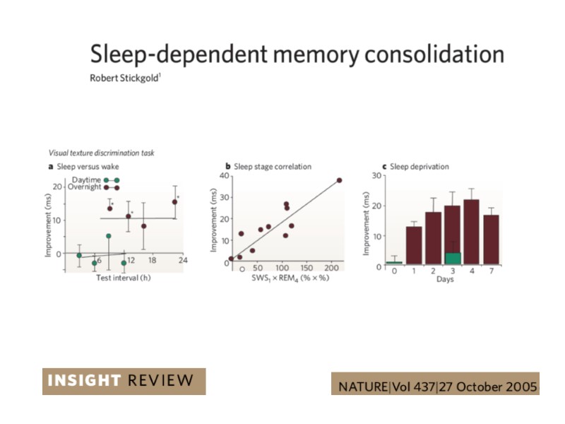
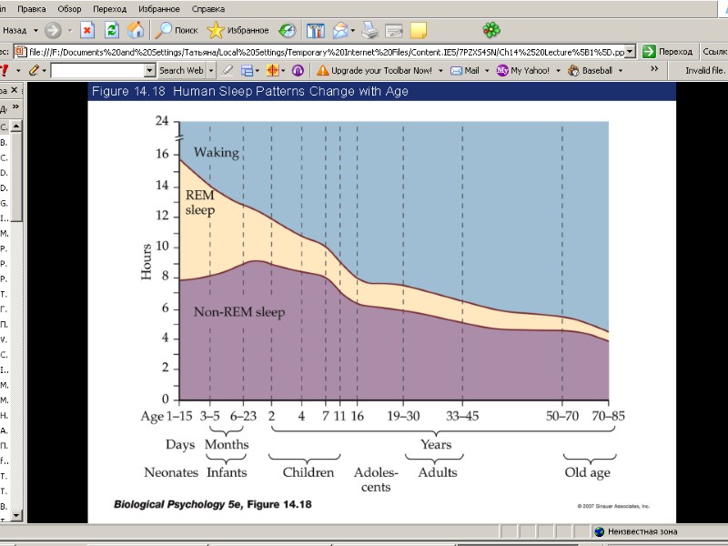
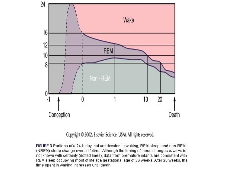
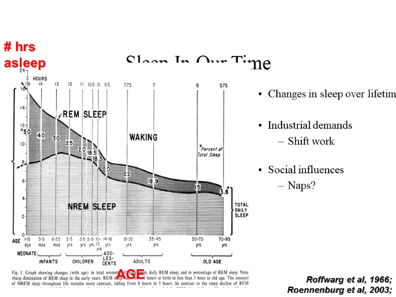 Sleep In Our Time Changes in sleep over lifetime Industrial demands Shift work Social influences Naps? Roffwarg et al, 1966; Roennenburg et al, 2003;

