

1709bf25e0a5ce57974feaf555ba1491.ppt
- Количество слайдов: 59
 ECRR International Conference May 4 -6 2009. Lesvos Greece In Utero exposure to Chernobyl accident radiation and the health risk assessment. Professor Angelina Nyagu Physicians of Chernobyl, Kiev Ukraine
 Introduction to the problem • What do we know about the qualitative and quantitative effects of ionizing radiation on the developing embryo?
 Specific radiation effects on foetus: mental retardation , microcephaly - Japanese study Those exposed at a gestational age of 8– 15 weeks were most at risk. Survivors of the atomic bombing in Japan who were exposed in utero during this sensitive period show a linear increase in the frequency of mental retardation with radiation dose (40% per Gy). There were 2, 800 people in this study.
 • • • Average New Percent") Tabulations of CNR (J. Goffman Japanese’ investigation criticism) • • • Average New Percent / Percent Increase in Rate Fetal Dose Old Percent of Mental Retardation 4 rads (2. 63 / 2. 28) = 1. 15 15 % increase 10 rads (3. 13 / 2. 28) = 1. 37 37 % increase (at the optimum "hormetic" dose) 15. 4 rads (3. 77 / 2. 28) = 1. 65 65 % increase 23. 0 rads (4. 75 / 2. 28) = 2. 08 2. 08 -fold increase 30. 8 rads (6. 00 / 2. 28) = 2. 63 2. 63 -fold increase 46. 2 rads (9. 12 / 2. 28) = 4. 00 4 -fold increase 61. 5 rads (13. 36 / 2. 28) = 5. 86 5. 86 -fold increase 72. 0 rads (16. 85 / 2. 28) = 7. 39 7. 39 -fold increase
Tabulations of CNR (J. Goffman Japanese’ investigation criticism) • • • Average New Percent / Percent Increase in Rate Fetal Dose Old Percent of Mental Retardation 4 rads (2. 63 / 2. 28) = 1. 15 15 % increase 10 rads (3. 13 / 2. 28) = 1. 37 37 % increase (at the optimum "hormetic" dose) 15. 4 rads (3. 77 / 2. 28) = 1. 65 65 % increase 23. 0 rads (4. 75 / 2. 28) = 2. 08 2. 08 -fold increase 30. 8 rads (6. 00 / 2. 28) = 2. 63 2. 63 -fold increase 46. 2 rads (9. 12 / 2. 28) = 4. 00 4 -fold increase 61. 5 rads (13. 36 / 2. 28) = 5. 86 5. 86 -fold increase 72. 0 rads (16. 85 / 2. 28) = 7. 39 7. 39 -fold increase
 How Many children are Mentally Retarded? • In-utero irradiation during the vulnerable period causes the brilliant to become less brilliant, the average to become "below average, " and the retarded to become more retarded. And by pushing more people over the heavy vertical line into the realms of mental retardation and sever retardation, such exposure automatically increases the percent of a population-sample which is retarded and severely retarded. (Gohn Goffman, 1994)
 The foetus. Recent overview • Typical effects of radiation on embryo: Ø Intrauterine growth retardation (IUGR) Ø Embryonic, foetal, or neonatal death Ø Congenital malformations Ø Cancer
 Gestational Stage Radiogenic effects 0 -9 days Embryo contains only few cells which are not specialized. Preimplantation Before about 2 weeks gestation the health effect of concern from an exposure of > 0. 1 (Gy) or 10 rads is the death of the embryo. Because the embryo is made up of only a few cells, damage to one cell, the progenitor of many other cells, can cause the death of the embryo, and the blastocyst will fail to implant in the uterus. Embryos that survive, however, will exhibit few congenital abnormalities. If too many cells are damaged - embryo is resorbed. If only few killed - remaining pluripotent cells replace the cells loss within few cell divisions; Atomic Bomb survivors - high incidence of both - normal birth and spontaneous abortion. 10 days-6 weeks 40 weeks For all stages, one has to expect induction of childhood cancers, in particular, childhood leukaemia's. The risk of childhood leukaemia's can be shown to be increased down to doses of 10 m. Gy. The doubling dose is in the range of 30 m. Gy. One must keep in mind, however, that the spontaneous risk is small: about 5 per 100. 000 children per year. Thus, a very small risk is doubled by about 30 m. Gy. Organogenesis Radiation risks are most significant during organogenesis and in the early fetal period somewhat less in the 2 nd trimester and least in the third trimester Congenital anomalies, growth retardation, mental retardation foetal Growth retardation, microcephly, Most risk мental retardation
.") Stochastic threshold dose-response relationships of diseases produced by environmental agents (Brendt, 1987, 1990, 1999). Phenomenon Pathology Stochastic Threshold Site Diseases Risk Definition Damage to a DNA single cell may result in disease Cancer germ cell mutation Some risk exists at all doses; at low doses, risk is usually less than the spontaneous risk Incidence of the disease increases but the severity and nature of the disease increase with dose Multiple cell and tissue injury Birth defects, growth retardation, death, toxity, mental retardation etc. No increased risk below the threshold dose Both the severity and incidence of the disease increase with dose Multiple variable etiology, affecting many cellular and organ functions
Stochastic threshold dose-response relationships of diseases produced by environmental agents (Brendt, 1987, 1990, 1999). Phenomenon Pathology Stochastic Threshold Site Diseases Risk Definition Damage to a DNA single cell may result in disease Cancer germ cell mutation Some risk exists at all doses; at low doses, risk is usually less than the spontaneous risk Incidence of the disease increases but the severity and nature of the disease increase with dose Multiple cell and tissue injury Birth defects, growth retardation, death, toxity, mental retardation etc. No increased risk below the threshold dose Both the severity and incidence of the disease increase with dose Multiple variable etiology, affecting many cellular and organ functions
 Background Incidence of Conceptus Complications without Diagnostic Imaging Radiation Spontaneous incidence of major malformations Approximately 1% to 3% Intrauterine growth restriction 4% Spontaneous abortion At least 15% Genetic disease 8% to 10% Mental retardation (intelligence quotient less than 70) Approximately 3% Severe mental retardation (unable to care for self) 0. 5% Heritable effects 1% to 6% Spontaneous risk of childhood leukemia and cancer (ages 0 to 15) 0. 16% Children developing cancer up to age 15 (United Kingdom) 0. 15% Children developing leukemia only to age 15 (United Kingdom) 0. 03% Lifetime risk of contracting fatal cancer 20% Lifetime risk of contracting cancer 33%
 Fetal Effects of Ionizing Radiation: Severe Mental Retardation Organogenesis 2 to 8 weeks Diagnostic radiation No increased risk 100 m. Gy to 200 m. Gy Mental retardation 1, 000 m. Gy (100 rad) Reduction of IQ (25 to 30 points); Severe mental retardation in 40% of case 1, 500 m. Gy (150 rad) Severe mental retardation in 60% of cases Fetal development Early 8/9 to 15 weeks Diagnostic radiation No increased risk 200 m. Gy (20 rad) Malformation of forebrain producing mental retardation 1, 000 m. Gy (100 rad) Reduction of IQ (25 to 30 points); Severe mental retardation in 40% of cases 1, 500 m. Gy (150 rad) Severe mental retardation in 60% of cases Mild 16 to 25 weeks Diagnostic radiation No increased risk Above diagnostic radiation Less risk of IQ reduction and severe me retardation Late 26 to delivery Central nervous system is radioresistent
 • • Threshold dose for developmental effects approximately") Considerations for pregnancy termination (WHO, IAEA) • • Threshold dose for developmental effects approximately 0. 1 Gy. At 0. 1 Gy , increase of 0. 1 -1%. ICRP (1990 Recommendations of the International Commission on Radiological Protection. Report 60. ) recommends a limit of radiation exposure to a member of the general public as 100 mrem/y (1 m. Sv/y) and the limit for the fetus of an occupationally exposed individual to 200 mrem (2 m. Sv) during the gestation period. • There was a long-standing debate on whether a threshold dose exists for the weeks 8 to 15, whereas it was comparatively clear from the beginning that a threshold dose is present for weeks 16 to 25. Biology always pointed to threshold doses for both time periods, because many cells have to be killed or impaired in their migration behaviour in order to cause a severe mental retardation. ICRP meanwhile suggests a threshold dose of about 300 m. Gy for both time intervals. It is not clear whether a threshold dose exists for IQ reduction. This question will be hard to answer in any case, because even if one assumes a linear dose dependence without a threshold, the risk in the low dose range will be so low that it is impossible to detect it. ICRP estimates the risk to be a reduction of 21 IQ-points per Gray for the weeks 8 to 15, and 13 IQ-points per Gray for weeks 16 to 25. Both numbers do not include the cases of severe mental retardation The foetal absorbed dose > 0, 5 Gy at 7 -13 weeks: substantial risk of intrauterine growth retardation (IUGR) and central nervous system (CNS) damage.
Considerations for pregnancy termination (WHO, IAEA) • • Threshold dose for developmental effects approximately 0. 1 Gy. At 0. 1 Gy , increase of 0. 1 -1%. ICRP (1990 Recommendations of the International Commission on Radiological Protection. Report 60. ) recommends a limit of radiation exposure to a member of the general public as 100 mrem/y (1 m. Sv/y) and the limit for the fetus of an occupationally exposed individual to 200 mrem (2 m. Sv) during the gestation period. • There was a long-standing debate on whether a threshold dose exists for the weeks 8 to 15, whereas it was comparatively clear from the beginning that a threshold dose is present for weeks 16 to 25. Biology always pointed to threshold doses for both time periods, because many cells have to be killed or impaired in their migration behaviour in order to cause a severe mental retardation. ICRP meanwhile suggests a threshold dose of about 300 m. Gy for both time intervals. It is not clear whether a threshold dose exists for IQ reduction. This question will be hard to answer in any case, because even if one assumes a linear dose dependence without a threshold, the risk in the low dose range will be so low that it is impossible to detect it. ICRP estimates the risk to be a reduction of 21 IQ-points per Gray for the weeks 8 to 15, and 13 IQ-points per Gray for weeks 16 to 25. Both numbers do not include the cases of severe mental retardation The foetal absorbed dose > 0, 5 Gy at 7 -13 weeks: substantial risk of intrauterine growth retardation (IUGR) and central nervous system (CNS) damage.
 Chernobyl caused significantly lower external foetal doses, but it caused the high doses on the foetal thyroid by the incorporation of the radioiodine and other radionuclides in first stage of accident.
 Chernobyl investigations in Ukraine • • The objectives of the study was the psychometric, neurophysiological and neuropsychiatric (ICD-10) criteria) characterization of acutely prenatally irradiated children. This study involves acutely prenatally exposed children — born between April 26 th, 1986 and February 26 th 1987 from pregnant women at the time of the accident who had been evacuated from the 30 -kilometer zone surrounding the Chernobyl NPP to Kiev — and their classmates. This sample seems to be optimal for examination of possible distinguished effects of exposure in different periods of cerebrogenesis. During the first stage(1990 -1992) it was examined children five- six year-old. • At the second stage(1994 -1996) the epidemiological WHO project “Brain Damage in Utero” (IPHECA) was implemented. • At the third stage (2002 -2004) it was examined a cohort of 154 children born between April 26 th 1986 and February 26 th 1987 to mothers who had been evacuated from Chernobyl exclusion zone to Kiev and 143 classmates from Kiev. In the third stage reconstruction of individual doses of children born to mothers evacuated from the Chernobyl exclusion zone was carried out at taking internal and external exposure.
 METHOTODOLGY. Clinical examination • Children were profoundly medically examined by general paediatrist, paediatristpsychoneurologist, paediatrist-endocrinologist, paediatrist-Ear-Nose-Throat (ENT), paediatrist-ophtalmologist, paediatristcardiologist, paediatrist-haematologists, paediatrist-pulmonologists, paediatrist-gastroenterologists, paediatrist-surgeon, paediatrist-gynecologist (for girls), • general and biochemical blood tests, immunological tests, urine tests, coprogram, thyroid and visceral ultrasonography, • Electrocardiogram (ECG), electroencephalogram (EEG), rheoencephalogram (Rh. EG) as well as fibrogastoscopy, cardiac ultrasonography, and magnetoresonance imaging (MRI) for diagnostic reasons. It should be emphasised that neuropsychiatric assessments presented here are based on neurological and psychiatric examinations, psychometry of both children and their mothers.
 Estimation of Prenatal Age at Exposure • • • In order to avoid uncertainties concerning the estimation of prenatal age at the time of the Chernobyl accident we used the formulas offered for estimation of prenatal age at atomic bombing in Hiroshima and Nagasaki: Days of pregnancy (Y) = 280 — (date of birth — April 26 th, 1986), where the day of birth has been obtained by interviewing the mothers of the children. The mean duration of pregnancy is taken to be 280 days. The days from birth were counted back until the accident and subtracted from the 280 days, the duration of a pregnancy. Since the duration is calculated from the beginning of the last menstrual cycle, additionally 14 days have to be subtracted. Gestational weeks after fertilization at the time of the accident were thus calculated by the following equation: Gestational weeks (G) = (Y — 14 days) / 7 days, where G was taken to be zero if G<0. According to different radiosensitivity of the fetus the gestational time is divided into 4 periods in relation to the Chernobyl accident. In the exposed groups there are less children who were at the earliest stages of prenatal development. A possible explanation are increased numbers of abortions and miscarriages due to the Chernobyl accident.
 DOSIMETRY • Individual reconstruction of total fetal doses, fetal thyroid doses and fetal doses on the brain has been carried out using 2 methods: • 1) fetal thyroid dose is assumed to be equal to the thyroid dose of the mother, and 2) according to the model by ICRP Publication 88 (2001). • The main irradiation sources of the pregnant women were: 1) external -irradiation of the whole body; 2) irradiation of thyroid by radioactive iodine isotopes; 3) internal irradiation by inhaled radionuclides; 4) internal irradiation by ingestion of radioactively contaminated food. • The doses were reconstructed for the exposed children from Pripyat and also for the control group in Kiev by Professor Victor REPIN • (Laboratory of dosimetry RCRM in Ukraine)
 • There are 20 children from Pripyat") Dose on embryo and foetus distribution (ICRP-88) • There are 20 children from Pripyat (13. 2%) who had been exposed in utero >100 m. Sv – the threshold for medical abortion due to prenatal irradiation (European Commission, 1998; ICRP Publication 84, 2000).
Dose on embryo and foetus distribution (ICRP-88) • There are 20 children from Pripyat (13. 2%) who had been exposed in utero >100 m. Sv – the threshold for medical abortion due to prenatal irradiation (European Commission, 1998; ICRP Publication 84, 2000).
") Dose on embryo and foetus distribution (ICRP 88)
Dose on embryo and foetus distribution (ICRP 88)
 In UTERO Thyroid doses were estimated 0, 01 - 3, 34 Gy. 30 -km Zona • The mean doses according trimester of gestation: • • Contaminated territories Control % Until 8 weeks – 0, 0 Gy; of 8 to 15 week – 0, 31 Gy; of 16 to 25 week - 0, 8 Gy; More than 25 weeks – 0, 62 Gy. Gy
 There are (35. 5%) children from Pripyat") Dose on thyroid in utero distribution (ICRP-88) There are (35. 5%) children from Pripyat who got in utero thyroid doses >1 Sv.
Dose on thyroid in utero distribution (ICRP-88) There are (35. 5%) children from Pripyat who got in utero thyroid doses >1 Sv.
 Geometric means of the thyroid doses in utero related to the periods of cerebrogenesis at 26. 04. 1986 in exposed group in Pripyat According to the model by ICRP-88 there is a strong influence of gestational age on the thyroid doses in utero: later intrauterine period at the time of exposure — higher the thyroid doses in utero
 Main group (n=147) (Evacuees from Chernobyl 30") First Stage (children 5 -6 years old) Main group (n=147) (Evacuees from Chernobyl 30 -km Zone) Control group (n=101 -city. Kiev) а b(%) a b(%) 1 -st -healthy children 12 8, 2 2 -nd –dynamic diseases 68 46, 3 67 67 3 -rd –chronic relapsing diseases 55 37, 4 30 30 4 -th – chronic decompensate diseases, congenital defects, anomalies 7 4, 8 2 2 Health groups Average doses of gamma-irradiation varied as 7 m. Sv-13 m. Sv (Monte-Carlo dose reconstruction method for fetus). Individual dose of Thyroid – varied within 10 -120 c. Sv. It is concluded that multifactor impact of Chernobyl disaster unfavourable factors defined health deterioration in children irradiated in prenatal period shortening amount of practically healthy kids down to 5%. The results showed much 5%. more somatic diseases and neurovegetative mental disorders. At the same time it was clearly recognized the decrease of immunity (hypo immunoglobulin level and increase of T-lymphocytes, T-helpers), neurological, gastrointestinal and endocrine diseases.
First Stage (children 5 -6 years old) Main group (n=147) (Evacuees from Chernobyl 30 -km Zone) Control group (n=101 -city. Kiev) а b(%) a b(%) 1 -st -healthy children 12 8, 2 2 -nd –dynamic diseases 68 46, 3 67 67 3 -rd –chronic relapsing diseases 55 37, 4 30 30 4 -th – chronic decompensate diseases, congenital defects, anomalies 7 4, 8 2 2 Health groups Average doses of gamma-irradiation varied as 7 m. Sv-13 m. Sv (Monte-Carlo dose reconstruction method for fetus). Individual dose of Thyroid – varied within 10 -120 c. Sv. It is concluded that multifactor impact of Chernobyl disaster unfavourable factors defined health deterioration in children irradiated in prenatal period shortening amount of practically healthy kids down to 5%. The results showed much 5%. more somatic diseases and neurovegetative mental disorders. At the same time it was clearly recognized the decrease of immunity (hypo immunoglobulin level and increase of T-lymphocytes, T-helpers), neurological, gastrointestinal and endocrine diseases.
 It was also established in this cohort that starting with the") Thyroid-stimulating hormone (TSH) It was also established in this cohort that starting with the 0. 3 Gy threshold dose thyroid-stimulating hormone (TSH) level grew along with fetal thyroid dose increase. Thereupon the radiation-induced malfunction of the thyroid-pituitary system on this stage was suggested as important biological mechanism in the genesis of health risk assessment and mental disorders of prenatally irradiated children.
Thyroid-stimulating hormone (TSH) It was also established in this cohort that starting with the 0. 3 Gy threshold dose thyroid-stimulating hormone (TSH) level grew along with fetal thyroid dose increase. Thereupon the radiation-induced malfunction of the thyroid-pituitary system on this stage was suggested as important biological mechanism in the genesis of health risk assessment and mental disorders of prenatally irradiated children.
 Distribution of the psychic development level disturbances in children irradiated in utero during the pregnancy 1 -3 trimesters: a – the norm; b- below the norm ; c- psychic development delay. The levels of separate psychic functions development ( 6 years old): 1 - notions; 2 -psychomotor system; 3 attention; 4 -memorizing. a , b - 1 trimester, b, c - 3 trimester (control and irradiated children accordingly). Was used the Kern-Jeracika and verbal tests of willingness to teaching (school education). . Signs of mental progress retardation were met in 77% of kids from Pripyat city exposed in utero within first pregnancy trimester, in 69% of those exposed in second trimester and in 45% — in third one. Among kids resident in Kiev city the percent of persons with decreased mental development value was substantially lower (p<0. 05). In 25. 5% of cases in Pripyat group the brain organic pathology signs were revealed. Brain circulation disorders according to the rheoencephalography data were observed a bit more often in children from Pripyat city exposed to radiation within first pregnancy trimester. The low induces of psychic development in utero irradiated children are largely determined by the irradiation factor.
. WHO project “Brain Damage in Utero” (IPHECA).") 2 -th stage (children 11 year old). WHO project “Brain Damage in Utero” (IPHECA).
2 -th stage (children 11 year old). WHO project “Brain Damage in Utero” (IPHECA).
 Mental health in children exposed to radiation in prenatal period and their mothers Groups Number of children with revealed mental retardation IQ<70. A decrease in high (IQ>110), as well as statistically significant higher prevalence of mental retardation (IQ<70) in Ukrainian prenatally irradiated children compared to the controls: 21 (3. 9%) vs. 12 (1. 6%) correspondingly (χ2=6. 27; df=1; P<. 05). Note: Number of kids with emotional, behavioural and nondifferentiated disorders Indices characterisi ng degree of mental health in mothers (A)* - IQ<70 through Raven Coloured Matrices and BPVS ; (B)** - IQ<70 through any two intellectual tests Methods Non-verbal intellect (Draw-a. Man) Verbal intellect (BPVS) Nonverbal intellect (Raven Coloured Matrices) General intellect lowering (A)* General intellect lowerin g (B)** Ratter scale À(2) Ratter scale (2) General Health Questionnaire GHQ-28 IQ «Experim ental» n=544 11 (2. 06 %) n=535 61 (11. 34 %) n=538 59 (10. 95 %) n=539 19 (3. 49 %) n=544 23 (4. 34%) n=544 152 (41. 76%) n=364 137 (34. 86%) n=393 24. 26± 0, 4 n=382 33. 6± 0, 6 n=377 P For c 2 or Student’s criteria >0. 05 <0. 01 >0. 05 <0. 01 <0. 05 Control, N=759 8 (1. 06 %) n=755 66 (8. 87 %) n=744 92 (12. 12 %) n=759 8 (1. 05 %) n =759 269 (38. 93 %) n=691 20. 73± 0, 5 n=639 43. 6± 0, 5 n=750 16 (2. 10 %) n=759 214 (28. 69 %) n=746
 IPHECA WHO PILOT PROJECT CONCLUSION • Analysis of the results in three countries (Belarus, Russian and Ukraine) has shown the following: • а) incidence of mild mental retardation in prenatally irradiated children is higher when compared with the control group; b) an upward trend was detected in cases of behavioural disorders and in changes in the emotional problems in children exposed in utero; c) incidence of borderline nervous and psychological disorders in the parents of prenatally irradiated children is higher than that of controls. • • • In the frame of the WHO Pilot Project «Brain Damage in Utero» we have previously revealed a significant increase of borderline and low range IQ, emotional and behavioural disorders. • Since possible dose correlations were not investigated and contradictory results of the mental health assessment of the in utero exposed children and the aetiology of the observed neuropsychiatric disorders were found.
 and non-exposed") Distribution of IQ scores in the prenatally irradiated children ( «Experimental» group) and non-exposed control children Non-verbal intelligence ( «Draw-a. Man» test) Verbal intelligence ( «British Picture Vocabulary Scale» ) Non-verbal intelligence ( «Raven Coloured Matrices» )
Distribution of IQ scores in the prenatally irradiated children ( «Experimental» group) and non-exposed control children Non-verbal intelligence ( «Draw-a. Man» test) Verbal intelligence ( «British Picture Vocabulary Scale» ) Non-verbal intelligence ( «Raven Coloured Matrices» )
 Conventional EEG of B-v 23. 12. 1998. Without anticonvulsants. The 3 rd minute of hyperventilation — paroxysmal activity (like «spike-waves» ) in the fronto-temporal area shifted to the left (leads F 7, T 3, T 4, T 5, T 6). MRI (1998&1999).
 EEG-pattern Children exposed to radiation in prenatal period n=50 p c 2 Children from control group n=50 Age norm: 8 (16. 0 %) <0. 05 24 (48. 0 %) Pathological: Disorganized slow 23 (46. 0 %) <0. 05 8 (16. 0 %) Disorganized with paroxysmal activity 14 (28. 0 %) <0. 05 2 (4. 0 %) Laterality: Left-hemisphere 20 (40. 0 %) <0. 05 6 (12. 0 %) Disorganized with activity predominance Hypersynchronous 5 (10. 0 %) >0. 05 16 (32. 0 %) Right-hemisphere 13 (26. 0 %) >0. 05 10 (20. 0 %) Symmetry 7 (14. 0 %) <0. 01 32 (64. 0 %) Brain bioelectrical activity patterns in prenatally irradiated children compared to the norm Increase of activity of EEG Depression of and physiological activity Increase of - pathological spectrum Low-voltage EEG (20– 25 μV) with excess of slow (δ) and fast (β) activity together with depression of normal physiological α- and θ-activity with paroxysmal activity; Disorganised slow EEG-pattern with δ-activity domination; Disorganised EEG-pattern with paroxysmal activity; Epileptiformal EEG with «spike» or «polyspike—wave» complexes in the fronto-temporal region, mainly of the left hemisphere.
 EEG
EEG
 Low-voltage EEG (20–") Abnormal EEG-patterns in the prenatally irradiated children as follows: • (1) Low-voltage EEG (20– 25 μV) with excess of slow (δ) and fast (β) activity together with depression of α- and θ-activity with paroxysmal activity shifted to the left fronto-temporal region was one of the most distinguished conventional EEG-pattern in the children of the acutely exposed group (31% vs. 8%, χ2=16. 85, P<. 001). • (2) Disorganised slow EEG-pattern with δ-activity domination characterised by disorganised activity of moderate (40– 55 μV) or high (70– 80 μV) amplitude with a mainly δ-range slow activity domination and non-regular α-activity where hyperventilation led to bilateral paroxysmal activity discharges, as well as. • (3) disorganised EEG-pattern with paroxysmal activity, similar in general to the one described above, but characterised by generalised paroxysmal discharges and bursts of acute, θ- and δ-waves of high amplitude where the hyperventilation led to the bilateral paroxysmal activity increase, were found equally in the both groups. • (4) Epileptiformal EEG with «spike» or «polyspike—wave» complexes in the fronto-temporal region, mainly of the left hemisphere, and bilateral paroxysmal activity in the form of δ-waves of very high amplitude (higher than 100 μV) was another of the most distinguished conventional EEG-pattern among the children of the acutely exposed groups.
Abnormal EEG-patterns in the prenatally irradiated children as follows: • (1) Low-voltage EEG (20– 25 μV) with excess of slow (δ) and fast (β) activity together with depression of α- and θ-activity with paroxysmal activity shifted to the left fronto-temporal region was one of the most distinguished conventional EEG-pattern in the children of the acutely exposed group (31% vs. 8%, χ2=16. 85, P<. 001). • (2) Disorganised slow EEG-pattern with δ-activity domination characterised by disorganised activity of moderate (40– 55 μV) or high (70– 80 μV) amplitude with a mainly δ-range slow activity domination and non-regular α-activity where hyperventilation led to bilateral paroxysmal activity discharges, as well as. • (3) disorganised EEG-pattern with paroxysmal activity, similar in general to the one described above, but characterised by generalised paroxysmal discharges and bursts of acute, θ- and δ-waves of high amplitude where the hyperventilation led to the bilateral paroxysmal activity increase, were found equally in the both groups. • (4) Epileptiformal EEG with «spike» or «polyspike—wave» complexes in the fronto-temporal region, mainly of the left hemisphere, and bilateral paroxysmal activity in the form of δ-waves of very high amplitude (higher than 100 μV) was another of the most distinguished conventional EEG-pattern among the children of the acutely exposed groups.
 Factors making impact on mental health in children exposed to radiation in prenatal period NON-RADIATION FACTORS Mother low education level RADIATION FACTORS h=0. 8 VERBAL IQ DECREASE h=0. 3 Residence territory 137 Cs – contamination level in prenatal period h=0. 8 Delivery complications h=0. 3 NON-VERBAL IQ DECREASE h=0. 4 Cerebrogenesis most critical period (8 – 15 weeks of prenatal progress) at the time of Chernobyl disaster (04. 26. 1986) Mental health worsening in mother h=0. 5 EMOTIONALBEHAVIOURAL DISORDERS h=0. 6 Internal radiation dose on thyroid h — correlation interrelations coefficient. — reliable effect (p<0. 05) of the factor.
. Methodology - Methods • • Intelligence Assessment by") The 3 -rd Stage (2002 -2004). Methodology - Methods • • Intelligence Assessment by the adapted and normalised version for the Ukrainian children of the Wechsler Intelligence Scale for Children, WISC (the verbal, performance and full scale IQs). Additional Psychological and Demographic Measurements – Russian translation of Achenbach’s Child Behaviour Checklist (CBCL) – Rutter A (2) Behaviour Rating Scale – General Health Questionnaire (GHQ-28) – The vocabulary subtest of the Wechsler Adult Intelligence Scales (WAIS) – Impact of Events Scale (IES) and Irritability, Depression Anxiety Scale (IDA) – Self-rating Depression Scale (Zung’s) – Questionnaire on stress-factors related to the Chernobyl accident – School performance – Demographic background, family history, educational level of the family, social and economical status as well as they completed a standardised questionnaire on radiation history Clinical Psychiatric and Neurological assessment according to ICD-10
The 3 -rd Stage (2002 -2004). Methodology - Methods • • Intelligence Assessment by the adapted and normalised version for the Ukrainian children of the Wechsler Intelligence Scale for Children, WISC (the verbal, performance and full scale IQs). Additional Psychological and Demographic Measurements – Russian translation of Achenbach’s Child Behaviour Checklist (CBCL) – Rutter A (2) Behaviour Rating Scale – General Health Questionnaire (GHQ-28) – The vocabulary subtest of the Wechsler Adult Intelligence Scales (WAIS) – Impact of Events Scale (IES) and Irritability, Depression Anxiety Scale (IDA) – Self-rating Depression Scale (Zung’s) – Questionnaire on stress-factors related to the Chernobyl accident – School performance – Demographic background, family history, educational level of the family, social and economical status as well as they completed a standardised questionnaire on radiation history Clinical Psychiatric and Neurological assessment according to ICD-10
 In exposed group there are less children who were at the earliest stages of prenatal development (0– 7 weeks after conception) that could be explained with abortions and miscarriages due to the Chernobyl accident
 HEALTH GROUPS
 Full scale IQ • Lower full scale IQ") Wechsler Intelligence Scale for Children (WISC) Full scale IQ • Lower full scale IQ There are significant (P<0. 001) differences on intelligence of exposed children:
Wechsler Intelligence Scale for Children (WISC) Full scale IQ • Lower full scale IQ There are significant (P<0. 001) differences on intelligence of exposed children:
 Verbal IQ • Lower verbal IQ There are significant (P<0.") Intelligence of children (WISC) Verbal IQ • Lower verbal IQ There are significant (P<0. 001) differences on intelligence of exposed children: the aptitude is more simple with low IQ and less with high IQ in exposed group.
Intelligence of children (WISC) Verbal IQ • Lower verbal IQ There are significant (P<0. 001) differences on intelligence of exposed children: the aptitude is more simple with low IQ and less with high IQ in exposed group.
 Performance IQ Differences on performance IQ are nonsignificant (P>0. 05)") Intelligence of children (WISC) Performance IQ Differences on performance IQ are nonsignificant (P>0. 05)
Intelligence of children (WISC) Performance IQ Differences on performance IQ are nonsignificant (P>0. 05)
. Discrepancies IQp–IQv There are significant (P<0. 001) differences on") Intelligence of children (WISC). Discrepancies IQp–IQv There are significant (P<0. 001) differences on intelligence of exposed children: • • Higher IQ discrepancies due to verbal IQ deterioration
Intelligence of children (WISC). Discrepancies IQp–IQv There are significant (P<0. 001) differences on intelligence of exposed children: • • Higher IQ discrepancies due to verbal IQ deterioration
 Fraction of control and exposed children below specific verbal and performance IQ
 Correlation between IQ discrepancy «performance IQ - verbal IQ» and fetal dose in children irradiated in utero, who have IQ discrepancy >25 points
 Relationships between Verbal IQ and Vocabulary subtest of WISC vs foetal thyroid dose, in children of the both groups (n=47) exposed at 16– 25 weeks after fertilisation
 Correlations between verbal IQ of children of both groups and dose on thyroid in utero (ICRP-88)
. Somatic complaints (T) • Somatization") Behavioral and emotional problems. Children. Achenbach test (Youth Self-Report). Somatic complaints (T) • Somatization Emotional and behavioural sphere of exposed children significantly (P<0. 01) more differs with:
Behavioral and emotional problems. Children. Achenbach test (Youth Self-Report). Somatic complaints (T) • Somatization Emotional and behavioural sphere of exposed children significantly (P<0. 01) more differs with:
. Total score (T) Children of the exposed group show an") Achenbach test (Youth Self-Report). Total score (T) Children of the exposed group show an increased level of emotional and behavioural problems in comparison with children from Kiev (P<0. 05)
Achenbach test (Youth Self-Report). Total score (T) Children of the exposed group show an increased level of emotional and behavioural problems in comparison with children from Kiev (P<0. 05)
") Mother’s stress events Mothers of children evacuated from Pripyat experienced much more (P<0. 001) real stress events (evacuation, lack of information about relatives, migration, difficulties of medical care, etc. ) There is a significant (P<0. 001) mental health problems in mothers of children evacuated from Pripyat: PTSD (0, 001) Depression (0, 001) Somatoform disorders (0, 001) anxiety, insomnia (0, 001) Social dysfunction (0, 04) Severe depression(0, 01)
Mother’s stress events Mothers of children evacuated from Pripyat experienced much more (P<0. 001) real stress events (evacuation, lack of information about relatives, migration, difficulties of medical care, etc. ) There is a significant (P<0. 001) mental health problems in mothers of children evacuated from Pripyat: PTSD (0, 001) Depression (0, 001) Somatoform disorders (0, 001) anxiety, insomnia (0, 001) Social dysfunction (0, 04) Severe depression(0, 01)
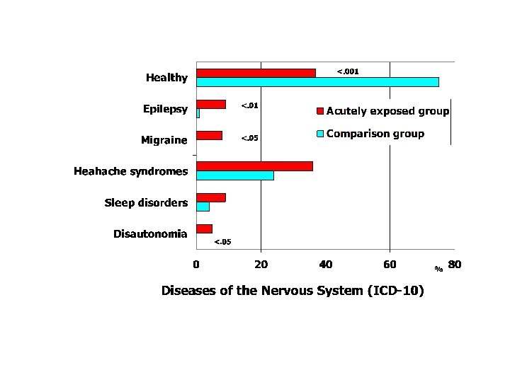
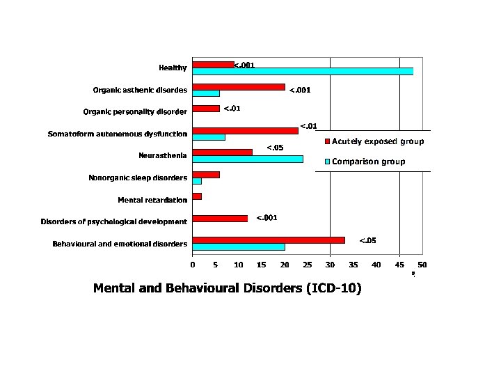
 FINALLY Thus, the neuromental health of the acutely prenatally irradiated children at the • • Chernobyl exclusion zone is deteriorated in comparison with the non-evacuee classmates living in Kiev due to more frequency of episodic and paroxysmal disorders, organic, including symptomatic, mental disorders, somatoform autonomic dysfunction, disorders of psychological development, and behavioural and emotional disorders with onset usually occurring in childhood and adolescence. Obviously, their neuromental health disorders are etiologically heterogeneous including psycho-social and economic factors, medical problems in their families, however an effect of real stress events (but not only their perception) during pregnancy together with prenatal irradiation cannot be excluded. Intelligence of the acutely prenatally irradiated children is deteriorated due to reduction of full scale and verbal IQ, as well as WISC performance/verbal discrepancies, with verbal decrements. In spite of the children’s intelligence is multifactorial, the contribution of prenatal irradiation was revealed. Characteristic neurophysiological changes of the acutely prenatally irradiated children are also etiologically heterogeneous, but the dose—effect relationship, especially at critical periods of cerebrogenesis, can testify the impact of prenatal irradiation. This study suggests that prenatal exposure to ionising radiation at thyroid foetal dose 0. 2 – 2 Gy and foetal dose 11– 92 m. Sv can result in detectable brain damage. The data obtained reflect great importance, interdisciplinarity, and complexity of such problem as brain damage in utero following radioecological disaster and a necessity to integrate international efforts to its solving.
 Finally • Thus this integrate research conducted in this area has made a valuable contribution to radiological protection by reinforcing the view that functionally significant radiation effects on the developing brain are most likely to occur at the low doses. • TSH level grows with foetal thyroid dose increase with a 0. 3 Sv threshold. Probably, these children had been affected by intrauterine hypothyroidism resulted in intelligence disturbances during the life. Obviously, an international psychoendocrine study should be organise for exploration of functions of the pituitary-thyroid system as a possible biological basis of mental health problem in children irradiated in utero as a result of the Chernobyl disaster. • Neurophysiological abnormalities together with intelligence disturbances, both doserelated, especially at 16– 25 weeks after fertilisation, as well as a «concentration» of the most severe neuropsychiatric disorders among the children exposed at the critical periods of cerebrogenesis, can testify to the developing brain abnormalities due to multiple factors with effects of prenatal irradiation. • Consideration must to given to deterministic effects prevail during the initial phase of damage may subsequently be modified by compensation within the brain.
 Finally • • • The results of this study agree with the Japanese studies concerning: 1) critical periods of cerebrogenesis — 8– 15 and, especially, 16– 25 week after week fertilisation. 2) dose related full scale IQ reduction, 3) an increase of paroxysmal disorders. The highest vulnerability of the brain under exposure at 16– 25, but not 8– 15 weeks after fertilisation as in the Japanese sample, we can explain by: 1) maximal radioiodine transfer rate in foetal thyroid at about 20– 25 weeks, 2) more «delicate» examination of intelligence disturbances that corresponds exactly to the events of the brain creation at 16– 25 weeks after fertilisation (apoptosis and its underlying molecular mechanisms; growth factor gene expression, cell formation and migration; neuronal differentiation, gross anatomical parameters in cortical and commissural diameters, synaptogenesis and synaptic remodeling limbic system and brain asymmetry forming, etc. ). An absence of dramatic increase of mental retardation, especially its severe form, as well as microcephalia obviously can be explained by significantly lower foetal doses of irradiation that in the atomic bomb survivors and lack of information about all in utero survivors. It is need the strong epidemiological investigation of the whole of this cohort. The «dose—effects» relationships concerning both intelligence and EEG-parameters, which are the most marked at the critical periods of cerebrogenesis, testify to significant contribution of prenatal irradiation into the organic brain damage.
 RECOMENDATIONS In this view this study should be continued as follows: view • the large-scale epidemiological investigation on this cohort only will give us the answer on open question on low dose risk after in utero radiation. • It seems, that the acutely prenatally exposed children at the Chernobyl exclusion zone is an unique sample that should be used for reassessment of risks of prenatal irradiation at radiation accidents on nuclear reactors. • to study of the whole of this cohort of children irradiated in utero in Ukraine; identification of further children irradiated in utero and children exposed at the age of 0– 1 years is necessary; • identification and forming of cohorts of age-, gender- and urban/ruralmatched children from radioactively clean areas of the Ukraine; • the verification and development of the currently available dosimetric models; • the assessment and verification of neuropsychiatric disorders; • the risk analysis of the influence of radioiodine in prenatal period and during the 1 st year of life on brain development; • the risk assessment of other stochastic and non- stochastic diseases on this base;
 QUESTION! Is there or is there not a threshold to the biological effects after in utero irradiation? At present, neither the experimental nor the epidemiological data are compelling in this regard. This matter is central to radiation protection and warrants more exhaustive study. More research it is needed (EC, 2002).
 Acknowledgements I am very grateful to all the staff in the Neurology, Radiology Departments of Research Center of Radiation Medicine AMS of Ukraine (Kiev), Psychiatry Department of Children of Research Institute of Neurology and Psychiatry AMS of Ukraine (Kharkov); Research NGO Center “Endopolimed” (Ukraine) I am very grateful to our scientific consultants. I greatly appreciate their expert advice and constructive criticism. • • Professor William Jack Shull (USA, Japan); Professor James N. Yamazaki (Japan); Professor Leonid Prilipko (WHO); Professor A. Kellerer (Germany); Dr. R. Pott-Born (Germany) Professor W. Yule (United Kingdom) Professor J. Stevenson (United Kingdom)

 “Life is a risk process – so risk that none of us will escape it alive” (Hendee, 1991).
 Contact details: Professor Angelina Nyagu, MD Ph. D & Dr Med Sci President of Association “PHYSICIANS of CHERNOBYL”. Editor of Int. J. Radiat. Med. Kiev Ukraine Tel/ Fax : +38 044 484 07 34 Email: anyagu@ukr. net Web: www. physiciansofchernobyl. org. ua