
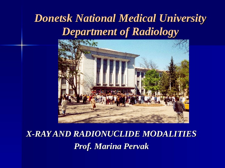
Donetsk National Medical University Department of Radiology X-RAY

pervak_x-ray-rn_lecture_1.ppt
- Размер: 5.3 Mегабайта
- Количество слайдов: 20
Описание презентации Donetsk National Medical University Department of Radiology X-RAY по слайдам
Donetsk National Medical University Department of Radiology X-RAY AND RADIONUCLIDE MODALITIES Prof. Marina Pervak
4 modalities (or 4 groups of methods) of modern Diagnostic imaging (Radiology): — — X-ray examination ; ; — — Nuclear medicine imaging ; ; — Di agnostic ultrasound ; ; — Magnetic resonance imaging ( ( MRI). . X-ray and radionuclide examinations use ionizing radiation Biological effect is tissue ionization.
X-ray examination methods: — radiography, — fluoroscopy, — fluorography, — tomography, — computed tomography. All X-ray examinations are based on the detection of radiation passed through the patient (transmitted radiation).
X-rays are generated by X-ray tube ( ( generator of radiation). NB!NB! Different tissues provide different degrees of X-ray attenuation. Attenuation is a loss of X-ray energy. Different tissues allow the transmission of different amounts of X-ray imaging is the imaging of shadows. X-rays penetrate tissues and are detected on the other side of the patient by different detectors. Main groups of X-ray techniques: I) I) direct analogue techniques, 2) 2) indirect analogue techniques, 3) 3) digital techniques.
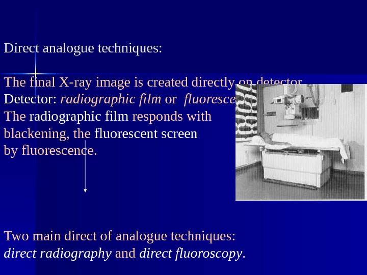
Direct analogue techniques: The final X-ray image is created directly on detector. Detector: radiographic film or fluorescent screen. The radiographic film responds with blackening, the fluorescent screen by fluorescence. Two main direct of analogue techniques: direct radiography and direct fluoroscopy.
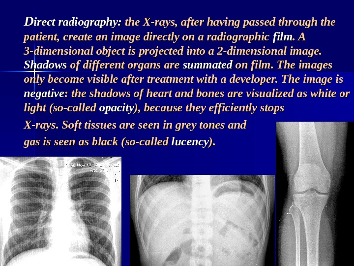
DD irect radiography: the X-rays, after having passed through the patient, create an image directly on a radiographic film. A. A 3 -dimensional object is projected into a 2 -dimensional image. Shadows of different organs are summated on film. The images only become visible after treatment with a developer. The image is negative : the shadows of heart and bones are visualized as white or light (so-called opacity ), because they efficiently stops X-rays. Soft tissues are seen in grey tones and gas is seen as black (so-called lucency ). ).
DD irect fluoroscopy (screening): the transmitted X-ray beam fall on a fluorescent screen , resulting in a dynamic projection light image. The image is positive. The shadows of bones and heart are black (( real opacity ), and gas is seen as white or light ( real lucency ). ). NB!NB! The image can be observed directly by the radiologist on screen! Indirect fluoroscopy employs X-ray image intensifier and TV-technique. Primary projection image is created on a fluorescent screen, but screen image is not observed directly. The screen is part of an X-ray image intensifier that enhances the brightness of the primary image. The intensified image may be recorded via lenses by a TV camera and shown on a monitor. The image is positive. .
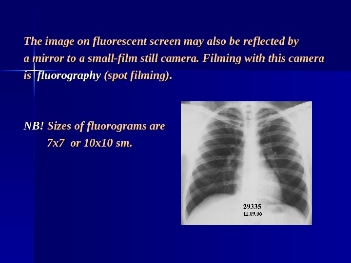
The image on fluorescent screen may also be reflected by a mirror to a small-film still camera. Filming with this camera is is fluorography (spot filming). NB!NB! Sizes of fluorograms are 7 x 7 or 10 x 10 sm.
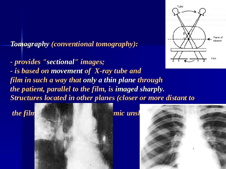
Tomography (conventional tomography): — provides » sectional » images; — is based on movement of X-ray tube and film in such a way that only a thin plane through the patient, parallel to the film, is imaged sharply. . Structures located in other planes (closer or more distant to the film) are blurred due to dynamic unsharpness.
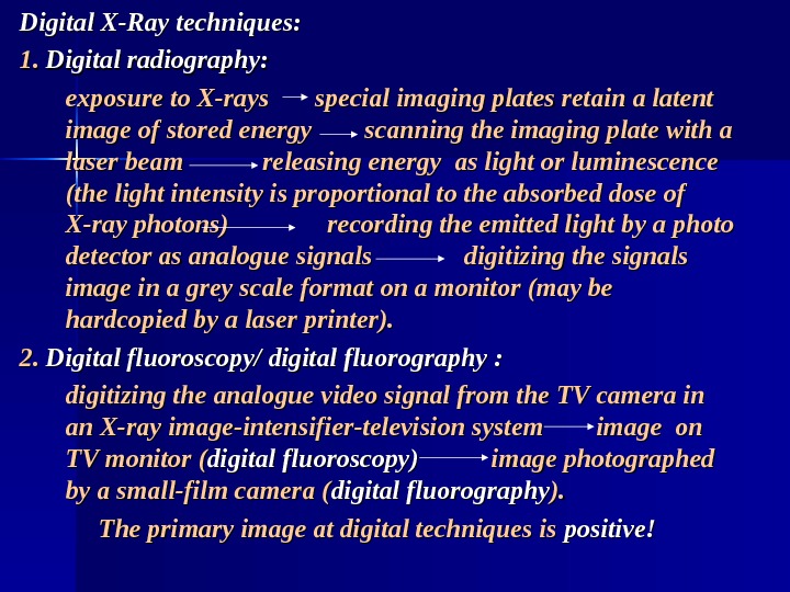
Digital X-Ray techniques: 1. 1. Digital radiography: exposure to X-rays special imaging plates retain a latent image of stored energy scanning the imaging plate with a laser beam releasing energy as light or luminescence (the light intensity is proportional to the absorbed dose of X-ray photons) recording the emitted light by a photo detector as analogue signals digitizing the signals image in a grey scale format on a monitor (may be hardcopied by a laser printer). 2. 2. Digital fluoroscopy/ digital fluorography : : digitizing the analogue video signal from the TV camera in an X-ray image-intensifier-television system image on TV monitor ( digital fluoroscopy) image photographed by a small-film camera ( digital fluorography ). ). The primary image at digital techniques is positive!
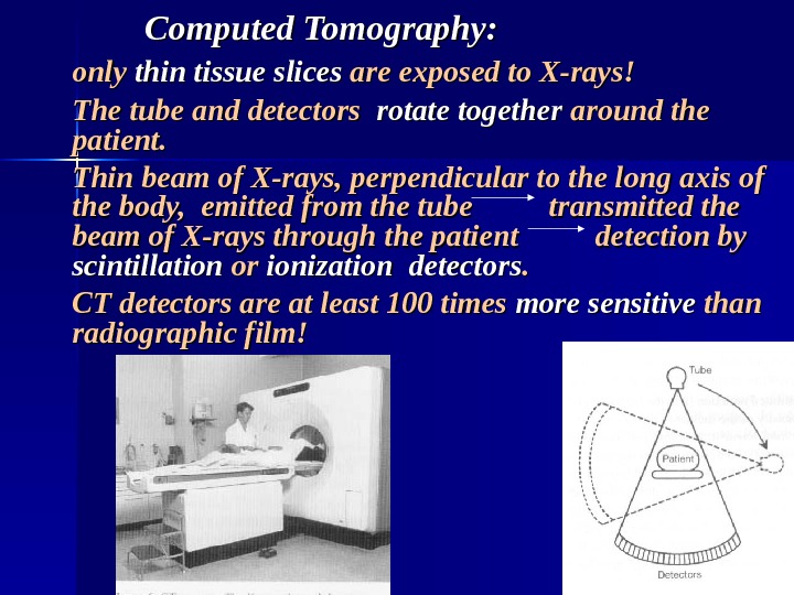
Computed Tomography: only thin tissue slices are exposed to X-rays! The tube and detectors rotate together around the patient. Thin beam of X-rays, perpendicular to the long axis of the body, emitted from the tube transmitted the beam of X-rays through the patient detection by scintillation or or ionization detectors. . CT detectors are at least 100 times more sensitive than radiographic film!
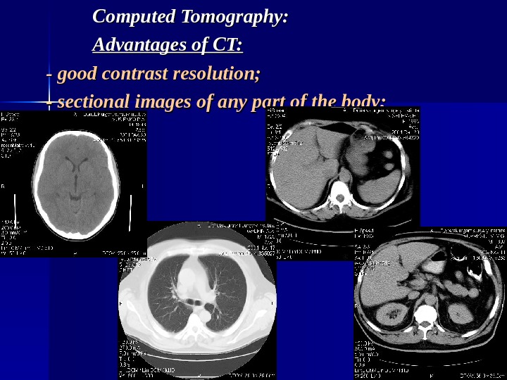
Computed Tomography: Advantages of CT: — good contrast resolution; — sectional images of any part of the body;
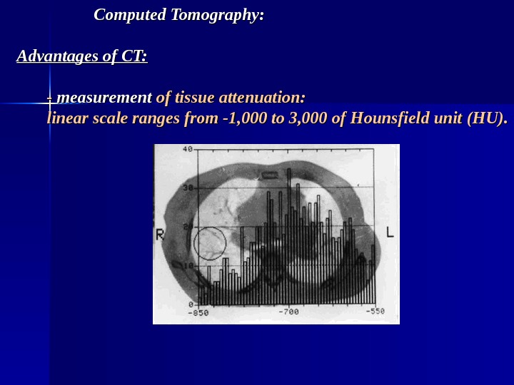
Computed Tomography: Advantages of CT: — — measurement of tissue attenuation: linear scale ranges from -1, 000 to 3, 000 of Hounsfield unit (HU).
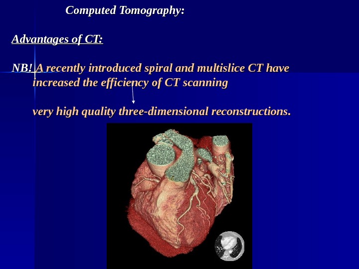
Computed Tomography: Advantages of CT: NB! A recently introduced spiral and multislice CT have increased the efficiency of CT scanning very high quality three-dimensional reconstructions.
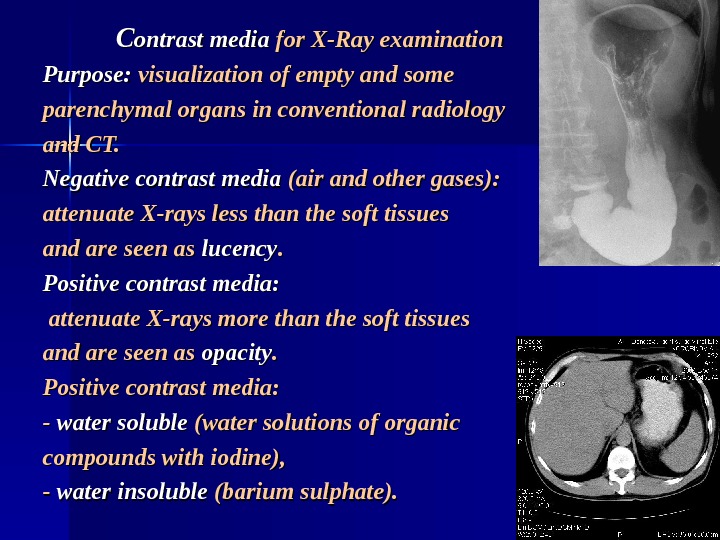
CC ontrast media for X-Ray examination Purpose: visualization of empty and some parenchymal organs in conventional radiology and CT. Negative contrast media (air and other gases): attenuate X-rays less than the soft tissues and are seen as lucency. . Positive contrast media: attenuate X-rays more than the soft tissues and are seen as opacity. . Positive contrast media: — — water soluble (water solutions of organic compounds with iodine), — — water insoluble (barium sulphate).
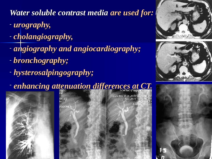
Water soluble contrast media are used for: — urography, — cholangiography, — angiography and angiocardiography; — bronchography; — hysterosalpingography; — enhancing attenuation differences at CT.
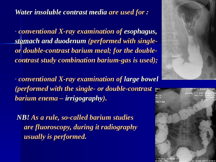
Water insoluble contrast media are used for : — conventional X-ray examination of esophagus, stomach and duodenum (performed with single- or double-contrast barium meal; for the double- contrast study combination barium-gas is used); — conventional X-ray examination of large bowel (performed with the single- or double-contrast barium enema – irrigography ). ). NB!NB! As a rule, so-called barium studies are fluoroscopy, during it radiography usually is performed.
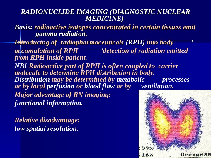
RADIONUCLIDE IMAGING (DIAGNOSTIC NUCLEAR MEDICINE) Basis: radioactive isotopes concentrated in certain tissues emit gamma radiation. . Introducing of radiopharmaceuticals (RPH) into body accumulation of RPH detection of radiation emitted from RPH inside patient. NB! Radioactive part of RPH is often coupled to carrier molecule to determine RPH distribution in body. Distribution may be determined by metabolic processes or by local perfusion or blood flow or by ventilation. Major advantage of RN imaging: functional information. Relative disadvantage: low spatial resolution.
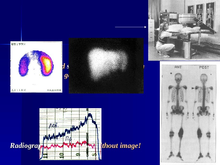
Detector: large, disk-shaped scintillation crystal (main component of the gamma camera) scintigraphy. Radiography: only curves – without image!
Tomographic techniques of RN imaging: I) I) single photon emission computed tomography (SPECT): gamma camera rotates around patient recording radioactivity at numerous angles reconstruction of sectional images; 2) 2) positron emission tomography (PET): using positrons emitting radionuclides and special detectors. NB!NB! PET allows estimation of RN concentration and study of metabolic processes at various disease states, especially in heart and brain.