

c5efb347be044d099fb919cbfaf27d31.ppt
- Количество слайдов: 103
 DIENCEPHALON David Kachlík Petr Zach
 diencephalon • • • epithalamus subthalamus metathalamus hypothalamus opticus
 diencephalon - development Alar plate → thalamus, subthalamus Bazal plate → hypothalamus canalis centralis → 3 rd ventricle fissura telodiencephalica sulcus hypothalamicus
 – ncl. habenularis med. + lat. • commissura habenularum") Epithalamus • habenula (trigonum habenulare) – ncl. habenularis med. + lat. • commissura habenularum • commissura posterior – Commisural fibers • Posterior thalamic nuclei, colliculi sup. , ncll. pretectales – Non commissural fibers • ncl. interstitialis Cajali + ncl. commissurae posterioris Darkschewitzi → fasciculus longitudinalis medialis from other side
Epithalamus • habenula (trigonum habenulare) – ncl. habenularis med. + lat. • commissura habenularum • commissura posterior – Commisural fibers • Posterior thalamic nuclei, colliculi sup. , ncll. pretectales – Non commissural fibers • ncl. interstitialis Cajali + ncl. commissurae posterioris Darkschewitzi → fasciculus longitudinalis medialis from other side
 Epithalamus
 Epithalamus Habenulae connections • AF: stria medullaris thalami – septum verum → habenula • EF: tractus habenulo-interpeduncularis (fasciculus retroflexus Meynerti) – ncll. habenulares → ncl. interpeduncularis → stem commissura habenularum
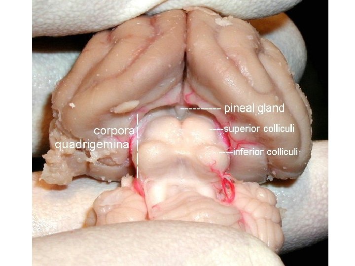
 Glandula pinealis; Corpus pineale „Epiphysis; “ • Developmental relation to parietal eye • Hateria New Zealand (Sphenodon punctatus) • Reaction to polarized light (monthly biorhytms)
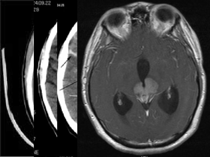
 Epiphysis • Behind upper posterior end of 3 rd ventricle • Part of epithalamus • Rudimentary endocrine gland with supressive efect on sexual glands pubertas praecox • Dorsally extends above brain stem (above lamina quadrigemina of mid brain) • melatonin change of level during day • acervulus cerebri (= calcium concrements in adults) – CT, MRI
 Subthalamus • Positioned below thalamus – separated by Forels field H 1 • Externally to hypothalamus – w/o visible border • zona incerta • nucleus subthalamicus (= corpus Luysi) • Forels fields = campi perizonales = H zone (Haubenfelder)
 Subthalamus • zona incerta – Composition resembles RF – Integration of inputs from cortex and stem – GABA inhibits ncll. intralaminares and association nuclei of thalamus (similarly to ncll. reticulares thalami) • nucleus subthalamicus (= corpus Luysi) – Connected to basal ganglia system (Glu into globus pallidus) – lesion: hemibalismus (rough non coordinated movements of contralateral cingulum muscles) after CMP, non ketonic hyperglycemia • Forels fields = campi perizonales = H zones (Haubenfelder) – H = ansa lenticularis • H 1 • H 2 = fasciculus thalamicus = fasciculus lenticularis
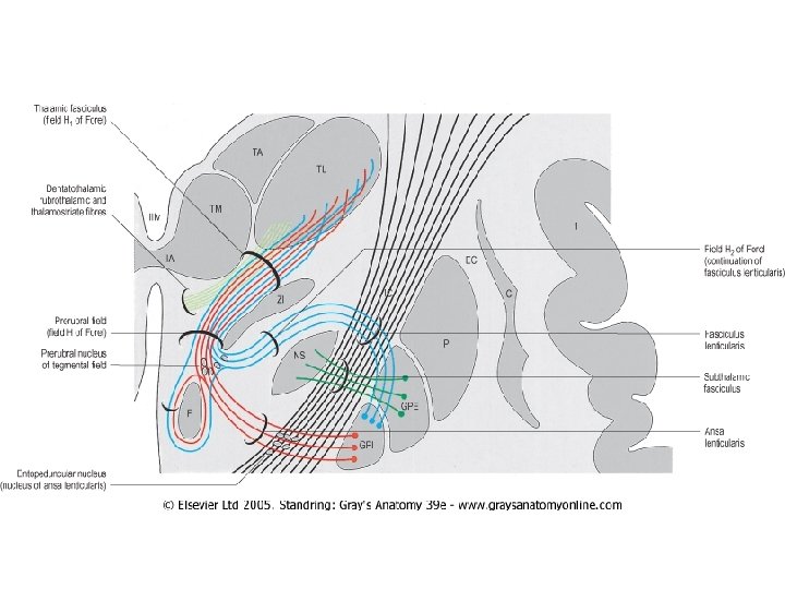
 Nuclear groups and fiber tracts associated with the subthalamus include the subthalamic nucleus, zonal incerta, and the fields of Forel and their associated fiber bundles. AL, ansa lenticularis; CP, cerebral peduncle; FF, fields of Forel; GPe, globus pallidus externus; GPi, globus pallidus internus; H 1, H 1 field of Forel (thalamic fasciculus); IC, internal capsule; LF, lenticular fasciculus (H 2); PPN, pedunculopontine nucleus; Put, putamen; SN, substantia nigra; STN, subthalamic nucleus; Thal, thalamus; ZI, zona incerta. H, corresponding to the nucleus of the medial field is not shown. Used with permission from Hamani et al. , Brain 127: 4 -20, 2004.
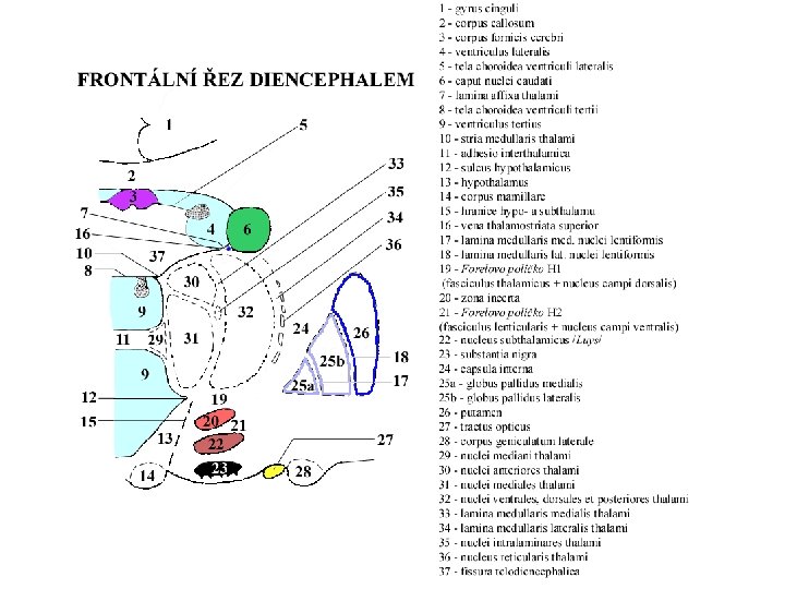
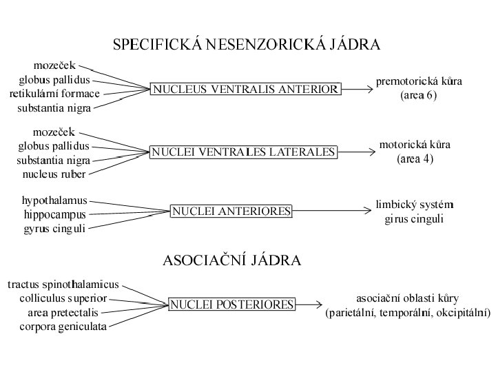
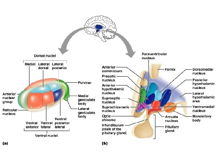
 • • • „secretary of brain“ all except for smell pulvinar") Thalamus (thalamus dorsalis) • • • „secretary of brain“ all except for smell pulvinar (dorsally) tuberculum anterius (ventrally) lamina medullaris medialis + lateralis thalami adhesio interthalamica (80 %) – w/o notion Nuclei parcellated according to position or connection – nuclei anteriores, dorsales, intralaminares, mediani, mediales, posteriores, ventrales, reticularis – specific sensory nuclei – specific non sensory nuclei – Non specific nuclei – Association nuclei
Thalamus (thalamus dorsalis) • • • „secretary of brain“ all except for smell pulvinar (dorsally) tuberculum anterius (ventrally) lamina medullaris medialis + lateralis thalami adhesio interthalamica (80 %) – w/o notion Nuclei parcellated according to position or connection – nuclei anteriores, dorsales, intralaminares, mediani, mediales, posteriores, ventrales, reticularis – specific sensory nuclei – specific non sensory nuclei – Non specific nuclei – Association nuclei
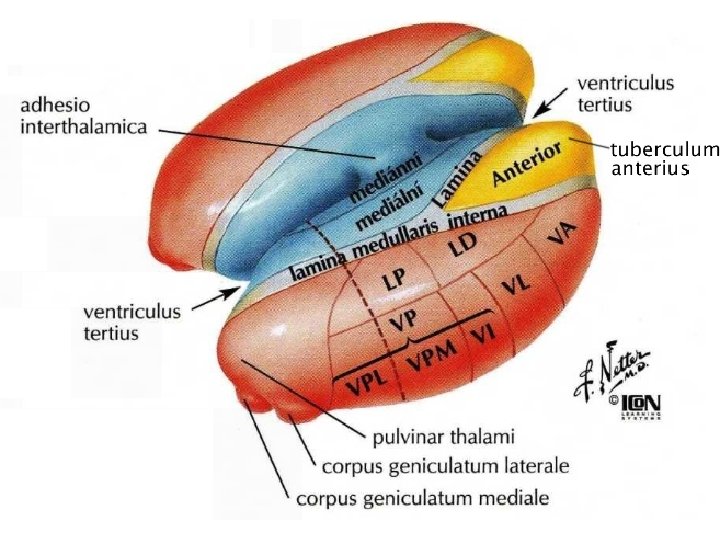
 Parcellation of thalamic nuclei acc to position nuclei anteriores, dorsales, intralaminares, mediani, mediales, posteriores, ventrales, reticularis
 Thalamic connections AF: • sensitive and sensory – Pain, proprioception, touch, taste, balance, hearing, vision • motoric – cerebellum, BG • RF - ARAS • limbic system – corpus mammillare, hippocampus EF: cortex + hypothalamus Reciprocal connections: BG, RF, cortex, stem, cerebellum, spine
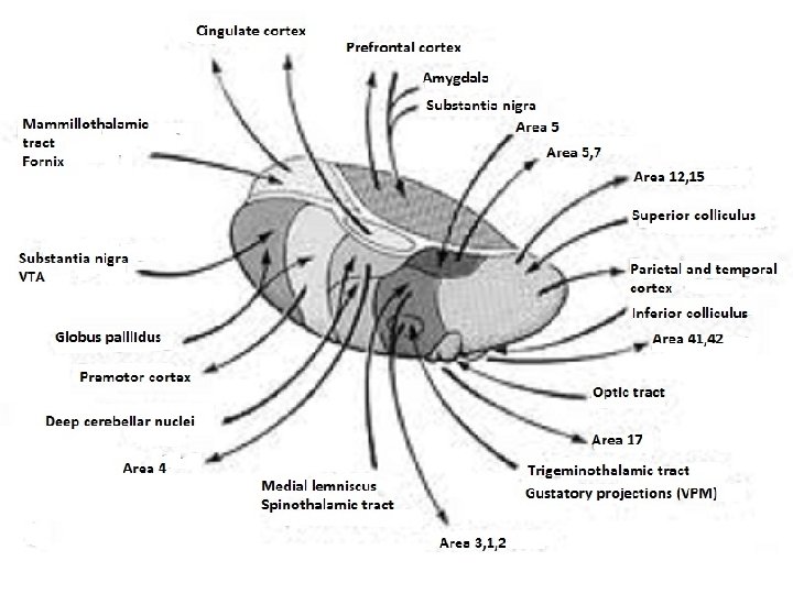
 Specific nuclei • tractus mamillothalamicus ncl. anterior gyrus cinguli – reverberation enforces emotions • globus pallidus ncl. VA prefrontal cortex • globus pallidus ncl. VL supplementary motoric cortex • nucleus dentatus cerebelli ncl. VL motoric cortex • lemniscus medialis et spinalis ncl. VPL senz. cortex • lemniscus trigeminalis ncl. VPM senzitive cortex
 area cingularis posterior • Olfactory and") Association nuclei • ncl. LD (lat. dors. ) area cingularis posterior • Olfactory and limbic brain ncl. MD (mediodors. ) prefrontal cortex (thinking, reasoning, mood, mind state – integration with sensory inputs) • colliculus superior ncl. LP (lat. post. ) + pulvinar visual and parietal association cortex (draws attention to objects on the periphery of visual field) • ncll. P (pulvinar) frontal, temporal, parietal and occipital association cortex (integration of visual, auditory, tactile and proprioceptive inputs)
Association nuclei • ncl. LD (lat. dors. ) area cingularis posterior • Olfactory and limbic brain ncl. MD (mediodors. ) prefrontal cortex (thinking, reasoning, mood, mind state – integration with sensory inputs) • colliculus superior ncl. LP (lat. post. ) + pulvinar visual and parietal association cortex (draws attention to objects on the periphery of visual field) • ncll. P (pulvinar) frontal, temporal, parietal and occipital association cortex (integration of visual, auditory, tactile and proprioceptive inputs)
 Non specific nuclei • ncll. intralaminares – Slow pain – ARAS • ncll. mediani • Limbic system (according to efferentation)
 Ncll. reticulares • GABA • Excitatory collaterals from all specific nuclei of thalamus and cortex • Inhibitory efferentation back to thalamus • Similarly as zona incerta • Function: labels new inputs and differentiates them from regular inputs from environment
 Thalamic connection Cortex tractus thalamocorticalis ↑ ↓ tractus corticothalamicus – strong tract inhibiting thalamus „cleaning further incoming informations“
 – Special") Thalamic connection • senzitive + sensory inputs – senzitivity (pain, proprioception, touch) – Special sensory (taste, balance, hearing, vision) • motoric inputs – Cerebellum, basal ganglia • Reticular formation • limbic system – corpus mammillare – Hippocampal formation • ncll. reticulares only do not have efferentation to other thalamic nuclei
Thalamic connection • senzitive + sensory inputs – senzitivity (pain, proprioception, touch) – Special sensory (taste, balance, hearing, vision) • motoric inputs – Cerebellum, basal ganglia • Reticular formation • limbic system – corpus mammillare – Hippocampal formation • ncll. reticulares only do not have efferentation to other thalamic nuclei
 Pain – processing in thalamus • fast – acute – ncl. VPL + VPM • slow – chronical – nuclei intralaminares • ncl. centri mediani (CM) • ncl. parafascicularis (PF)
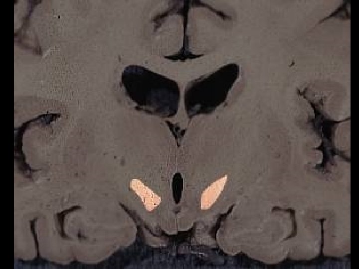
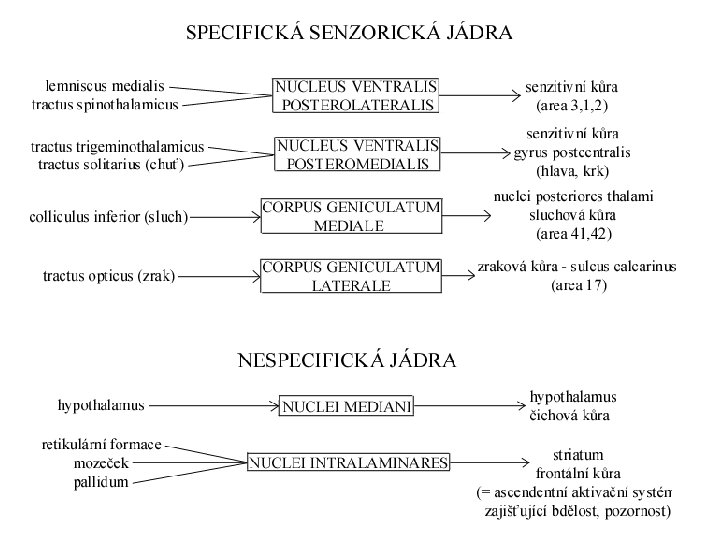
 Metathalamus • corpus geniculatum laterale – visual center • corpus geniculatum mediale – auditory center
 Corpus geniculatum laterale Visual center • pars magnocellualris: movement, depth and perspective • pars parvocellularis: diameters, volume, shape and colors
 Thalamus opticus 1. nervus opticus 2. chiasma opticum 3. tractus opticus 4. corpus geniculatum laterale 5. radiatio optica 6. Visual cortex
 hemiparesis") Thalamic syndrom „ 6 hemi“ • • • hemihypestezia /hemianestezia hemiataxia (+ hemiapraxia) hemiparesis hemialgia (+ hemipathia) hyperkinesis choreatic and athetoid hemianopsia homonymous contralateral • Consciousness problems / epilepsy / cataplexy
Thalamic syndrom „ 6 hemi“ • • • hemihypestezia /hemianestezia hemiataxia (+ hemiapraxia) hemiparesis hemialgia (+ hemipathia) hyperkinesis choreatic and athetoid hemianopsia homonymous contralateral • Consciousness problems / epilepsy / cataplexy
 Clinical talamic syndromes posterolateral talamic syndromes • senzitive and senzoric lesions • talamic syndrom = Dejerine-Roussy syndrome – ncl. VPL, VPM – talamic pain Joseph Jules Dejerine (18491917) Gustave Roussy (18741948)
 Clinical thalamic syndromes medial talamic syndromes • Consciousness lesions • „thalamic neglect“, talamic amnesia, akinetic mutizm anterolateral talamic syndromes • Motoric lesions • palsy, ataxia, motoric non coordination, dysfagia
 Hypothalamus
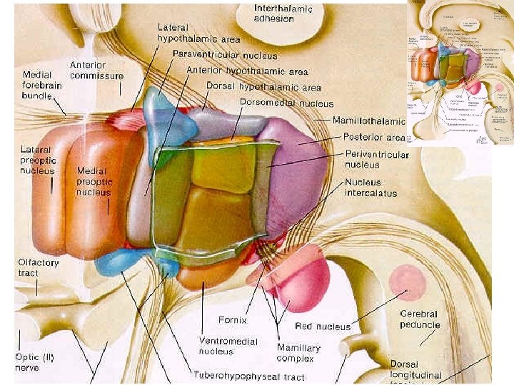
 Hypothalamus • • Is derivative of visceromotor zone of basal plate Highest autonomous center infundibulum + hypophysis tuber cinereum (eminentia mediana) + corpus mammillare • area preoptica + chiasma et tractus opticus
 Hypothalamus • infundibulum • tuber cinereum • corpora mammillaria • recessus infundibuli • recessus opticus
 Hypothalamus – borders • • up: sulcus hypothalamicus down: base of brain front: lamina terminalis back: continues into tegmentum mesencephali • Medially: 3 rd ventricle • laterally: capsula interna
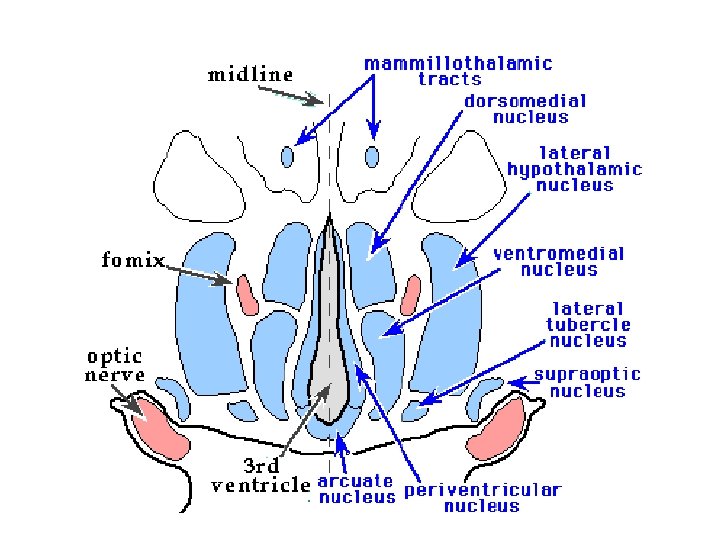
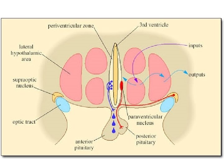
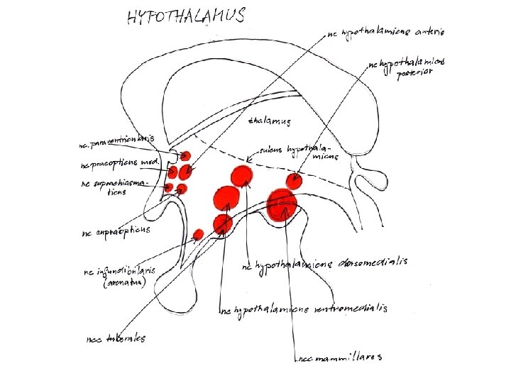
 Hypothalamus 3 longitudinal zones: periventricular, medial, lateral zones 3 horizontal zones: anterior, middle, posterior hypothalamus Nuclei • ventral hypothalamus (area hypothalamica rostralis) - nucleus paraventricularis, supraopticus, suprachiasmaticus • middle hypothalamus (area hypothalamica intermedia et dorsalis) nuclei tuberales laterales et ventromediales • posterior hypothalamus (area hypothalamica posterior) - nuclei mammillares, nucleus h. posterior, nucleus tberomammillaris
 Hypothalamus – function Receives inputs from almost all receptors – especially from RF, prefrontal cortex and hippocampus former hypothesis: • anterior hypothalamus - parasympaticus • Middle hypothalamus - sympaticus • posterior hypothalamus – limbic system
, nerves, CSF HYPOTHALAMUS Endocrine + autonomic system HOMEOSTASIS emotions") Hypothalamus – fyziology Hormones (blood), nerves, CSF HYPOTHALAMUS Endocrine + autonomic system HOMEOSTASIS emotions (= LIMBIC SYSTEM)
Hypothalamus – fyziology Hormones (blood), nerves, CSF HYPOTHALAMUS Endocrine + autonomic system HOMEOSTASIS emotions (= LIMBIC SYSTEM)
 Hypothalamus – function Hypothalamus • termoregulation – center hyperthermia – anterior h. – center cold – posterior h. • • lateral h. : center hunger, thirst and anger medial h. : center satiety and passivity anterior h. : center sleep and wake sex – ♂ nucleus preopticus – ♀ nucleus ventromedialis
 Hypothalamus – function Hypothalamus • ncl. suprachiasmaticus – center of circadian rhytms • ncl. supraopticus + paraventricularis (magnocellular neurons) – ADH (vazopresin) + oxytocin • ncl. arcuatus (infundibularis) and around (parvocellular neurons) – statins and liberins • ncl. tuberomamillaris – histamine to brain and spine („arousal“) • Activated by orexin from lat. hypothalamus • Lack in narcolepsia
 Anterior hypotalamus • ncl. paraventricularis - oxytocin, ADH • ncl. supraopticus – oxytocin, ADH • ncl. preopticus medialis – blood pressure down, puls too • ncl. hypothalamicus anterior – termoregulation, swetting, inhibition of TSH • ncl. suprachiasmaticus – circadian rhytm
 – in area") Sexually dimorphic areas of anterior hypothalamus • SDN (sexually dimorphic nucleus) – in area preoptica bigger in males comp to females (? testosterone effect) • INAH 3 in humans, o. SDN in sheeps, SDNPOA in rats, AHdc in macaques, POM in quails • Affects sexual behavior in animals
Sexually dimorphic areas of anterior hypothalamus • SDN (sexually dimorphic nucleus) – in area preoptica bigger in males comp to females (? testosterone effect) • INAH 3 in humans, o. SDN in sheeps, SDNPOA in rats, AHdc in macaques, POM in quails • Affects sexual behavior in animals
 • ncl. infundibularis = ncl. arcuatus –") Middle hypothalamus = tuberal hypothalamus (tuber cinereum) • ncl. infundibularis = ncl. arcuatus – statins and liberins • ncll. tuberales – hunger and thirst • ncl. hypothalamicus ventromedialis - hunger • ncl. hypothalamicus dorsomedialis – increase of blood pressure and puls
Middle hypothalamus = tuberal hypothalamus (tuber cinereum) • ncl. infundibularis = ncl. arcuatus – statins and liberins • ncll. tuberales – hunger and thirst • ncl. hypothalamicus ventromedialis - hunger • ncl. hypothalamicus dorsomedialis – increase of blood pressure and puls
") Posterior hypotalamus • ncll. mammillares – memory, connection to limbic system (ncl. anterior thalami) • ncll. hypothalamicus posterior – increase of blood pressure, mydriasis, tremor
Posterior hypotalamus • ncll. mammillares – memory, connection to limbic system (ncl. anterior thalami) • ncll. hypothalamicus posterior – increase of blood pressure, mydriasis, tremor
 Hypothalamus
 → tractus") Hypothalamus – white matter • fornix → corpus mammillare (nuclei corporis mammillaris) → tractus mammillaris princeps /splits into/ → tractus mamillothalamicus → ncl. anteriores thalami → tractus mammillotegmentalis RF of stem (ncl. Guddeni) • stria terminalis corpus amygdaloideum → hypothalamus • stria medullaris thalami hypothalamus habenula
Hypothalamus – white matter • fornix → corpus mammillare (nuclei corporis mammillaris) → tractus mammillaris princeps /splits into/ → tractus mamillothalamicus → ncl. anteriores thalami → tractus mammillotegmentalis RF of stem (ncl. Guddeni) • stria terminalis corpus amygdaloideum → hypothalamus • stria medullaris thalami hypothalamus habenula

 Hypothalamus – white matter • fasciculus medialis telencephali = fasciculus prosencephalicus medialis = medial forebrain bundle (MFB) – between medial and laterl zone of nuclei – connects hypotalamus with cortical limbic system + limbic system of brain stem (+ RF) • pedunculus mammillaris - connects corpora mammillaria and nucleus dorsalis tegmenti Guddeni (in RF of mid brain) and fasciculus longitudinalis posterior Schützi ncl. in medial zone of hypotalamus → autonomic nuclei, nuclei of cranial nerves (eventually into spine)
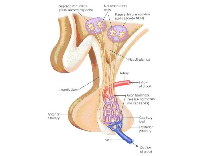
 • adenohypophysis (= lobus anterior) – Development from Rathke pouch from") Hypophysis (pituitary gland) • adenohypophysis (= lobus anterior) – Development from Rathke pouch from roof of pharynx – hormones (ACTH, TSH, FSH, LH, STH, MSH) – Influenced by hypotalamic releasing and inhibiting hormones – transport from ncl. arcuatus via tractus tuberoinfundibularis (= neurokrinie) → hypotalamohypofyzoportal system – Sheehan syndrome • neurohypophysis (= lobus posterior) – – – Development as diencephalic pouch nucleus supraopticus (vazopressin = ADH) ncl. paraventricularis (oxytocin) axonal transport from hypotalamus Reacts to changes of osmolality via organum subfornicale
Hypophysis (pituitary gland) • adenohypophysis (= lobus anterior) – Development from Rathke pouch from roof of pharynx – hormones (ACTH, TSH, FSH, LH, STH, MSH) – Influenced by hypotalamic releasing and inhibiting hormones – transport from ncl. arcuatus via tractus tuberoinfundibularis (= neurokrinie) → hypotalamohypofyzoportal system – Sheehan syndrome • neurohypophysis (= lobus posterior) – – – Development as diencephalic pouch nucleus supraopticus (vazopressin = ADH) ncl. paraventricularis (oxytocin) axonal transport from hypotalamus Reacts to changes of osmolality via organum subfornicale
 Hypotalamus – summary • Part of limbic system = preserve of species and individual – reproduction – Growth and metabolism – Intake of food and water – Attack and defense – termoregulation – Cycle of wake - sleep – memory
 Optional reading Neuroanatomy of sleep cycle
 Why sleep? • You need almost as much sleep after a day of sitting around the house as after a day of intense physical or mental activity (Horne and Minard, 1985, Shapiro et al. , 1981). • You feel tired at the end of the day because inhibitory processes in your brain force you to become less aroused and less alert.
 Functions of sleep • • • Rest our muscles Decrease metabolism Rebuild proteins in the brain Reorganize synapses Strenghten memories
 Why sleep? • You need almost as much sleep after a day of sitting around the house as after a day of intense physical or mental activity (Horne and Minard, 1985, Shapiro et al. , 1981). • You feel tired at the end of the day because inhibitory processes in your brain force you to become less aroused and less alert.
 Why sleep? • You need almost as much sleep after a day of sitting around the house as after a day of intense physical or mental activity (Horne and Minard, 1985, Shapiro et al. , 1981). • You feel tired at the end of the day because inhibitory processes in your brain force you to become less aroused and less alert.
 Search for primary reason • All species sleep, not just vertebrates with big brain and complex memories • Bacteria have circadian rhytms • Conservation theory: • -Simple way of conserving energy • NASA´s Rover built to explore Mars had mechanisms to make it „sleep“ at night to conserve its batteries.
 Conservation theory • We might not guess original function for which it was evolved. • Consider computer analogy: • Important functions today include writing papers, sending e-mail, searching Internet, playing games, storing photographs, playing movies etc. • Previously served for mathematical operations
 Search for primary reason • Sleep conserves energy during the inefficient time • Mammal´s body temperature is decreased by 1°C or 2°C during sleep • Animals increase sleep duration during food shortages • Sleep is therefore analogous to hibernation
 Hibernation • Hibernating animals decrease their body temperature to that of the environment (but do not let it drop to freeze ) • Hamsters sometimes hibernate, if you let your favorite pet during winter in cold place, make sure it is not hibernating before you bury it • Hibernating animals come out of hibernation for few hours every few days, raising temperature to normal
 Hibernation • Hibernating retards the ageing process • Hibernation is also a period of relative invulnerability to infection and trauma. Procedures that would ordinarily damage brain, such as inserting a needle into it, produce little if any harm (Zhou et al. , 2001) • Na-K dependent ATPase on cell membrane
 Hibernation • Herbivores graze many hours per day – sleep is briefer and often interrupted • Dolphins and other aquatic mammals have asleep one hemisphere at time, other is awake and control swimming and breathing • Birds during migration decrease need for sleep – in primates drugs exciting glutamate receptor can decrease sleep need • Even if you keep migratory bird in cage during migratory period, it does not sleep
 Hibernation • The fact that it is possible to decrease the need for sleep argues that sleep is not necessary for repair and restoration, or at least that sleep usually last longer than required for repair and restoration Hazel dormouse hibernating
 Swift example
 Swift example • Question: when a baby swift first takes off from its nest, how long would you guess its first flight lasts, until it lands again? • Answer: up to 2 years! • It spends both night and days in the air, except huge storms • Perhaps it switches brain hemispheres, but we do not know – EEG during flight is problem
 Other functions of sleep • • Restorative functions Sleep and memory 600 hrs of REM per year REM – eyeball movement for cornea to get enough oxygen by shaking eyeballs • Strenghtening consolidation of different type of memory
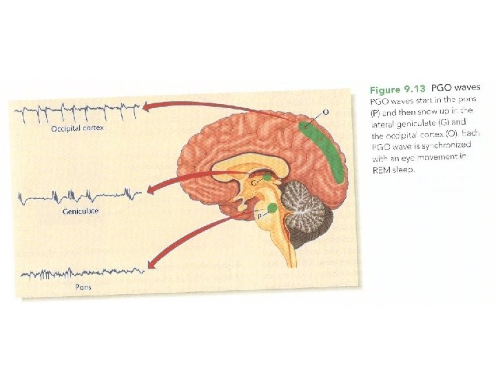
 Biology of dreaming • Activation-synthesis hypothesis – dreams begin with periodic bursts of spontaneous activity in the pons – the PGO waves, which partly activates many but not all parts of the cortex. Cortex combines this haphazard input with whatever other activity was already occuring and does its best to synthetize a story that makes sense of all this informations
 Biology of dreaming • Because activity is supressed in the primary visual cortex and somatosensory cortex, normal sensory information cannot compete with the self generated stimulation and hallucinations result • Input from pons usually activates amygdala – emotional processing • Prefrontal cortex is inactive during PGO waves, memory is weak
 Biology of dreaming • Patients with pons lesion still have dreams, eventhough they do not move eyeballs and other features of REM • Paradox: brain produces dreams, but do not percieve them as self-produced. Some people can tickle themselves and actually feel it as a tickling sensation, at least slightly
 Biology of dreaming • Clinico-anatomical hypothesis • Similar to activation-synthesis theory • Inferior part of parietal cortex is active during dreaming – patients with damage have no dreams • www. dreamresearch. net
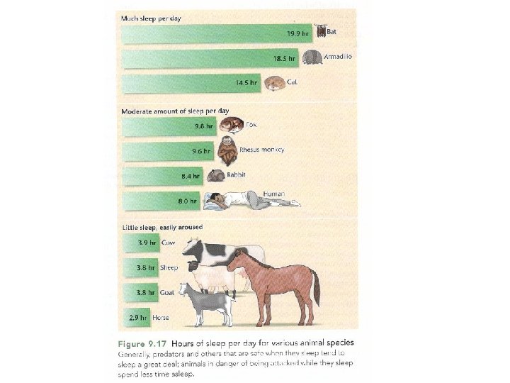
 Variation in sleep • Cats and bats eat rich food and face little threat – they sleep many hours per day
 Biological clock • Richter 1967 introduced idea that the brain generates its own rhythms • Disruption of BC occurs after damage to hypothalamic area – suprachiasmatic nucleus • After disruption rhythms are less consistent and no longer synchronized to light and dark pattern
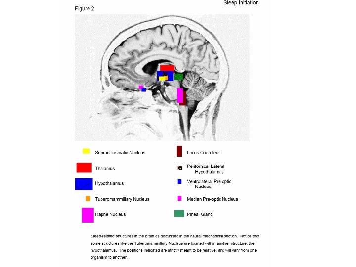
 Suprachiasmatic nucleus
 Suprachiasmatic nucleus • It generates rhythm in a genetically controlled, unlearned manner • From Drosophilla – isolated genes period (per) and timeless (tim), producing proteins Per and Tim • In the morning their levels are low, in the evening high • Interact with protein Clock to induce sleepiness
 SCN
SCN
 Suprachiasmatic nucleus • Pulse of light during night inactivates protein Tim, so extra light during evening resets biological clock • People having mutation in per gene have odd circadian rhythms: run faster • Mutation in per is closely linked to clinical depression
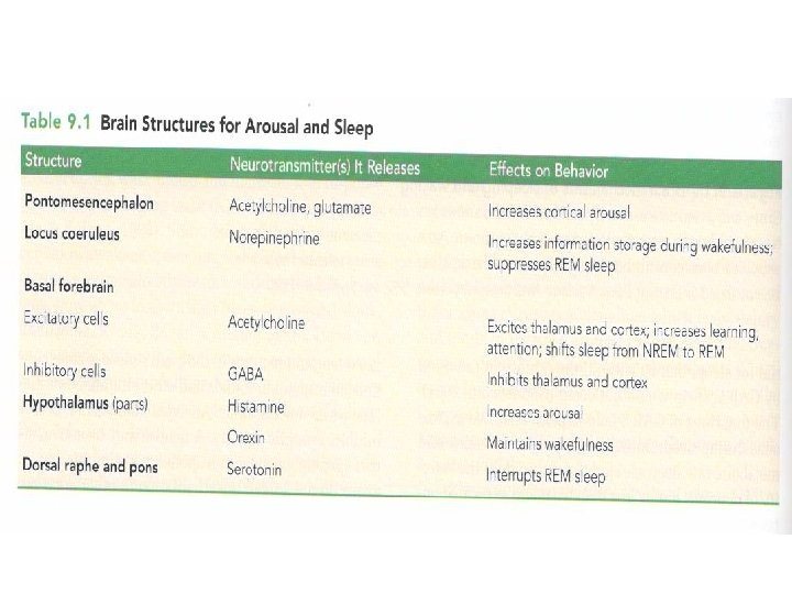
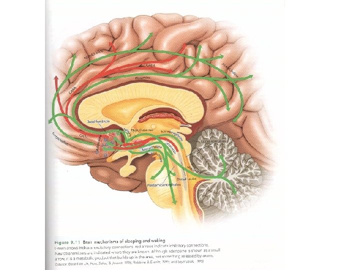
 • Sleep antagonist • Extra DRN path (DRN SC nucleus)") Dorsal raphe nucleus (DRN) • Sleep antagonist • Extra DRN path (DRN SC nucleus) • Intra DRN path (medial and lateral DRN nc. )
Dorsal raphe nucleus (DRN) • Sleep antagonist • Extra DRN path (DRN SC nucleus) • Intra DRN path (medial and lateral DRN nc. )
 • Does not communicate with SC nc. as DRN, but through") Locus coerulaeus (LC) • Does not communicate with SC nc. as DRN, but through dorsal medial hypothalamus • The only adrenergic nucleus involved in sleep • Arousal function, but according to some also most decreased firing rate during REM phase sleep
Locus coerulaeus (LC) • Does not communicate with SC nc. as DRN, but through dorsal medial hypothalamus • The only adrenergic nucleus involved in sleep • Arousal function, but according to some also most decreased firing rate during REM phase sleep
 • Histamine neurotransmission (the only place in brain, where histamine is") Tuberomammillary nucleus (TMN) • Histamine neurotransmission (the only place in brain, where histamine is produced) • Maximum firing rate during waking state
Tuberomammillary nucleus (TMN) • Histamine neurotransmission (the only place in brain, where histamine is produced) • Maximum firing rate during waking state
 • Hypocretin causes arousal • Hypocretin") Perifornical lateral hypothalamus • Source of Hypocretin (Orexin) • Hypocretin causes arousal • Hypocretin has effect on LC and TMN, but not with SCN
Perifornical lateral hypothalamus • Source of Hypocretin (Orexin) • Hypocretin causes arousal • Hypocretin has effect on LC and TMN, but not with SCN
 and increases") Melatonin • Melatonin is produced by pineal gland (is outside blood barrier) and increases sleepiness • Tumor of pineal gland – sleepiness • Melatonin secretion starts usually 2 -3 hours prior to bedtime • Moderate dose of melatonin in the afternoon phase-advances the clock • Single dose in the morning has little effect
Melatonin • Melatonin is produced by pineal gland (is outside blood barrier) and increases sleepiness • Tumor of pineal gland – sleepiness • Melatonin secretion starts usually 2 -3 hours prior to bedtime • Moderate dose of melatonin in the afternoon phase-advances the clock • Single dose in the morning has little effect
 Anti Alzheimer disease Anti ageing factor Prolonging") Melatonin • • Dim-Light Melatonin Onset (DLMO) Anti Alzheimer disease Anti ageing factor Prolonging REM phase of sleep Its level with ageing decreases Scavenger function Drowsiness and hypothermia = circadian r.
Melatonin • • Dim-Light Melatonin Onset (DLMO) Anti Alzheimer disease Anti ageing factor Prolonging REM phase of sleep Its level with ageing decreases Scavenger function Drowsiness and hypothermia = circadian r.
 Biological clock
 Triggers • Light is dominant „zeitgeber“ for land animals • Retinohypothalamic path – from special population of retinal ganglion cells, having its own photopigment – melanopsin, unlike the ones found in rods and cones • These cells respond directly to light and do not require any input from rods and cones
 Jet lag • Disruption of circadian rhythms due to crossing time zones • Going west we stay awake later at night – phase-delay • Going east we have to go to sleep earlier and awake earlier – phase-advance • Adjusting to jet lag - ↑ cortisol levels – hippocampal shrinkage
 Jet lag memory loss • Female flight attendants – 5 years on Chicago-Italy route with less than 6 days interval – smaller hippocampal volume and memory impairments (Cho, 2001) • To treat the jet lag, the recommended dose of melatonin is 0. 3– 0. 5 mg, to be taken the first day of traveling
 • Parasomnias (Bruxism, sleep sex, sleep enuresis,") Sleep disorders • Dyssomnias (insomnia, hypersomnia, narcolepsia…) • Parasomnias (Bruxism, sleep sex, sleep enuresis, pavor nocturnus, somnambulism, exploding head syndrome…)
Sleep disorders • Dyssomnias (insomnia, hypersomnia, narcolepsia…) • Parasomnias (Bruxism, sleep sex, sleep enuresis, pavor nocturnus, somnambulism, exploding head syndrome…)