

26980e36448366688b39b68569a65602.ppt
- Количество слайдов: 77
 Department of Otorhinolaryngology
 COMPLICATIONS of Suppurative Otitis Media Ossama Mahmoud Professor of Otorhinolaryngology Ain Shams University
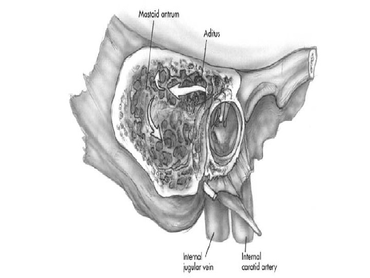
 Complications of Otitis Media The temporal bone is a complex anatomic region with close proximity to a variety of critical structures. These structures are at risk during both acute and chronic suppurative otitis media.
 Complications of Otitis Media • Due to antibiotics, the incidence of complications has greatly declined. (also treating surgical problems with antibiotics alone or giving incomplete courses that mask the infection lead to complications) • Complications are usually associated with granulation tissue formation and/or the presence of a cholesteatoma (bone erosion).
 Complications of Otitis Media Complications arise mostly due to: -- Infection spreading by direct extension from the middle ear or mastoid cavity to adjacent structures. - Thrombophlebitis (haematogenous)
 Complications of Otitis Media • Patients appear more ill than expected – fever, new onset vertigo, sensorineural hearing loss, fetid drainage, facial nerve weakness, proptotic ear – lethargy and mental status changes • CT and MRI are indicated – CT is superior for evaluating the bony details of the middle ear and mastoid space – MRI is more sensitive for diagnosing suspected intracranial complications.
 Complications of Otitis Media Treatment is: Parentral Broad Spectrum Antibiotics and Surgery are required
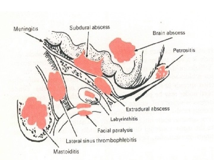
 complications: 1 - Acute Mastoiditis.") Complications of Suppurative O. M. Cranial (or Temporal bone) complications: 1 - Acute Mastoiditis. 2 - Acute Petrositis. 3 - Otitic Facial paralysis. 4 - Acute Labyrinthitis.
Complications of Suppurative O. M. Cranial (or Temporal bone) complications: 1 - Acute Mastoiditis. 2 - Acute Petrositis. 3 - Otitic Facial paralysis. 4 - Acute Labyrinthitis.
 Intracranial Complications 1 - Extra-dural (epidural) abscess.") Complications of Suppurative O M (cont. ) Intracranial Complications 1 - Extra-dural (epidural) abscess. 2 - Meningitis. 3 - Brain abscess (cerebral or cerebellar). 4 - Lateral sinus thrombosis. Extracranial complications 1 - External otitis. 2 - Jugular vein thrombophlebitis 3 - Bezold’s abscess 4 -Retropharyngeal abscess.
Complications of Suppurative O M (cont. ) Intracranial Complications 1 - Extra-dural (epidural) abscess. 2 - Meningitis. 3 - Brain abscess (cerebral or cerebellar). 4 - Lateral sinus thrombosis. Extracranial complications 1 - External otitis. 2 - Jugular vein thrombophlebitis 3 - Bezold’s abscess 4 -Retropharyngeal abscess.
 Acute Mastoiditis
 Acute Mastoiditis Extension of the suppurative inflammatory process beyond the mucous membrane lining of the mastoid air cells leading to osteitis of the bony septa. N. B. At this early stage resolution is possible without surgery, if proper medical treatment is given.
 The bony inter-cellular septa will break down with coalescence of") Acute Mastoiditis (cont. ) The bony inter-cellular septa will break down with coalescence of the infected cells to form one cavity full of pus leading to Coalescent Mastoiditis or Mastoid Abscess.
Acute Mastoiditis (cont. ) The bony inter-cellular septa will break down with coalescence of the infected cells to form one cavity full of pus leading to Coalescent Mastoiditis or Mastoid Abscess.
 In early Coalescent Mastoiditis the outer cortex of mastoid is") Acute Mastoiditis (cont. ) In early Coalescent Mastoiditis the outer cortex of mastoid is intact but with extension of the disease pus may erode outer cortex of mastoid leading to Subperiosteal Mastoid Abscess which can extend by perforating the periosteium to became Subcutaneous Mastoid Abscess. If it opens through the skin Mastoid Fistula will result.
Acute Mastoiditis (cont. ) In early Coalescent Mastoiditis the outer cortex of mastoid is intact but with extension of the disease pus may erode outer cortex of mastoid leading to Subperiosteal Mastoid Abscess which can extend by perforating the periosteium to became Subcutaneous Mastoid Abscess. If it opens through the skin Mastoid Fistula will result.
 1 - Tenderness over") Clinical Picture Exaggerated symptoms of ASOM (fever, pain and HL) 1 - Tenderness over mastoid antrum and 2 -External swelling A- Post-auricular abscess - Auricle is displaced outwards, forwards and downwards (erect auricle). - Post-auricular groove is preserved but if the abscess ruptures through periosteum and becomes subcutaneous , the groove will be obliterated. - DD. Post auricular lymphadenitis 2 ry to Furunculosis of external auditory meatus.
Clinical Picture Exaggerated symptoms of ASOM (fever, pain and HL) 1 - Tenderness over mastoid antrum and 2 -External swelling A- Post-auricular abscess - Auricle is displaced outwards, forwards and downwards (erect auricle). - Post-auricular groove is preserved but if the abscess ruptures through periosteum and becomes subcutaneous , the groove will be obliterated. - DD. Post auricular lymphadenitis 2 ry to Furunculosis of external auditory meatus.
 Clinical Picture Early stage of Mastoiditis Mastoid fistula
 Mastoid Abscess
 Clinical Picture B- Zygomatic abscess ; It is due inflammation of the zygomatic air cells. The swelling is above and in front of the ear. C- Bezolds abscess; Pus pierces the tip or inner surface of mastoid and form abscess in the sternomastoid muscle In the neck. D- Retropharyngeal abscess; Pus tracking from the peritubal cells along the Eustachian tube.
 Clinical Picture 3 - Internal swelling Sagging of posterosuperior bony meatal wall, due to periostitis and edema over the anterior antral wall. 4 - Ear discharge usually profuse , "Mucopurulent or purulent and may be pulsating with reservoir sign “ rapid re-accumulation " 5 - Drum membrane perforated (small with pulsating discharge) or intact and bulging.
 Investigations 1 - C&S of ear discharge 2 - CT scan of the temporal bone to detect any additional cranial or intracranial complications
 Treatment of Acute Mastoiditis 1 - Conservative treatment is to be tried for 48 hours in mild cases without evidences of abscess formation; parentral broad spectrum antibiotics. Myrigotomy if DM found intact and bulging. 2 - Cortical Mastoidectomy operation is the standard treatment if the patient is not responding to conservative treatment, or if a mastoid abscess is evident or if other complications are suspected to be present.
 Masked Mastoiditis It Is the result of INCOMPLETE TREATMENT of ASOM with antibiotics leading to masking of the acute symptoms while the pathological process is progressing in the mastoid. Clinical picture: - Slight pain and tenderness over the mastoid. - Intra-cranial complications may occur and may be the presenting symptom.
 Chronic Mastoiditis • There is thick unhealthy chronically inflamed mucosa with granulation tissue and osteitis with sclerosis of mastoid air cells. (sclerosed mastoid in X-Ray) • It is condition which may be present in CSOM (tubo-tympanic type and attico-antral types). • Persistent ear discharge is the main presenting symptom
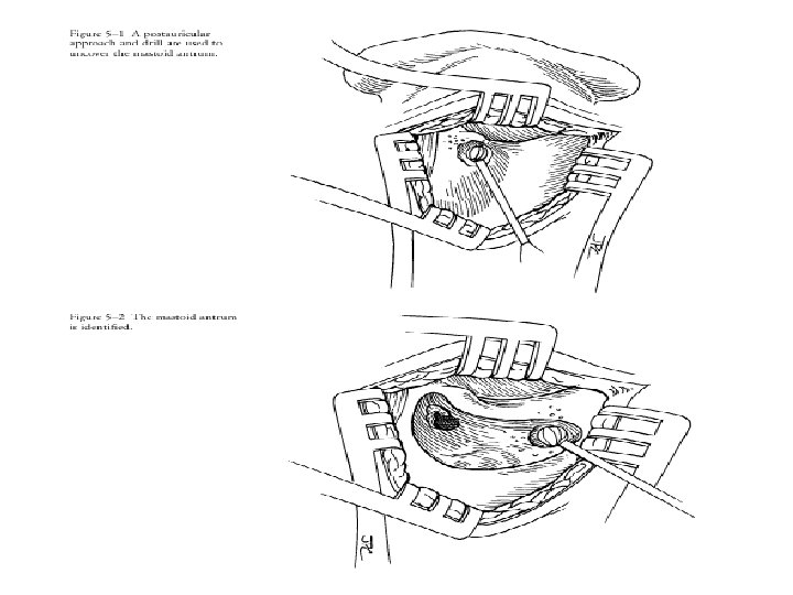
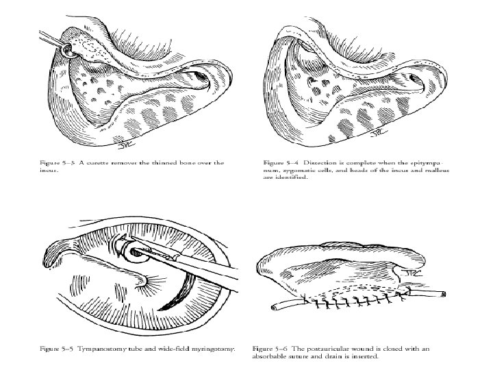
 Cortical Mastoidectomy Operation • It is a drainage operation in which exentration of the mastoid air cells is done. • It is a preliminary step in most of ear surgeries
 INDICATIONS 1 - Acute Mastoiditis with failure of medical treatment (persistent pain, tenderness and fever , etc , … for more than 2 days). 2 - Subperiosteal Mastoid abscess. 3 - Mastoid fistula. 4 - Mastoiditis with complications as facial paralysis, meningitis or lateral sinus thrombosis.
 for") INDICATIONS 5 - Persistent ear discharge in cases of ASOM or CSOM (tubo-tympanic) for more than one month despite proper conservative treatment 6 -Resistant cases of OME. 7 - Part of ear surgeries (e. g. Sac operations in Meniere‘s disease ------- etc. ).
INDICATIONS 5 - Persistent ear discharge in cases of ASOM or CSOM (tubo-tympanic) for more than one month despite proper conservative treatment 6 -Resistant cases of OME. 7 - Part of ear surgeries (e. g. Sac operations in Meniere‘s disease ------- etc. ).
 Petrositis It is inflammation of the air cells in the petrous apex of the temporal bone , the 6 th (abducent) and 5 th (trigeminal) cranial nerves are affected as they are closely related to the petrous apex.
 Clinical Picture The condition is called “GRADINIGO SYNDROME” Triade of :") Petrositis (cont. ) Clinical Picture The condition is called “GRADINIGO SYNDROME” Triade of : 1 - Diplopia with convergent squint due to 6 th nerve paralysis. 2 - Trigeminal neuralgia (retro-orbital pain and headache) due to irritation of the trigeminal ganglion. 3 - Discharging ear.
Petrositis (cont. ) Clinical Picture The condition is called “GRADINIGO SYNDROME” Triade of : 1 - Diplopia with convergent squint due to 6 th nerve paralysis. 2 - Trigeminal neuralgia (retro-orbital pain and headache) due to irritation of the trigeminal ganglion. 3 - Discharging ear.
 Investigations: 1 - CT scan of temporal bone 2 - C&S") Petrositis (cont. ) Investigations: 1 - CT scan of temporal bone 2 - C&S of ear discharge Treatment : 1 - Conservative in mild and early cases 2 - Mastoidectomy with exentration of petrous apex air cells or subtotal petrosectomy
Petrositis (cont. ) Investigations: 1 - CT scan of temporal bone 2 - C&S of ear discharge Treatment : 1 - Conservative in mild and early cases 2 - Mastoidectomy with exentration of petrous apex air cells or subtotal petrosectomy
 Otitic Labyrinthitis It is a complication of ASOM or more common CSOM. Types: 1. Circumscribed Labyrinthitis. (labyrinthine fistula). 2. Diffuse serous Labyrinthitis. 3. Diffuse suppurative Labyrinthitis.
 Circumscribed Labyrinthitis “Labyrinthine Fistula/ Para-labyrinthitis” It results from erosion of the bony wall of one of the SSC (usually the lateral) , or less commonly the promontory by cholesteatoma. The inflammatory process is outside the endosteal lining of the labyrinth (intact inner ear function).
 Labyrinthine Fistula Clinical Picture In addition to the clinical picture of OM new symptoms appear in the form of • Intermittent attacks of vertigo • Usually not accompanied by nausea and vomiting and usually precipitated by pressure on the tragus or sudden head movement.
 Labyrinthine Fistula Clinical Picture Nystagmus accompanies the vertigo and usually horizontal with rapid component to the affected side (irritant lesion).
 Labyrinthine Fistula Clinical Picture Fistula test is positive (pressure on tragus, use of pneumatic otoscope or manipulating an aural polyp induces vertigo and nystagmus).
 Diffuse serous Labyrinthitis “Catarrhal Labyrinthitis” It is a serous inflammation of the membranous labyrinth (inflamatory cells in the peri-lymph without organisms). Clinical Picture: 1. That of ASOM or CSOM. 2. Vertigo, nausea & vomiting are severe. 3. Nystagmus is usually horizontal with rapid component to affected side (irritant lesion). 4. Deafness becomes severe and mixed (Conductive & SNHL).
 Diffuse purulent Labyrinthitis • At first the previous symptoms increase markedly and HL may be severe or total. • Nystagmus is beating first towards the affected side (irritant) but changes to the other side (dead labyrinth) when destruction of the labyrinth becomes complete. Nystagmus will disappear later as it will be compensated by the healthy side.
 Diffuse Purulent Labyrinthitis Absent or minimal toxic manifestations as the surface area of the inner ear is small so there is no or little diffusion of toxins. Presence of fever and other toxic manifestations may suggest occurrence of meningitis.
 Treatment of Labyrinthitis Conservative Treatment - Antibiotics that cross the BBB to guard against meningitis. - Labyrinthine sedatives and anti-emetics : as Dramamine , stugeron, diazepam “valium” and zofran (4 mg) amp. . Surgical Treatment either; - Cortical mastoidectomy for control of suppurative otitis media, or - Radical mastoidectomy and labyrinthectomy in cases of supprative labyrinthitis with dead labyrinth to prevent intracranial extension of infection
 Otitic Facial Nerve Paralysis As a complication of ASOM facial nerve paralysis occurs in children if there is congenital dehiscence in the bony canal of the nerve (20% of population). Paralysis is usually incomplete and is due to inflammation of the nerve sheath and compression by pus. Treatment: 1. Early myringotomy (usually with Grommet’s tube) 2. Antibiotics (parentral) and steroids. 3. Cortical mastoidectomy if the paralysis persist in spite of other lines of treatment or if there is acute mastoiditis.
 Facial Nerve Paralysis as a complication of CSOM Destruction of the bony canal and pressure on the nerve is either by: 1) Cholesteatoma 2) Osteomylitis of the mastoid. 3) Tuberculous OM. (Multiple Drum M. perforations & pale mucosa).
 Facial Nerve Paralysis as a complication of CSOM Treatment 1 - Mastoidectomy operation with exposure and decompression of the facial nerve. 2 - In case of tuberculous OM Antituberculous ttt usually gives cure of the paralysis. Surgical ttt is only for cases showing no recovery after the disease has been cured.
 1. Immediate after the operation is due to direct") Post operative Facial Paralysis (Iatrogenic) 1. Immediate after the operation is due to direct trauma to the nerve. Treatment : • If Partial: corticosteroids & antibiotics. • If Complete: Immediate exploration of the nerve and remove any bone specule compressing the nerve or do nerve suturing or nerve graft if needed (from Greater Auricular nerve).
Post operative Facial Paralysis (Iatrogenic) 1. Immediate after the operation is due to direct trauma to the nerve. Treatment : • If Partial: corticosteroids & antibiotics. • If Complete: Immediate exploration of the nerve and remove any bone specule compressing the nerve or do nerve suturing or nerve graft if needed (from Greater Auricular nerve).
 usually due to") Post-operative facial paralysis 2. Delayed (few hours or days after recovery) usually due to pressure on the nerve by edema , haematoma or tight pack. Treatment: 1) Removal of the pack. 2) Antibiotics & Cortisone.
Post-operative facial paralysis 2. Delayed (few hours or days after recovery) usually due to pressure on the nerve by edema , haematoma or tight pack. Treatment: 1) Removal of the pack. 2) Antibiotics & Cortisone.
 Extradural Abscess It is collection of pus and /or granulation tissue between skull bone and dura.
 Extradural Abscess Clinical Picture The condition is usually symptomless and accidentally discovered during mastoidectomy. Presentations : There may be persistent 1 - Earache or headache. 2 - Low grade Fever (about 37. 5 - 38°C). 3 - Pulsating ear discharge.
 that cross BBB. 2 - Cortical Mastoidectomy") Extradural Abscess Treatment 1 - Antibiotics (Injection) that cross BBB. 2 - Cortical Mastoidectomy operation , abscess must be evacuated and bone must be removed until healthy dura is reached.
Extradural Abscess Treatment 1 - Antibiotics (Injection) that cross BBB. 2 - Cortical Mastoidectomy operation , abscess must be evacuated and bone must be removed until healthy dura is reached.
 Diffuse Leptomeningitis It is diffuse inflammation of the arachnoid, subarachnoid space & pia mater. Symptoms 1) Symptoms of infection e. g. high fever, malaise……. etc. 2) Symptoms of increased intracranial tension: - Severe headache. - Vomiting. - Blurring of vision. 3) Symptoms of meningeal irritation Irritability , Photophobia , neck rigidity and retraction.
 High fever (> 39 C) and tachycardia. 2) Neck Rigidity.") Diffuse Leptomeningitis Signs 1) High fever (> 39 C) and tachycardia. 2) Neck Rigidity. 3) Signs of meningeal irritation: a- Kernig’s sign Flex hip and knee , then trying to extend the knee will produce severe pain and will be resisted by the patients. b- Brudziniski’s sign Flex the neck , hip and knee will become flexed. 4 - Papilloedema (edema of optic disc) on fundus examination.
Diffuse Leptomeningitis Signs 1) High fever (> 39 C) and tachycardia. 2) Neck Rigidity. 3) Signs of meningeal irritation: a- Kernig’s sign Flex hip and knee , then trying to extend the knee will produce severe pain and will be resisted by the patients. b- Brudziniski’s sign Flex the neck , hip and knee will become flexed. 4 - Papilloedema (edema of optic disc) on fundus examination.
 Investigations of Meningitis A- CT Temporal Bone & Brain (To detect probable intracranial complication if any). B- Lumbar Puncture: 1 - CSF examination. 2 - Culture & Sensitivity.
 Aspect Pressure Cells Proteins Sugar Chloride Organisms C. S. F. In meningitis normal CSF Turbid. Clear. High. 60 -180 mm Of CSF. Thousands, mainly 1 -5 lymphocytes polymorphs. per c mm. Increased (due to 40 mg/100 ml. the bacteria). Decreased 80 mg/100 ml. ( nutrition of bacteria). Decreased 750 mg/l 00 ml. (due to vomiting). Can be cultured. Absent.
 Treatment of Meningitis 1 - Antibiotics: i- Intrathecal injection of crystalline penicillin ii- Intravenous injection of drugs crossing BBB as, 3 rd generation cephalosporins & Flagyl for anerobes 2 - Measures to reduce the increased intracranial tension: i- Repeated lumbar punctures. ii- Hypertonic glucose solution IV & Diuretics. iii- Dexamethason injections.
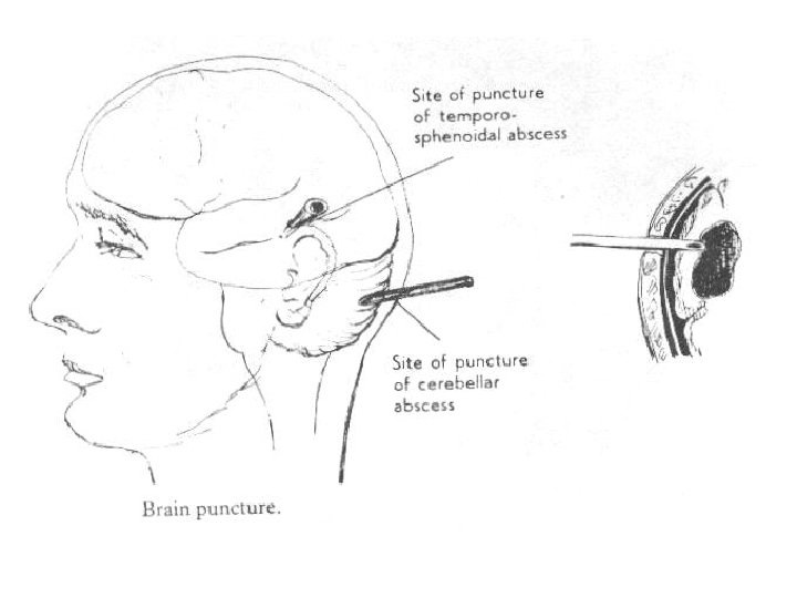
 Brain Abscess It is either Temporal or Cerebellar
 Brain Abscess Clinical Picture I- Stage of encephalitis: 1 - High fever & rapid pulse. 2 - Rigors or convulsions specially in children. 3 - Headache.
 II- Latent Stage: (weeks to months) Due to") Brain Abscess Clinical Picture (cont. ) II- Latent Stage: (weeks to months) Due to localization of the abscess with diminished brain. Most of symptoms disappear and patient may feel some headache and lack of concentration.
Brain Abscess Clinical Picture (cont. ) II- Latent Stage: (weeks to months) Due to localization of the abscess with diminished brain. Most of symptoms disappear and patient may feel some headache and lack of concentration.
 Brain Abscess Clinical Picture III- Manifest Stage: Due to increase in the size of the abscess. A- Manifestations of Toxaemia: i. Anorexia and loss of weight. ii. Mental dullness , slow cerebration and delirium. iii. Leucocytosis which may reach 20. 000 or more.
 Brain Abscess Clinical Picture B- Manifestations of Increased Intracranial Tension: 1 - Headache which is severe and not relieved by analgesics. 2 - Projectile vomiting (not preceded by nausea and not related to meals). 3 - Blurring of vision due to papilloedema.
 Brain Abscess Clinical Picture Prolonged increased ICT may lead to - Slow full pulse(40/min. ) - Subnormal temperature - Slow cerebration - Slow deep respiration
 Brain Abscess C- Manifestations of Localization: Temporal Lobe Abscess - Nominal Aphasia (inability to name objects due to - pressure on Broca’s area) Homonymous hemi-anopia( defect in field of vision) Uncinate fits (epileptic fits preceeded by aura) Hemiplegia Hemianesthesia
. 2. Slurred") Brain Abscess Clinical Picture Cerebellar Abscess: 1. Tremors with muscle weekness (hypotonia). 2. Slurred speech 3. Incoordination of movements (asynergia and dysmetria) can be shown by finger nose test. 4. Ataxia: unsteadiness of gait with deviation to the side of lesion. 5. Vertigo and nystagmus. 6. Dysdiadokokinesis: ( patient is unable to do rapid pronation and supination ).
Brain Abscess Clinical Picture Cerebellar Abscess: 1. Tremors with muscle weekness (hypotonia). 2. Slurred speech 3. Incoordination of movements (asynergia and dysmetria) can be shown by finger nose test. 4. Ataxia: unsteadiness of gait with deviation to the side of lesion. 5. Vertigo and nystagmus. 6. Dysdiadokokinesis: ( patient is unable to do rapid pronation and supination ).
 Brain Abscess Clinical Picture IV- Terminal Stage: Due to rupture of the abscess resulting in either: 1) Diffuse encephalitis. or 2) Diffuse meningitis. Coma and death will occur.
 Brain Abscess Investigations 1. CT scan with contrast &/ or MRI show site , size of abscess and whether acute or chronic 2. Fundus examination show Papilloedema. 3. Field of vision examination show homonymmous hemianopia. 4. CBC show marked leucocytosis (20000). 5. C/S from pus from abscess after drainage or from ear discharge. N. B. Never do Lumber Puncture as CONIZATION of medulla may occur due to marked rise of I. C. T.
 Brain Abscess
 Brain Abscess Treatment Acute Abscess 1. Antibiotics that cross BBB. 2. Measures to Lower the increased ICT. 3. Repeated Tapping of abscess through burr holes by neurosurgery or through mastoidectomy (N. B. Repeated CT must be done to ensure complete drainage). 4. Mastoidectomy of the affected ear as a treatment for otitis media when the condition of the patient allows.
. 3. Mastoidoidectomy") Brain Abscess Treatment B. Chronic Abscess 1. Excision 2. Antibiotics (Parentral-crossing BBB). 3. Mastoidoidectomy for affected ear when the condition of the patient allows.
Brain Abscess Treatment B. Chronic Abscess 1. Excision 2. Antibiotics (Parentral-crossing BBB). 3. Mastoidoidectomy for affected ear when the condition of the patient allows.
 venous sinus.") Lateral Sinus Thrombophlebitis It is infective thrombosis of the lateral (sigmoid) venous sinus.
Lateral Sinus Thrombophlebitis It is infective thrombosis of the lateral (sigmoid) venous sinus.
 is formed as") Lateral Sinus Thrombophlebitis Pathogenesis • Peri-sinus abscess (type of extra-dural abscess) is formed as an extension from infected mastoid • Infection extends into the sinus wall and lumen causing thrombophlebitis. • Infected thrombus may be fragmented with detachment of septic emboli in blood stream • Extension of thrombosis to cavernous , supermay take placeior petrosal, superior sagittal sinus or to the internal jugular vein may occur
Lateral Sinus Thrombophlebitis Pathogenesis • Peri-sinus abscess (type of extra-dural abscess) is formed as an extension from infected mastoid • Infection extends into the sinus wall and lumen causing thrombophlebitis. • Infected thrombus may be fragmented with detachment of septic emboli in blood stream • Extension of thrombosis to cavernous , supermay take placeior petrosal, superior sagittal sinus or to the internal jugular vein may occur
 - Remittent fever") Lateral Sinus Thrombosis Clinical Picture 1 - Pyaemic Type (Malarial like) - Remittent fever and rigors occurring at irregular intervals, between them temp. reach near the base line ( remains above 37°C). - Multiple pyaemic abscesses in different parts of the body due to separation of septic emboli.
Lateral Sinus Thrombosis Clinical Picture 1 - Pyaemic Type (Malarial like) - Remittent fever and rigors occurring at irregular intervals, between them temp. reach near the base line ( remains above 37°C). - Multiple pyaemic abscesses in different parts of the body due to separation of septic emboli.
 Lateral Sinus Thrombosis Clinical Picture D. D. from Malaria a- Fever and rigors in malaria occurs at regular intervals and between them temp. can reach 37°C. b- Leucopenia in malaria instead of leucocytosis in thrombosis. c- Blood film will show malaria parasites. (during the attack)
 2 - Septicaemic or Typhoid Type Continuous") Lateral Sinus Thrombosis Clinical Picture (cont. ) 2 - Septicaemic or Typhoid Type Continuous fever without remissions or rigors. D. D. from Typhoid fever: a- Widal test Is positive in typhoid. b- Leucopenia in typhoid. 3 - Latent Type Condition may be asymptomatic and discovered only during Mastoid Operation for acute mastoiditis.
Lateral Sinus Thrombosis Clinical Picture (cont. ) 2 - Septicaemic or Typhoid Type Continuous fever without remissions or rigors. D. D. from Typhoid fever: a- Widal test Is positive in typhoid. b- Leucopenia in typhoid. 3 - Latent Type Condition may be asymptomatic and discovered only during Mastoid Operation for acute mastoiditis.
 Lateral Sinus Thrombosis Clinical Picture 4 - If Septic thrombosis extend to the Jugular vein in the neck. a- Cord like mass in the neck. b- Torticollis. c- Cervical lymphadenitis may occur.
 Antibiotics (according to blood culture? ? ? ) 2)") Lateral Sinus Thrombosis Treatment 1) Antibiotics (according to blood culture? ? ? ) 2) Antipyretic analgesics, light diet, fluids. 3) Anticoagulants as heparin may be given in cases with extension of the thrombus ? ? . 4) Mastoidectomy operation and exposure of the sinus with removal of bone until healthy dura is reached. Incision of the sinus and evacuation of the infected clot is done until unclotted blood is reached.
Lateral Sinus Thrombosis Treatment 1) Antibiotics (according to blood culture? ? ? ) 2) Antipyretic analgesics, light diet, fluids. 3) Anticoagulants as heparin may be given in cases with extension of the thrombus ? ? . 4) Mastoidectomy operation and exposure of the sinus with removal of bone until healthy dura is reached. Incision of the sinus and evacuation of the infected clot is done until unclotted blood is reached.
 Lateral Sinus Thrombosis Treatment Ligation of the internal Jugular vein can be done if we cannot reach the lower limit of the thrombus and it must be ligated below the level of common facial vein which must be ligated also to avoid cross thrombosis to the cavernous sinus.
 Lateral Sinus Thrombosis Treatment N. B. During operation we must differentiate between thrombosed sinus and healthy one by the following: Thrombosed sinus is: 1)Grayish and dull instead of bluish and glistening. 2)Firm and pulsating instead of soft and not pulsating.