

Renal Cystic Disease.pptx
- Количество слайдов: 34
 Cystic Kidney Disease Philadelphia University Physician Assistant Program
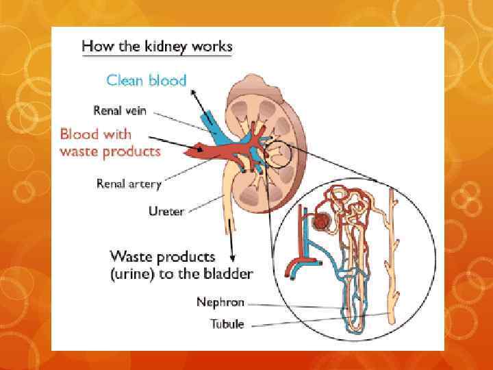
 Cysts How would you define them? Where do you find them?
 Cysts Simple Renal Cysts Complex Renal Cysts Polycystic Kidney Disease
 Simple Renal Cysts Observed frequently in normal kidneys. Up to 50% of persons over 50 may have asymptomatic renal cysts (CMDT p. 934) Most common renal masses, accounting for roughly 65 to 70 percent of cases. These cysts most often occur in patients over 50 years of age.
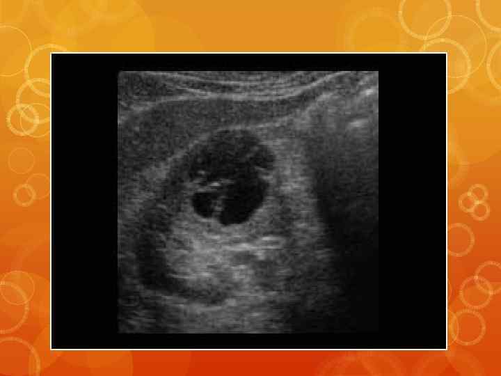
 Simple Renal Cysts
 Simple Renal Cysts May be solitary, or multiple and bilateral. They typically produce no symptoms or signs. Little clinical significance. Rarely, however, they can be associated with rupture (hemorrhage), hematuria, pain, abdominal mass, infection, and/or hypertension.
 Simple Renal Cysts Usually diagnosed via ultrasound or CT scan. Often an incidental finding. CT scan should be performed if: Ultrasound is equivocal. calcifications or septae are seen. multiple cysts are clustered in a pattern that could mask an underlying carcinoma.
 Simple Renal Cysts The major concern with simple renal cysts is differentiating them from more serious disorders: polycystic kidney disease complex cysts solid masses (i. e. renal carcinoma or abscess)
 Simple Renal Cysts Treatment: Generally, no treatment required.
 —Schematic diagrams of shapes of cystic lesions. “Smooth shape with septation” was defined as a septate simple closed curve with borders of the same circle and internal septation. Kim S Y et al. AJR 2006; 187: 1192 -1198 © 2006 by American Roentgen Ray Society


 Complex Renal Cysts may harbor neoplastic masses A cyst’s complexity is rated based on structure and density.
 Complex Renal Cysts If an ultrasound finds that a cyst may be complex, the lesion is often evaluated by CT scan. The cyst’s complexity is rated according to the Bosniak renal cyst classification system.
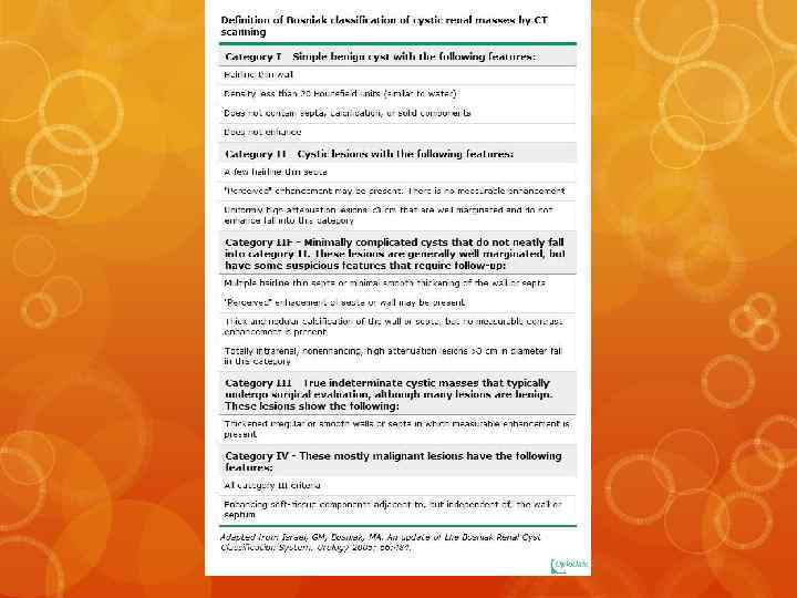
 Complex Renal Cysts Cyst complexity dictates whether continued radiographic observation and/or surgery is required.
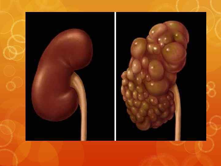
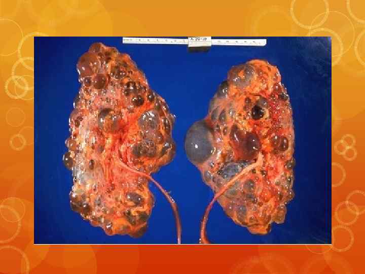
 Common") Polycystic Kidney Disease We will focus on Autosomal dominant polycystic kidney disease (ADPKD) Common disorder approximately 1 in every 800 -1000 live births
Polycystic Kidney Disease We will focus on Autosomal dominant polycystic kidney disease (ADPKD) Common disorder approximately 1 in every 800 -1000 live births
 Less than one-half of those affected will be") Autosomal Dominant Polycystic Kidney Disease (ADPKD) Less than one-half of those affected will be diagnosed during their lifetime the disease is often clinically silent
Autosomal Dominant Polycystic Kidney Disease (ADPKD) Less than one-half of those affected will be diagnosed during their lifetime the disease is often clinically silent
 Presentation Abdominal Mass Hypertension (50% at time of") Autosomal Dominant Polycystic Kidney Disease (ADPKD) Presentation Abdominal Mass Hypertension (50% at time of presentation) Gross Hematuria H/O UTIs H/O nephrolithiasis (20%) Additional Associations 40 -50% have hepatic cysts
Autosomal Dominant Polycystic Kidney Disease (ADPKD) Presentation Abdominal Mass Hypertension (50% at time of presentation) Gross Hematuria H/O UTIs H/O nephrolithiasis (20%) Additional Associations 40 -50% have hepatic cysts
 If genetic test existed, would you take it?") Autosomal Dominant Polycystic Kidney Disease (ADPKD) If genetic test existed, would you take it? What factors must you consider?
Autosomal Dominant Polycystic Kidney Disease (ADPKD) If genetic test existed, would you take it? What factors must you consider?
 Genetic Testing 85 percent with ADPKD have an") Autosomal Dominant Polycystic Kidney Disease (ADPKD) Genetic Testing 85 percent with ADPKD have an abnormality on chromosome 16 (PKD 1 locus) The remaining patients have a different defect that involves a gene on chromosome 4 (the PKD 2 locus) Patients with PKD 2 have a less severe phenotype than those with PKD 1, but neither disorder is benign
Autosomal Dominant Polycystic Kidney Disease (ADPKD) Genetic Testing 85 percent with ADPKD have an abnormality on chromosome 16 (PKD 1 locus) The remaining patients have a different defect that involves a gene on chromosome 4 (the PKD 2 locus) Patients with PKD 2 have a less severe phenotype than those with PKD 1, but neither disorder is benign
 If one has a family history of ADPKD,") Autosomal Dominant Polycystic Kidney Disease (ADPKD) If one has a family history of ADPKD, if they are asymptomatic, genetic screening is not routinely recommended.
Autosomal Dominant Polycystic Kidney Disease (ADPKD) If one has a family history of ADPKD, if they are asymptomatic, genetic screening is not routinely recommended.
 Diagnosis is usually made via imaging Ultrasound CT") Autosomal Dominant Polycystic Kidney Disease (ADPKD) Diagnosis is usually made via imaging Ultrasound CT MRI Complex criteria exist for diagnosis based on radiographic findings, that take age and family history into consderation.
Autosomal Dominant Polycystic Kidney Disease (ADPKD) Diagnosis is usually made via imaging Ultrasound CT MRI Complex criteria exist for diagnosis based on radiographic findings, that take age and family history into consderation.
 Common complications Pain Hematuria with cyst rupture Infection") Autosomal Dominant Polycystic Kidney Disease (ADPKD) Common complications Pain Hematuria with cyst rupture Infection Nephrolithiasis (20%) HTN – 50% at presentation; most end up with HTN Vascular Problems: Aneurysms: aortic, cerebral MVP (25%) and aortic valvulopathy Colonic diverticula
Autosomal Dominant Polycystic Kidney Disease (ADPKD) Common complications Pain Hematuria with cyst rupture Infection Nephrolithiasis (20%) HTN – 50% at presentation; most end up with HTN Vascular Problems: Aneurysms: aortic, cerebral MVP (25%) and aortic valvulopathy Colonic diverticula
 Treatment Mainstay is treatment of complications ACE-I and") Autosomal Dominant Polycystic Kidney Disease (ADPKD) Treatment Mainstay is treatment of complications ACE-I and ARB therapy may help in cases with significant proteinuria Ongoing research regarding c. AMP role and use of vasopressin-2 receptor antagonists Caffeine limitation may prevent cyst formation? Avoidance of contact sports for some (to minimize risk of rupture).
Autosomal Dominant Polycystic Kidney Disease (ADPKD) Treatment Mainstay is treatment of complications ACE-I and ARB therapy may help in cases with significant proteinuria Ongoing research regarding c. AMP role and use of vasopressin-2 receptor antagonists Caffeine limitation may prevent cyst formation? Avoidance of contact sports for some (to minimize risk of rupture).
 Medullary Sponge Kidney
 Medullary Sponge Kidney Common and typically benign. Often diagnosed in 30’s or 40’s. AD mutations in MCKD 1 (chromosome 1) or MCKD 2 (chromosome 16).
 Medullary Sponge Kidney Common Presentation: - Hematuria - Recurrent UTIs - Nephrolithiasis - decreased urinary concentrating ability
 Medullary Sponge Kidney Imaging: - Calcinosis may be seen on radiographs - linear and rounded medullary calcifications - CT scan for diagnosis. - cystic dilatation of the collecting tubules in 1 or more renal pyramids
 Medullary Sponge Kidney Treatment Treat associated conditions. Nephrolithiasis prevention. Prognosis No significant problems typically develop in those that have associated conditions treated.