

e45b05e86ef1424ebb7c8f8a9b2f410d.ppt
- Количество слайдов: 33
 CT Urography and applications in uroephithelial tumors Orith Portnoy Dept. of Diagnostic Radiology Sheba Medical Center, Sakler School of Med. Tel-Aviv University, Israel
 v. Initial modality for upper tract imaging in hematuria, flank pain") IVP (intravenous pyelography) v. Initial modality for upper tract imaging in hematuria, flank pain & others for 7 decades. v. Less sensitive than CT for: v v Renal masses (21% for 2 cm mass) Urinary tract stones Renal inflammation Renal trauma
IVP (intravenous pyelography) v. Initial modality for upper tract imaging in hematuria, flank pain & others for 7 decades. v. Less sensitive than CT for: v v Renal masses (21% for 2 cm mass) Urinary tract stones Renal inflammation Renal trauma
 v. Single detector MDCT volumetric acquisition high resolution reconstructions v. Both") CT Urography (CTU) v. Single detector MDCT volumetric acquisition high resolution reconstructions v. Both renal parenchyma and urothelium shown in a single examination v. Shortening schedule for diagnostic evaluation (hematuria)
CT Urography (CTU) v. Single detector MDCT volumetric acquisition high resolution reconstructions v. Both renal parenchyma and urothelium shown in a single examination v. Shortening schedule for diagnostic evaluation (hematuria)
 CTU at Sheba v Since 6/2004 v ~ 500 studies v GE MDCT 16/64 slice, Philips MDCT BR 40/64 slice
 v") CTU Protocol v Monitored by a radiologist v Non contrast phase (low dose) v Nephrographic phase (100 s delay) saline IV v Excretory phase (400 -800 slices) tailored v Reconstructions on a 4. 1 or 4. 2 GE workstation
CTU Protocol v Monitored by a radiologist v Non contrast phase (low dose) v Nephrographic phase (100 s delay) saline IV v Excretory phase (400 -800 slices) tailored v Reconstructions on a 4. 1 or 4. 2 GE workstation
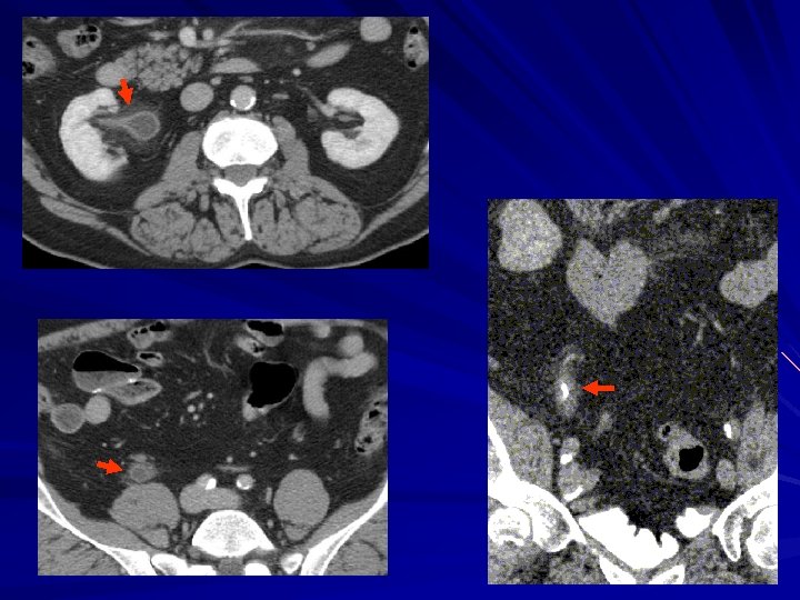
 Axial images – Normal CTU
 MPR MIP
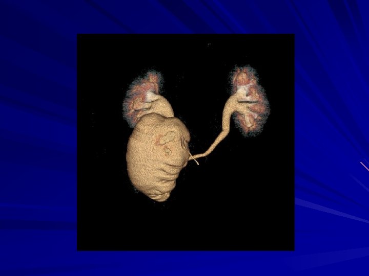
 3 D volume rendering
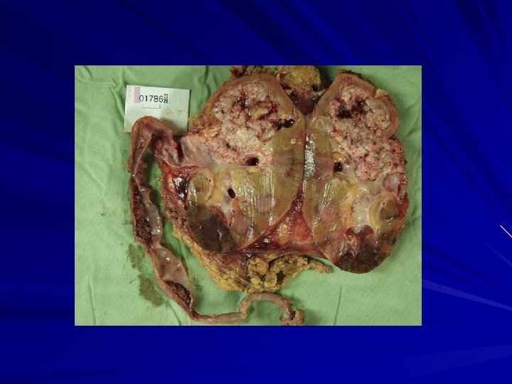
 CTU – Rec. bladder TCC 80 Y. O. man Macrohematuria S/P 17 operations for bladder TCC
 Staging - Lymphadenopathy
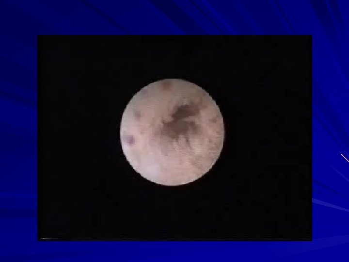
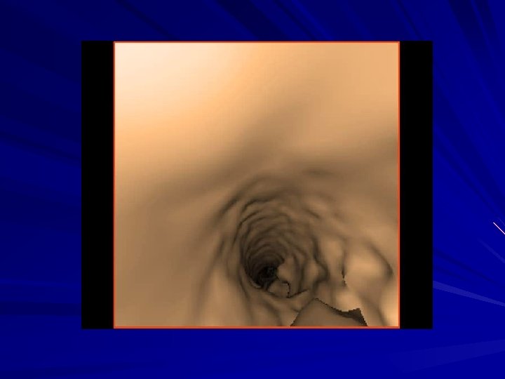
 CTU – virtual cystoscopy
 56 Y. O. man macrohematuria Rec. bladder TCC seen at cystoscopy Posterior view
 CTU and US 46 Y. O. women 1 event of macrohematuria
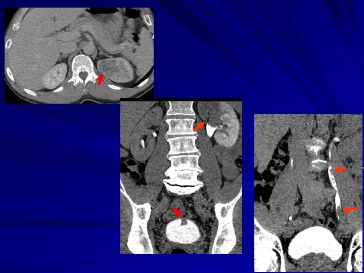
") CTU and IVP v 68 Y. O. man v Left flank pain US (stone) lithothripsy hematuria post 3 w IVP cystoscopy (susp. tumor)
CTU and IVP v 68 Y. O. man v Left flank pain US (stone) lithothripsy hematuria post 3 w IVP cystoscopy (susp. tumor)
 61 Y. O. man Recurrent macrohematuria 6 mo. before – US, IVP, cystoscopy
 CTU and PET CT Bladder TCC and CLL Retrograde pyelography – narrowed ureter
 Sensitivity v Detection of upper tract urothelial tumors by CTU – 91 -94% in relation to biopsy (Dillman Abd Imaging 2008) v Detection of bladder tumors: microhematuria – 40% vs. cystoscopy, macrohematuria high risk – 93% sens. , 99% spec. (Albani J Urol 2007, Turney BJU 2006) v High risk: >40 y, macrohematuria, smoking, GU tumor P/H, occupational exposure
 v Continent cutaneous") Types of Urinary Diversion after Cystectomy v Incontinent diversion (ileal, colonic) v Continent cutaneous catheterizable reservoir v Orthotopic neobladder
Types of Urinary Diversion after Cystectomy v Incontinent diversion (ileal, colonic) v Continent cutaneous catheterizable reservoir v Orthotopic neobladder
 Imaging after bladder reconstruction v Complications v Recurrence v. Understanding the reconstruction anatomy helps diagnose complications v US , IVP , cystography/lupography antegrade/retrograde pyelography, CT, nuclear medicine v CT-UROGRAPHY
 Bladder reconstructin FU
 v 68 Y. O. man v 6 years post bladder replacement d/t TCC v 6 months intermittent macrohematuria
 Posterior view
 CT 18 mo. before
 CTU - Disadvantages v Radiation dose Mean effective dose: 23 -35 m. Sv ØCTU 1. 5 more than standard IVP Ø Nawfel et al Radiology 2004 v v Time consuming processing, reviewing Lack large scale research on cost-effectiveness
 CTU - summary v Useful diagnostic examination that allows comprehensive evaluation of urinary tracts v. Problem solving tool with other modalities v Becoming the primary imaging study for the work -up of patients with hematuria and other genitourinary conditions v Shorter diagnostic evaluation, decrease need for ureteroscopies v Tailored examination can save radiation v Referrals should be limited (urologists)
 THANK YOU!
 CTU and “regular” CT 66 Y. O. man 1 year post partial nephrectomy for POST. RCC. VIEW New hydronephrosis on CT, suspect rec. obstructing tumor.