

c89f43e9df51e1affa441627b3ae205b.ppt
- Количество слайдов: 64
 Cranial Nerves Pundit Asavaritikrai, Ph. D, MD. Department of Anatomy, Faculty of Science Mahidol University neuronum@yahoo. com
 Overview • Brain Stem – Ascend. /Descend. P’w – Vital centres • Consciousness • Respiration • CVS – Cranial nerves
 Cranial Nerves & Cranial Nerve Reflexes • • • CN III, IV, & VI CN VII, CN VIII CN IX & X CN XII
 Memorize 2 -3 sections/division
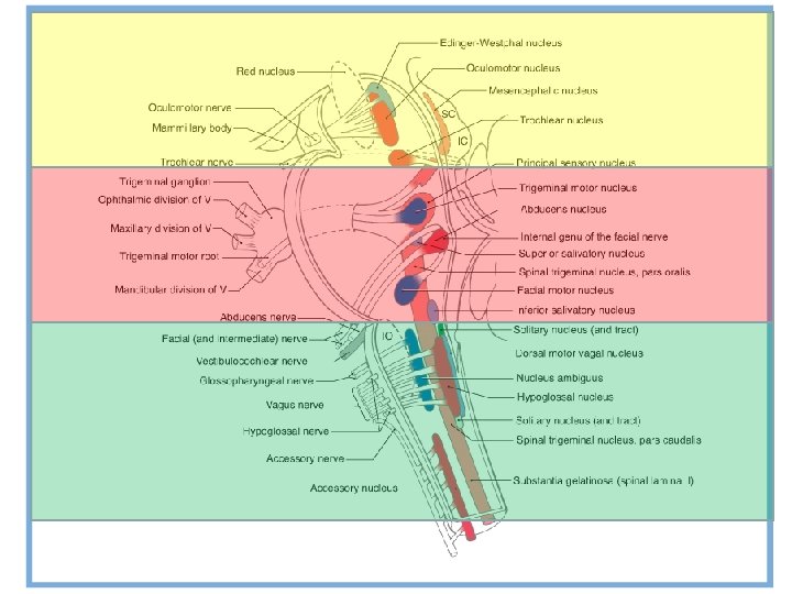
 Midbrain
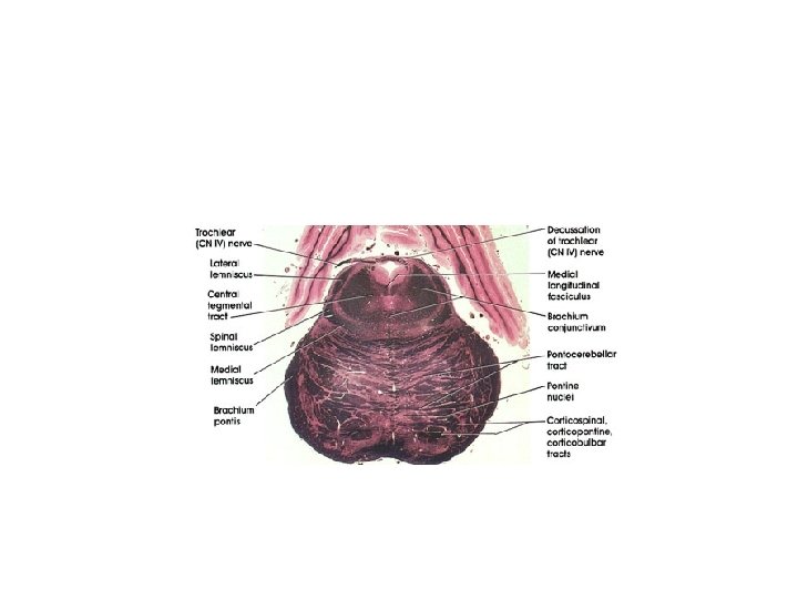
 Pons
Pons
 Open Medulla
 Closed Medulla
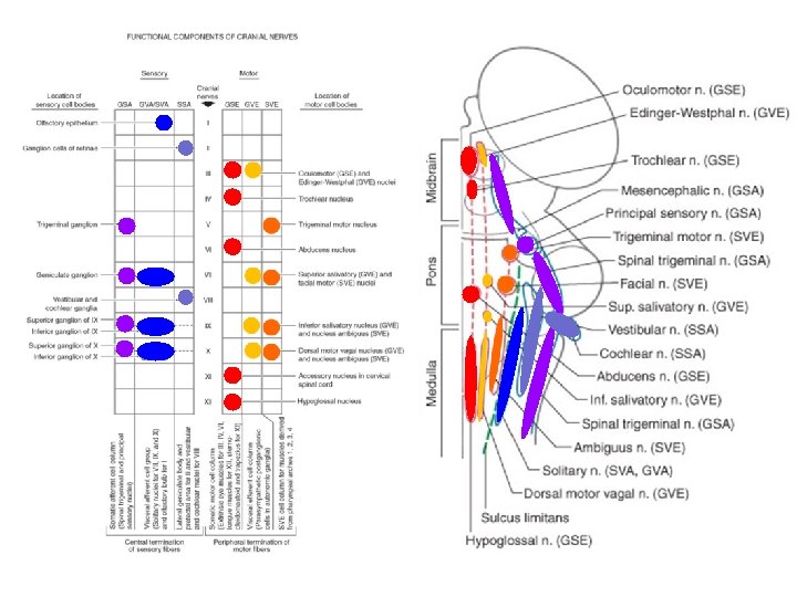
 CN I & II • CN I & II – brain extension – not real nerves – Special sensory afferents
 CN I Olfactory Nerve • Olfaction • Memory and Behavior • Pheromones • • Anterior olfactory nucleus Amydala Piriform cortex Enthorhinal cortex
 CN II Optic Nerve • Vision • Intraocular movement (+ III( • Blinking (+ V & VII( • Circadian rhythm
 The III, IV & VI
 CN III Oculomotor Nerve • Intraocular movement – Autonomic • Lens shape • Pupil size • Extrinsic Eye movement – Coordinate with CN IV & VI
 Control of Pupil Size • Parasympathetic • #1 = Edinger-Westphal nuc. • #2 = ciliary ganglion – pupillary constrictor – fibers travel in outer margin of CN III
 Pupillary Light Reflex • In: CN II – Pretectal area – Posterior Com. • Out: CN III-EW nuc.
 (CN II CN III)") Relative Afferent Pupillary Defect (RAPD) (CN II CN III)
Relative Afferent Pupillary Defect (RAPD) (CN II CN III)
 Adie’s Pupil • Abnormally dilated pupil • Can be tonic, sectional, vermiform iris • Abnormal postganglionic parasympathetic fibers
 Argyll-Robertson’s Pupil • Associated with Syphillis – Normal pupil accommodation – Does not constrict to light – Pretectal area damage • Prostitute’s pupil = Accommodate but does not react
 Sympathetic Control of Pupil • Sympathetic • #1 = T 1 lateral neurons • #2 = SCG – Pup. dilator, tarsus m, sweat gl. • Defects: Horner’s syndrome (เลก แหง ตก ไมงอก ) • Causes: – pulmonary apex – lateral medulla (+vestibular defects; vertigo) = Wallenberg syndrome
 • Sympathetic – Superior") Ptosis • Abnormal CN III – LPS – NMJ (Myasthenia) • Sympathetic – Superior tarsal m. • Does not involve CN VII (ปดไมสนท )
Ptosis • Abnormal CN III – LPS – NMJ (Myasthenia) • Sympathetic – Superior tarsal m. • Does not involve CN VII (ปดไมสนท )
 CN III, IV, & VI
") CN III, IV, VI • Function • Coordination • Control of coordination (conjugation)
CN III, IV, VI • Function • Coordination • Control of coordination (conjugation)
 • Internuclear connection • Nonvestibular pathways (among CN nuclei) –") MLF (medial longitudinal fasciculus) • Internuclear connection • Nonvestibular pathways (among CN nuclei) – VI-contralateral III – III-VII, VII-V, V-XII, XII-VII • Vestibular pathways: – – Eye Ear Neck Limb extensors p 389
MLF (medial longitudinal fasciculus) • Internuclear connection • Nonvestibular pathways (among CN nuclei) – VI-contralateral III – III-VII, VII-V, V-XII, XII-VII • Vestibular pathways: – – Eye Ear Neck Limb extensors p 389
 Disorders of the MLF • Internuclear Ophthalmoplegia
 CN III, IV, & VI: Coordination of Eye Movements
") Coordination of Eye Movements • Conjugate eye movement • Dysconjugate eye movement (vergence)
Coordination of Eye Movements • Conjugate eye movement • Dysconjugate eye movement (vergence)
 Dysconjugate Eye Movement • Vergence – ‘dysconjugate but still coordinate’ – involving vergence center in the midbrain, no MLF • Near triad (Accommodation) – Stimulus: Near object – Executor: cerebral cortex SC pretectal area • Ocular vergence (midbrain RF, both sides( • Lens rounding up (EW, both sides( • Pupil constriction (EW, both sides)
 CN III, IV, & VI: Supranuclear Control of Eye Movements
 Supranuclear Control Idea there must be some control above III, IV, VI (= supranuclear control) • 1. Gaze – Saccades (quick) – Smooth persuit (slow) – Foveation • 3. Vestibulo-ocular reflex • 4. Nystagmus
 Dysconjugated Eye Movement • No MLF • Near vision – Accommodation – Pupil constriction – Vergence
 Conjugate Eye Movements • Yoking mechanism • Via MLF E. g. CN VI contralat. CN III • Clinical use: e. g. Internuclear ophthalmoplegia
 1. Smooth Persuit • Conjugate movement that maintains foveation of a moving object • Can be Voluntary or Involuntary • Mechanisms – Stimuli = retinal slip – Processor = Area 19 & 39 (Angular gyrus) – Executor = Area 8 CN VI contralateral CN III ipsilateral
 • Rapid jerky involuntary conjugate movement • (Faster") 2. Reactive gaze (Saccadic eye movement) • Rapid jerky involuntary conjugate movement • (Faster than smooth persuit) • Stimuli = changing point of fixation, light, noise, noxious stimuli – Processor = Area 7 (parietal) – Executor = Area 8 & SC contralat. PPRF paramedian pontine reticular formation (pontine gaze centers) PPRF excites CN VI LR e. g. Lt. Frontal eye field excites contralateral CN VI • Clinical use – eye movements towards the side of lesion (ตามองฟองลชน ) p 394
2. Reactive gaze (Saccadic eye movement) • Rapid jerky involuntary conjugate movement • (Faster than smooth persuit) • Stimuli = changing point of fixation, light, noise, noxious stimuli – Processor = Area 7 (parietal) – Executor = Area 8 & SC contralat. PPRF paramedian pontine reticular formation (pontine gaze centers) PPRF excites CN VI LR e. g. Lt. Frontal eye field excites contralateral CN VI • Clinical use – eye movements towards the side of lesion (ตามองฟองลชน ) p 394
 • Conjugate movement that maintains eye position while head moves") 3. Vestibulo-Ocular Reflex (VOR) • Conjugate movement that maintains eye position while head moves • ~ involuntary/reflexive smooth persuit – Stimuli = warm water, head turning to that side – Processor & Executor = vestibular nuc. inhibit ipsilateral CN VI inhibit MLF contralateral CN III
3. Vestibulo-Ocular Reflex (VOR) • Conjugate movement that maintains eye position while head moves • ~ involuntary/reflexive smooth persuit – Stimuli = warm water, head turning to that side – Processor & Executor = vestibular nuc. inhibit ipsilateral CN VI inhibit MLF contralateral CN III
 • Ex. Stimulation of Rt. Vest. Nuc. inhibit Rt. CN") 3. Vestibulo-Ocular Reflex (VOR) • Ex. Stimulation of Rt. Vest. Nuc. inhibit Rt. CN VI & LR eyes deviate to left • Ex. Inhibition of Rt. Vest. Nuc by: – cold water in the Rt. – turning head to the Lt. – lesion of Rt. vestibular input Rt LR turns the eye to the Rt • Clinical use: – Doll’s eye reflex
3. Vestibulo-Ocular Reflex (VOR) • Ex. Stimulation of Rt. Vest. Nuc. inhibit Rt. CN VI & LR eyes deviate to left • Ex. Inhibition of Rt. Vest. Nuc by: – cold water in the Rt. – turning head to the Lt. – lesion of Rt. vestibular input Rt LR turns the eye to the Rt • Clinical use: – Doll’s eye reflex
 Vestibulo-ocular Reflex • Contralateral CN VI n. • From CN VI n – ipsi. CN III n
 Nystagmus • Vestibular • Optokinetic
, and – saccadic eye") Vestibular Nystagmus • Relationship between – smooth persuit (slow phase), and – saccadic eye movement (fast phase) ‘E. g. Right nystagmus refers to the fast phase of saccadic eye movement to the right’ • Types: – Physiologic nystagmus: • Optokinetic nystagmus • Vestibular nystagmus • Cold caloric testing* slow eye (VOR) will move the eyes to the side of cold water Saccades will move the eyes to opposite side of cold water (COWS) – Pathologic nystagmus: • Nystagmus at rest • Positional nystagmus • Vertical nystagmus • Pendular nystagmus
Vestibular Nystagmus • Relationship between – smooth persuit (slow phase), and – saccadic eye movement (fast phase) ‘E. g. Right nystagmus refers to the fast phase of saccadic eye movement to the right’ • Types: – Physiologic nystagmus: • Optokinetic nystagmus • Vestibular nystagmus • Cold caloric testing* slow eye (VOR) will move the eyes to the side of cold water Saccades will move the eyes to opposite side of cold water (COWS) – Pathologic nystagmus: • Nystagmus at rest • Positional nystagmus • Vertical nystagmus • Pendular nystagmus
 Nystagmus • VOR occurs – in slow phase • Fast phase – is mediated by – Superior collic.
 p 398
p 398
 Doll’s eye phenomenon & Caloric test
 The CN V • Facial sensation • Mastication • Jaw jerk reflex
 CN V: Sensory Distribution
 • Mesencephalic Nc • Out:") Jaw Jerk Reflex • In: CN V 3 (s) • Mesencephalic Nc • Out: CN V 3 (m) • Bilat. • Motor nuc. Of V
Jaw Jerk Reflex • In: CN V 3 (s) • Mesencephalic Nc • Out: CN V 3 (m) • Bilat. • Motor nuc. Of V
 CN VII Facial Nerve • GSA • SSE* • GVE
") Cranial Nerve Motor Nuclei = A group of Lower Motor Neurons (LMN)
Cranial Nerve Motor Nuclei = A group of Lower Motor Neurons (LMN)
 Taste: Gustation
 UMN lesion of Facial Nerve • Upper Face: – Dual innervation • Lower Face: – Contralateral Innervation • *UMN lesion of CN VII – Contralateral paralysis of (only) the lower face
 Corneal Blink Reflex
 CN VIII Vestibulo-Cochlear Nerve
 CN VII, IX, X Mixed Efferents: • SVE: – CN VII motor nuclei: Face • Bilat. & Contralat. Ctc. Innerv. • Defects: facial palsy – Ambiguus nuclei (IX & X): Pharynx & Larynx • Bilateral cortical innervation • Defects: dysphagia • GVE: – Sup. & Inf. Salivatory nucleus – Dorsal motor nucleus of X
 CN VII, IX, X Afferents: • GSA: pharynx/ear • SVA: taste – Solitary nucleus & tract (VII, IX, X( • GVA: pressure receptor, thoracic, abdomen – Medullar reticular formation • IX baroreceptors (carotid a(. • X baroreceptors (LV, aortic arch)
 CN IX Glossopharyngeal Nerve
 CN X Vagal Nerve & XI Spinal Accessory Nerve
 Gag Reflex
 CN XI, XII
 CN XII Hypoglossal Nerve
 References • Nadeau SE, et al, Medical Neuroscience 1 st Ed. , 2004: pp 358 -418 (Cycle 8), Saunders. • Haines DE, et al, Fundamental Neuroscience for Basic and Clinical Application, 3 rd Ed. , 2006: pp 209 -228 Elsevier.
 Santiago Ramon y Cajal (1852 -1934)") Fathers of Neuroscience Camillo Golgi (1843 -1926) Santiago Ramon y Cajal (1852 -1934)
Fathers of Neuroscience Camillo Golgi (1843 -1926) Santiago Ramon y Cajal (1852 -1934)
 Jean-Martin Charcot") Father of Neurosurgery & Father of Neurology Harvey Williams Cushing (1869 -1939) Jean-Martin Charcot (1825 -1893)
Father of Neurosurgery & Father of Neurology Harvey Williams Cushing (1869 -1939) Jean-Martin Charcot (1825 -1893)
 A CLINICAL LESSON AT "LA SALPETRIERE. " Joseph Babinski, Georges Gilles de la Tourette, Henri Parinaud Pierre Janet, William James, Pierre Marie, Albert Londe, Sigmund Freud, Charles-Joseph Bouchard, Axel Munthe, and Alfred Binet