
Concept about nervous system. Principles of the organization


6_lekciya_concept_about_nervous_system..ppt
- Размер: 11.1 Mегабайта
- Количество слайдов: 29
Описание презентации Concept about nervous system. Principles of the organization по слайдам
Concept about nervous system. Principles of the organization of nervous system. Simple and complex reflex arches. Formation of a cortex of a brain. Concept about analyzers.
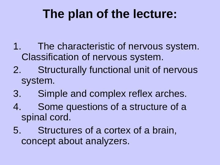
The plan of the lecture: 1. The characteristic of nervous system. Classification of nervous system. 2. Structurally functional unit of nervous system. 3. Simple and complex reflex arches. 4. Some questions of a structure of a spinal cord. 5. Structures of a cortex of a brain, concept about analyzers.
NEUROLOGY The dependence of organisms upon sources of environmental energy and the essentially dynamic nature of their life processes have been em phasized elsewhere. Metabolic processes within the organism itself create a number of stimuli to which the organism must react. The nervous sys tem also includes the specific apparatus of all conscious experience. It is the dominant, although not the only mechanism for the regulation of con duct and the maintenance of unity of the personality. The entire mass of nervous tissue in the body is called the nervous system. The function of the nervous system is based on two fundamental properties. The first is the ability to react to various external physical and other agents. The second is the ability to transmit the excitations thus elicited from one locality to another. The first property is called irritability, the second, conductivity. The activity of the nervous system is based on the reflex. The basic element of the nervous system is the nerve cell which, together with all the processes arising from it, is called the neuron. A long axial cylindrical process, called the axon, arises from the body of the cell in one direction. Short branched processes called dendrites lead in the other db rection. The central nervous system can be classified into central and peripheral systems. The central nervous system consists of the spinal cord and brain made up of gray and white matter. The peripheral nervous system includes all other components (the nerve roots, ganglions, plexus es, nerves, peripheral nerve endings). The gray matter is an accumulation of nerve cells together with the nearest branches and their processes.
The white matter consists of nerve fibres covered by a myelin sheath linking the different centers (an accumulations and cohesions of nerve cells). The nervous system is divided into two parts. The vegetative nervous system innervates the internal organs, the smooth muscles and vessels. The so matic (animal) nervous system controls the striated musculature of the body and primary innervates the organs of animal life. The basic anatomical element is the nerve cell (the neuron). A long axial process, called the axon or neurite, arises from the body of the neu ron in one direction. Short branched processes called dendrites lead in the other direction. Nervous impulses inside the neuron run from the den drites to the cell body and from there to the axon. The axons convey the nervous impulses away from the cell body. (The body uses a combination Telectrical impulses and chemical messengers to react and adjust to stim uli in order to maintain homeostasis. Afferent (sensory) neurons of the peripheral nervous system carry information from sensory receptor cells to the central nervous system. Efferent (motor) neurons of the peripheral nervous system convey information away from the central nervous sys tem to the effectors (muscles and glands). A nerve impulse travels alorig-an axon and eventually reaches the branching axon terminals in the trans-missive segment of the neuron. The junction between neurons is called a synapse. The transmition of a nerve impulse at a synapse can be either chemical or electrical, but chemical synapsis are far more common than electrical ones. In a chemical synapse, two cells communicate by way of a chemical agent called a neurotransmitter.
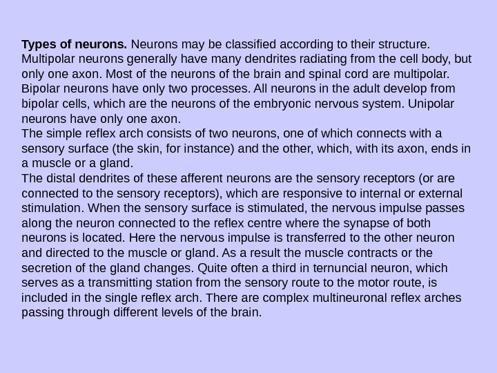
Types of neurons. Neurons may be classified according to their struc ture. Multipolar neurons generally have many dendrites radiating from the cell body, but only one axon. Most of the neurons of the brain and spinal cord are multipolar. Bipolar neurons have only two processes. All neurons in the adult develop from bipolar cells, which are the neurons of the embry onic nervous system. Unipolar neurons have only one axon. The simple reflex arch consists of two neurons, one of which connects with a sensory surface (the skin, for instance) and the other, which, with its axon, ends in a muscle or a gland. The distal dendrites of these afferent neurons are the sensory recep tors (or are connected to the sensory receptors), which are responsive to internal or external stimulation. When the sensory surface is stimulated, the nervous impulse passes along the neuron connected to the reflex cen tre where the synapse of both neurons is located. Here the nervous im pulse is transferred to the other neuron and directed to the muscle or gland. As a result the muscle contracts or the secretion of the gland changes. Quite often a third in ternuncial neuron, which serves as a transmitting station from the sensory route to the motor route, is included in the single reflex arch. There are complex multineuronal reflex arches passing through different levels of the brain.
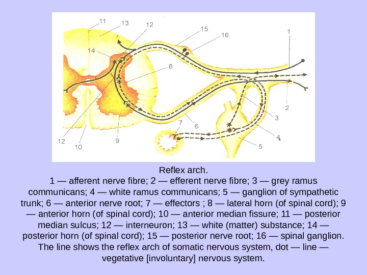
Reflex arch. 1 — afferent nerve fibre; 2 — efferent nerve fibre; 3 — grey ramus communicans; 4 — white ramus communicans; 5 — ganglion of sympathetic trunk; 6 — anterior nerve root; 7 — effectors ; 8 — lateral horn (of spinal cord); 9 — anterior horn (of spinal cord); 10 — anterior median fissure; 11 — posterior median sulcus; 12 — interneuron; 13 — white (matter) substance; 14 — posterior horn (of spinal cord); 15 — posterior nerve root; 16 — spinal ganglion. The line shows the reflex arch of somatic nervous system, dot — line — vegetative [involuntary] nervous system.
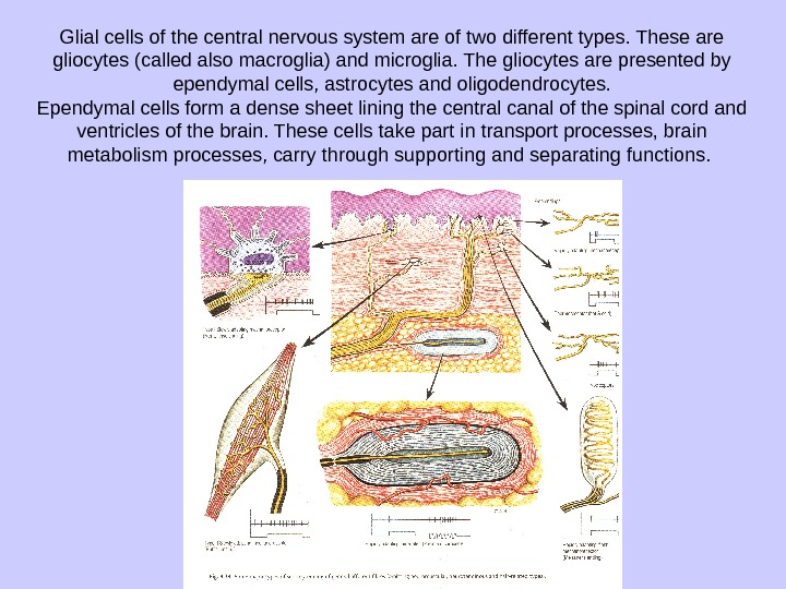
Glial cells of the central nervous system are of two different types. These are gliocytes (called also macroglia) and microglia. The gliocytes are presented by ependymal cells, astrocytes and oligodendrocytes. Ependymal cells form a dense sheet lining the central canal of the spinal cord and ventricles of the brain. These cells take part in transport processes, brain metabolism processes, carry through supporting and separating functions.
DEVELOPMENT OF THE HUMAN NERVOUS SYSTEM The nervous system develops from external embryonic layer (ecto derm). In the dorsal section of the germ trunk ectoderm cells form medul lary (nervous) plate. The medullary plate is gradually transformed into a groove (trough). The edges of the neural groove draw near to one another growing together, and the neural (nervous) tube arises. The neural (medullary) groove trans forms into a tube at first in the frontal portions of an embryo’s trunk and then — in the dorsal ones. The nervous tube gradually sinks into the ecto derm. On this stage of development, the walls of the nervous tube consist of the internal, the medial and the external layers. The internal (inner, ependymic)layer produces ependymocytes, which cover the cavity of the nervous tube, the central canal. The middle layer, which is formed by tight ly situated cells, during the further development gives rise to the graysubstance. Outside of the middle layer, there is a peripheral part of the nervous tube (outer, marginal layer), which thickens gradually at the expense of the neuritis (nervous fibers), which grow inside and form a connecting tracts of the white substance.
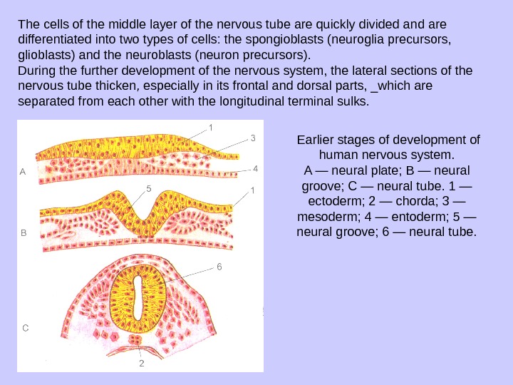
The cells of the middle layer of the nervous tube are quickly divided and are differentiated into two types of cells: the spongioblasts (neuroglia precursors, glioblasts) and the neuroblasts (neuron precursors). During the further development of the nervous system, the lateral sec tions of the nervous tube thicken, especially in its frontal and dorsal parts, _which are separated from each other with the longitudinal terminal sulks. Earlier stages of development of human nervous system. A — neural plate; В — neural groove; С — neural tube. 1 — ectoderm; 2 — chorda; 3 — mesoderm; 4 — entoderm; 5 — neural groove; 6 — neural tube.
During development and differentiation of the cell elements of the ner vous tube, its ventral end gradually thickens. Later this thickening trans forms into the cerebrum. The medial and dorsal parts of the nervous tube give rise to the spinal medulla. The terminal parts of the nervous tube gradually become thinner and form the so — called terminal thread of the spinal cord. During embryogenesis, the spinal cord grows slower than the vertebral column. Since the ventral terminus of the nervous tube is fixed in the cavity of the future scull, the lower (caudal) parts of the nervous tube delay in growth more the others. In this connection, the radicis of the de veloping spinal nerves, which took up their position in the intervertebral foramina, lengthen and change the orientation from the horizontal to the oblique and even the vertical. On the fourth week of the prenatal life three cerebral vesicles separated with the local narrowing of the nervous tube represent the future brain. During this stage of the development there is the ‘ anterior, or fore brain (prosencephalon), which develops from the anterior cerebral vesicle, the middle, or mid brain (mesencephalon), which de velops from the middle cerebral vesicle, posterior, or hind brain the rhomben cephalon or dorsal brain (rhombencephalon) formed from the third cerebral vesicle.
By the end of the fourth week of development of the anterior cerebral vesicle differentiates into two sections: the future cerebrum (telen-cephalon) and the future intermediate brain (diencephalon). The posterior cerebral vesicle also is subdivided into the dorsal brain (metencephalon) and the medulla (medulla oblongata). As a result, by the sixth week of development the brain consists of five cerebral vesicles. During the forma tion of the cerebral vesicles the ventral ending of the nervous tube bends in the sagittal plane; the prominence of the parietal bend is directed to the dorsal side in the region of the middle vesicle (cephalic flexure) occipital bend is formed at the border between the dorsal cerebral vesicle and the beginning of the spinal medulla (cervical flexure). Its prominence is also directed to the dorsal side. The pontile bend directed to the ventral side is ‘ formed in the region of rhombencephlon and divides the third cerebral I vesicle (rhombencephalon) into the dorsal brain and the medulla (pontine ‘ flexure) cavity of the rhombencephalon is transformed into the , fourth ventricle of the cerebrum.
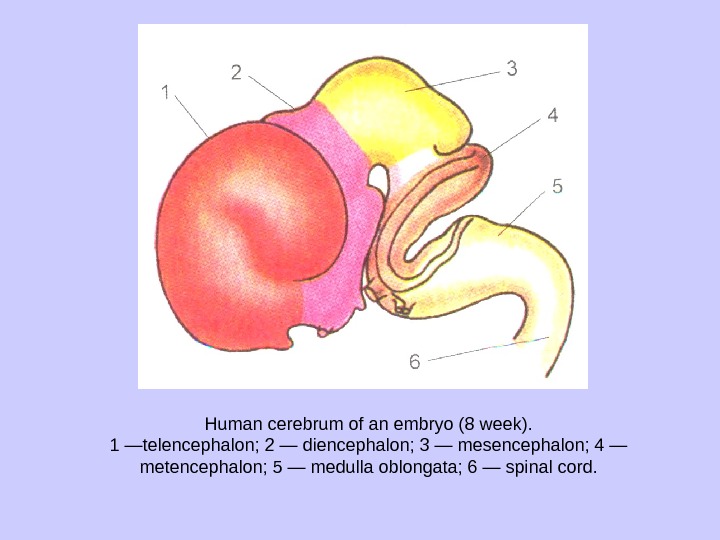
Human cerebrum of an embryo (8 week). 1 —telencephalon; 2 — diencephalon; 3 — mesencephalon; 4 — metencephalon; 5 — medulla oblon gata; 6 — spinal cord.
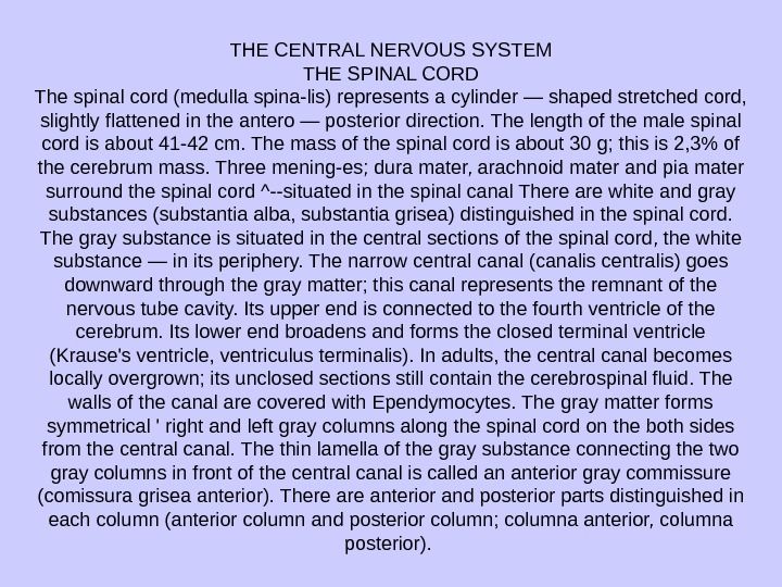
THE CENTRAL NERVOUS SYSTEM THE SPINAL CORD The spinal cord (medulla spina-lis) represents a cylinder — shaped stretched cord, slightly flattened in the antero — posterior direction. The length of the male spinal cord is about 41 -42 cm. The mass of the spinal cord is about 30 g; this is 2, 3% of the cerebrum mass. Three mening-es; dura mater, arachnoid mater and pia mater surround the spinal cord ^—situated in the spinal canal There are white and gray substances (substantia alba, substantia grisea) distin guished in the spinal cord. The gray sub stance is situated in the central sections of the spinal cord, the white substance — in its periphery. The narrow central canal (canalis centralis) goes downward through the gray matter; this canal represents the remnant of the nervous tube cavity. Its upper end is connected to the fourth ventricle of the cerebrum. Its low er end broadens and forms the closed terminal ventricle (Krause’s ventricle, ventriculus terminalis). In adults, the central canal becomes locally overgrown; its unclosed sections still contain the cerebrospinal fluid. The walls of the canal are covered with Ependymocytes. The gray matter forms symmetrical ‘ right and left gray columns along the spi nal cord on the both sides from the central canal. The thin lamella of the gray substance connecting the two gray columns in front of the central canal is called an anterior gray commissure (comissura grisea anterior). There anterior and posterior parts distin guished in each column (anterior column and posterior column; columna anterior, columna posterior).
From the 8 th cervical segment to the 2 d lum bar segment inclusive on each side the gray matter also forms a lateral bulging — the lateral or intermediate column (columna lateralis, columna intermedia). There are no lateral columns above and below this level. At their place on the cross — sections of the spinal cord the anterior, posteior and lateral horn (cornu anterior, cornu posterior, cornu laterale) of gray substance are distinguished. The anterior cornu is wider than the posterior one. The lateral horn topographically corresponds to the lateral column of the gray substance. Bodies of neurons, amyelinic and thin myelin fibers and neuroglia form the gray substance of the spinal cord. Bodies of the largest neurons of the spinal cord are situated in the anterior horn. They form five nuclei (clumps). Among these nuclei, there antero — and postero — lateral nuclei (n. anterolateralis, n. posterolateralis), and a central nucleus (n. centralis). Topography of the segments of spinal cord within the vertebral canal. 1 — cervical segments (CI — CVIU); 2 — thoracic seg ments (Th. I — Th. XII); 3 — lumbar segments (LI — LV); 4—sacral segments (SI — SV); 5 — coccygeal segments (Col—Co. III).
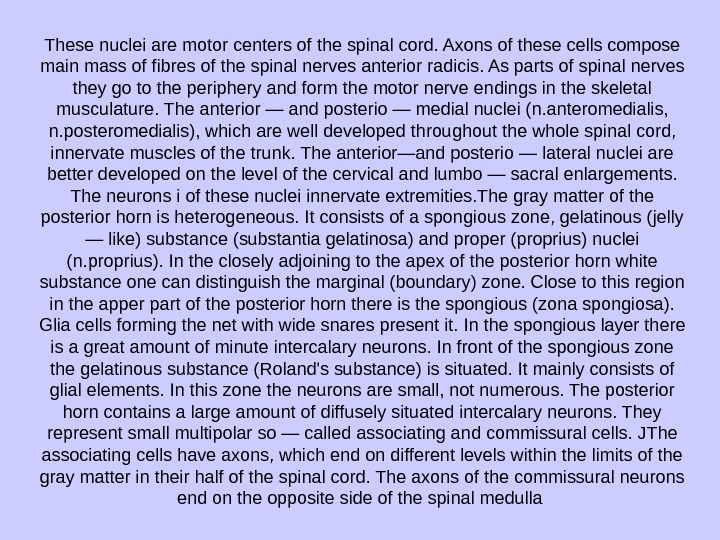
These nuclei are motor centers of the spinal cord. Axons of these cells compose main mass of fibres of the spinal nerves anterior radicis. As parts of spinal nerves they go to the periphery and form the motor nerve endings in the skeletal musculature. The anterior — and posterio — medial nuclei (n. anteromedialis, n. posteromedialis), which are well developed throughout the whole spinal cord, innervate muscles of the trunk. The anterior—and posterio — lateral nuclei are better developed on the level of the cervical and lumbo — sacral enlargements. The neurons i of these nuclei innervate extremities. The gray matter of the posterior horn is heterogeneous. It consists of a spongious zone, gelatinous (jelly — like) substance (substantia gelatinosa) and proper (proprius) nuclei (n. proprius). In the closely adjoining to the apex of the posterior horn white substance one can distinguish the marginal (boundary) zone. Close to this region in the apper part of the posterior horn there is the spongious (zona spongiosa). Glia cells forming the net with wide snares present it. In the spongious layer there is a great amount of minute intercalary neurons. In front of the spongious zone the gelatinous substance (Roland’s substance) is situated. It mainly consists of glial ele ments. In this zone the neurons are small, not numerous. The posterior horn contains a large amount of diffusely situated intercalary neurons. They represent small multipolar so — called associating and commissural cells. JThe associating cells have axons, which end on different levels within the limits of the gray matter in their half of the spinal cord. The axons of the commissural neurons end on the opposite side of the spinal medulla
In the basis of the posterior horn of the spinal cord, in its medial part, there is a dorsal thoracic nucleus (Clark’s nucleus), (n. thoracicus dorsa-lis). It consists of large intercalary neurons with well — developed inten sively branching dendrites (Schilling’s cells). Axons of cells of this nucle us go to the lateral cord of the white substance on the same side of the spinal cord and also form conducting cells (posterior spino — cerebellar (Flechsig’s) tract). In the lateral cornu of the spinal cord there are centers of the sympathetic part of the vegetative nervous system. These centers are presented by some groups of minute neurons gathered in the interme-diolateral nucleus (n. intermediolateralis). Axons of these neurons forming a vegetative nucleus in the spinal cord from 8 th cervical to 2 d lumbar segments, extend through the anterior cornu and leave the spinal cord, as parts of the anterior radicis of the spinal nerves.
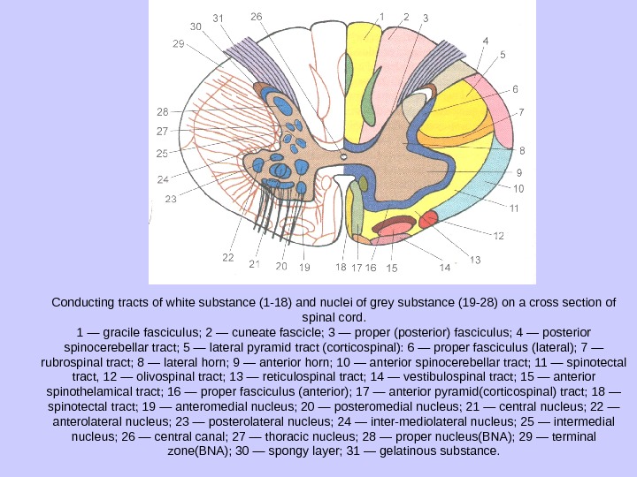
Conducting tracts of white substance (1 -18) and nuclei of grey substance (19 -28) on a cross section of spinal cord. 1 — gracile fasciculus; 2 — cuneate fascicle; 3 — proper (posterior) fasciculus; 4 — posterior spinocerebellar tract; 5 — lateral pyramid tract (corticospinal): 6 — proper fasciculus (lateral); 7 — rubrospinal tract; 8 — lateral horn; 9 — anterior horn; 10 — anterior spinocerebellar tract; 11 — spinotectal tract, 12 — olivospinal tract; 13 — reticulospinal tract; 14 — vestibulospinal tract; 15 — anterior spinothelamical tract; 16 — proper fasciculus (anterior); 17 — anterior pyramid(corticospinal) tract; 18 — spinotectal tract; 19 — anteromedial nucleus; 20 — posteromedial nucleus; 21 — central nucleus; 22 — anterolateral nucleus; 23 — posterolateral nucleus; 24 — inter-mediolateral nucleus; 25 — intermedial nucleus; 26 — central canal; 27 — thoracic nucleus; 28 — proper nucleus(BNA); 29 — terminal zone(BNA); 30 — spongy layer; 31 — gelatinous substance.
THE BRAIN The Brain (encephalon) together with membranes surrounding it is /located in the cranial cavity. That is why the convex surface of the brain I conforms to the internal concave surface of the arch of the cranium. The I inferior surface of the brain is contiguous to the interior base of the skull. THE TELENCEPHALON The Telencephalon (telencephalon) consists of two hemispheres separated by the longitudinal cerebral fissure. The corpus callosum, the anterior and the posterior commisures and the commissure of the fornix connect both hemispheres. The cavity of the telencephalon is presented by the right and the left lateral ventricles, which are placed inside corresponding hemispheres. A hemisphere of the telencephalon consists of an external coat — the cerebral cortex and the white matter that lies deeper. Thereare accumulations of the gray matter (the basal nuclei) in the white matter in the base of the telencephalon. The border between the telencephalon and the diencephalon passes in a region of the internal capsule, which adjoins the lateral side of the thalamus. A cerebral hemisphere (hemispherium cerebri) has a superolateral, a medial and inferior surfaces (faces). The superolateral surface (facies superolateralis) is convex, the medial (facies medialis) is flattened against the opposite hemisphere. The inferior surface (facies anterior) has the most complex irregular relief which conforms to the interior base of the cranium. The surfaces of the hemispheres are separated from each other by the superior, inferolateral and inferomedial margins (margo superior, margo infero-lateralis, margo inferomedialis).
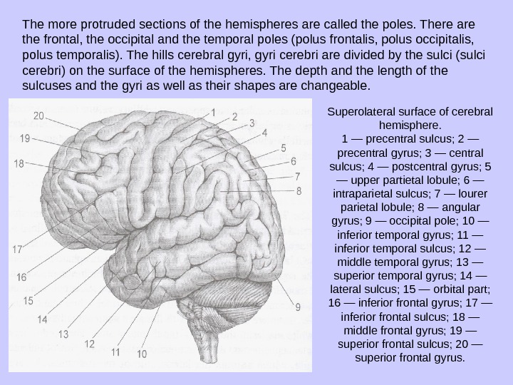
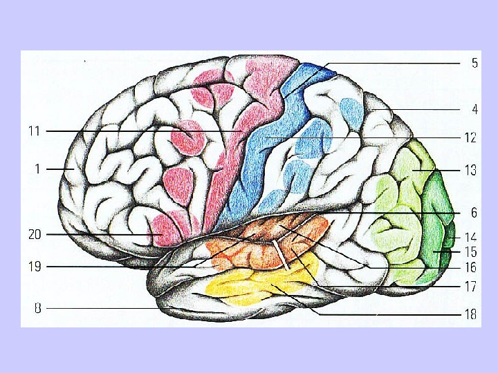
The more protruded sections of the hemi spheres are called the poles. There are the frontal, the occipital and the temporal poles (polus frontalis, polus occipitalis, polus temporalis). The hills cerebral gyri, gyri cerebri are divided by the sulci (sulci cerebri) on the surface of the hemispheres. The depth and the length of the sulcuses and the gyri as well as their shapes are changeable. Superolateral surface of cerebral hemisphere. 1 — precentral sulcus; 2 — precentral gyrus; 3 — central sulcus; 4 — postcentral gyrus; 5 — upper partietal lobule; 6 — intraparietal sulcus; 7 — lourer parietal lobule; 8 — angular gyrus; 9 — occipital pole; 10 — inferior temporal gyrus; 11 — inferior temporal sulcus; 12 — middle temporal gyrus; 13 — superior temporal gyrus; 14 — lateral sulcus; 15 — orbital part; 16 — inferior frontal gyrus; 17 — inferior frontal sulcus; 18 — middle frontal gyrus; 19 — superior frontal sulcus; 20 — superior frontal gyrus.
The texture of the Cortex of the Cerebral Hemispheres The superficial layer of the cerebral hemispheres is formed by the gray matter called the cerebral cortex (cortex cerebri, s. pallium). More than 90% of the surface of the cerebral hemispheres belong to the new cortex (neocortex). Phylogenetically the older parts of the cortex include the old cortex (archicortex) and the ancient cortex (paleocortex). The ar-chicortex is represented by the gyms dentatus and base of the hippocam pus; the paleocortex is represented by parts of the cortex in the region of the basomedial surface of the temporal lobe (the anterior perforated sub stance etc. ). The thickness of the cortex in different sections varies from 1, 3 to 5 mm. The most thick cortex is situated in the superior portions of the precentral and the postcentral gyri. The cortex of the convex surface of convolutions is thicker, than on their lateral aspects and in the depth of sulci. The area of the cortical surface of the cerebral hemispheres is 220000 mm 2. There about 10 -14 mlrd. neurons in the cortex and each of them forms synapses with 8 -10 thsnd other nerve cells. Approximately 4% of the volume of the cerebral cortex fall to the share of the neural cells. About 8% of the volume of the cortex appertain to the glial elements which fulfil auxiliary functions (supporting etc. ). The processes of the nerve cells (ax-ons and dendrites) and the pathways occupy 80 -85% of the volume of the cortex. The form and interlocation of the nerve cells are not the same in different sections of the cortex.
These parts differ in cytoarchitectonics (distribution of cells) and my-eloarchitectonics (distribution of fibers). For neocortex the six — layered structure is typical. The old and the ancient cortex form two — three lay ers. The neocortex is formed of six layers: 1) the molecular lamina (lamina molecularis, s. plexiformis), 2) the external granular lamina (lamina granu-laris externa), 3} the external lamina of pyramidal cells (lamina pyramidalis externa), 4) the internal granular lamina (lamina granularis interna), 5) the internal lamina of pyramidal cells (lamina pyramidalis interna), 6) the multi form (polyform) lamina (lamina multiformis). In the cerebral cortex the pyramidal cells of different sizes (from 10 to 140 mem) dominate. The small pyramidal cells which are placed in all layers of the cortex are com-missural and associative neurons. The bigger ones generate impulses for voluntary movements of muscules. These impulses pass through the motor nuclei of the brain and of the spinal cord to sceletal muscules. The external molecular layer of the cortex is characterized by disposi tion of the small multipolar associative neurons and the numerous fibres inside it. The internal granular lamina is formed by small star — shaped neurons. In the internal layer of pyramidal cells which is / the most developed in the precentral gyms large (giant) pyramidal cells dominate. The diameter of the pyramidal cells (cells of Betz) is 80 -125 mem, they are rich in chromatophilie substance. Their axons form pyrami dal paths. From the axons of these cells the collaterals move away to the \ cortex, the basal ganglia, the red nuclei, the reticular formation, the olivary I nuclei and the nuclei of the pons. In the polymorphic layer of the cortex I there are neurons size and form of which vary. Their axons run to the { white matter and their dendrites go to the molecular layer.
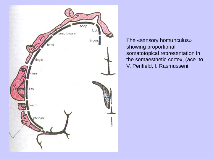
The distribution of functions in the cerebral cortex In the cortex of the brain the all the external and the internal irritations are being analysed. The centers which regulate the execution of certain functions are situated in the cortex, that is why the cortex can be considered as the aggregate of the cortical parts of analysators. An analysator is an intricate complex of anatomical structures. It consists of the peripheral receptive (perceptional) apparatus, conductors of nerve impulses and the center (cortical termination). The cortical termination of the analysator is not a strictly anatomically outlined zone. In the cerebral cortex one can distinguish the nucleus of the sensory system and the scattered elements. In the cortex of the postcentral gyrus (field 1 -3) and in the superpari-etal lobule (fields 5 and 7 ) the neurons of the nucleus of the cortical analy sator of general sensibility (senses of temperature, pain, touch and pres sure) and proprioceptive sensibility are situated. The postcentral gyrus of each hemisphere connects with the opposite side of a body. The highest position is occupied by the neurons responsible for the sensibility of the inferior parts of a trunk and lower limbs. The lowest receptive fields which are projected near the lateral sulcus are responsible for sensibility of the high parts of the trunk, the head and the upper extremities
The «sensory homunculus» showing proportional somatotopical representation in the somaesthetic cortex, (ace. to V. Penfield, I. Rasmusseni.
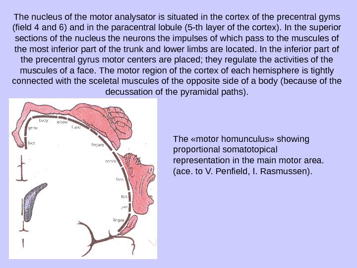
The nucleus of the motor analysator is situated in the cortex of the precentral gyms (field 4 and 6) and in the paracentral lobule (5 -th layer of the cortex). In the superior sections of the nucleus the neurons the impulses of which pass to the muscules of the most inferior part of the trunk and lower limbs are located. In the inferior part of the precentral gyrus motor centers are placed; they regulate the activities of the muscules of a face. The motor region of the cortex of each hemisphere is tightly connected with the sceletal muscules of the opposite side of a body (because of the decussation of the pyramidal paths). The «motor homunculus» showing proportional somatotopical representation in the main motor area. (ace. to V. Penfield, I. Rasmussen).
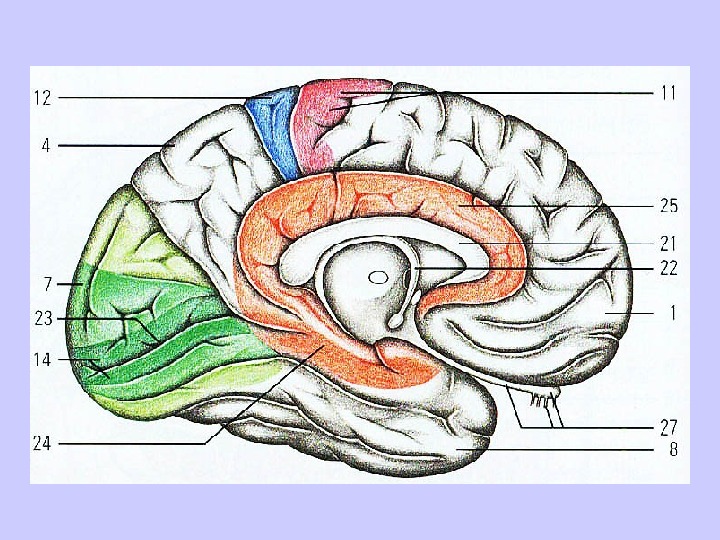
The nucleus of the analysator of the coordinated turning of the head and the eyes to the opposite side is situated in the posterior parts of the middle frontal gyrus (promoter zone, field 8). The coordinated turning of the eyes and the head is regulated not only when the proprioceptive impulses from the oculomotor muscules come to the cortex of the frontal gyrus. This motor act is provided as well by coming of impulses from the eye retina in the field 17 of the occipital lobe which is situated near the nucleus of the visual analysator. The nucleus of the motor analysator which controls the synthesis of all complex (combmated) voluntary movements is situated in the deep layers /of the inferior parietal lobule and the supramarginal gyrus. In the right — handers this nucleus is located in the left hemisphere; in the left — handers this nucleus is situated in the right one. The exercising of the voluntary movements is the result of the formation of temporary connections between the neurons of the nucleus of this analysator and the pre — central I gyrus. The nucleus of the analysator which is responsible for recognizing an object to the touch (the sense of stereognosia) is situated in the superfi-Vcial layers of the cortex of the superior parietal lobule. The nucleus of the auditory analysator is placed in the depth of the v lateral sulcus in the region of the transverse temporal gyri (Heschli’s con volutions). The nucleus of the olfactory analysator is situated on the inferior sur-/ face of the temporal lobe in the region of the uncus (fields A and E) and in vifesj’egion of the hippocampus The nucleus of the motor analysator of written speech, which is con nected with letters and other signs, is situated in the cortex of the posterior part of the middle frontal gyrus.
This nucleus tightly adjoins to the sections of the precentral gyms that is responsible for the motor func tions of the arm and the coordinated turning of the head and the eyes to the opposite side. The destruction of the field 40 leads to loss of the ability to do by the arm exact and fine movements when wrighting letters, signs and words (agraphy). The nucleus of the motor analysator of the verbal speech (speechmo- tor analysator) is situated in the posterior parts of the inferior frontal gyrus (Broca center, field 44). This nucleus borders on the portions of the pre central gyrus that are motor analysators for the head and the neck muscle movements. The injury of the field 44 leads to loss of the ability to utter words (aphasia). The nucleus of the vocalization speech analysator is locat ed in the central parts of the inferior frontal gyrus (field 45). The injury of this field leads to the inability to compose and repeat the musical phrases (amusia) and to the inability to compose the sensible sentences from the separated letters (agrammatismus). In this case the speech consists of a set of words which are not halted by sense. The nucleus of the auditory speech analysator is situated in the region of the superior temporal gyrus near the nucleus of the auditory analysator. The nucleus of the visual speech analysator is located in the cortex of the angular gyrus (field 39) near the nucleus of the visual analysator. When , the field 39 is affected one could observe loss of the ability to perceive the iwritten language and to read.