
Chronic Obstructive Pulmonary Disease (( COPD


chronic_obstructive_airways_disease_2.ppt
- Размер: 10.1 Mегабайта
- Количество слайдов: 109
Описание презентации Chronic Obstructive Pulmonary Disease (( COPD по слайдам
Chronic Obstructive Pulmonary Disease (( COPD ))
THE Guideline Global Initiative for Chronic Obstructive Lung Disease (GOLD), World Health Organization (WHO), National Heart, Lung and Blood Institute (NHLBI)
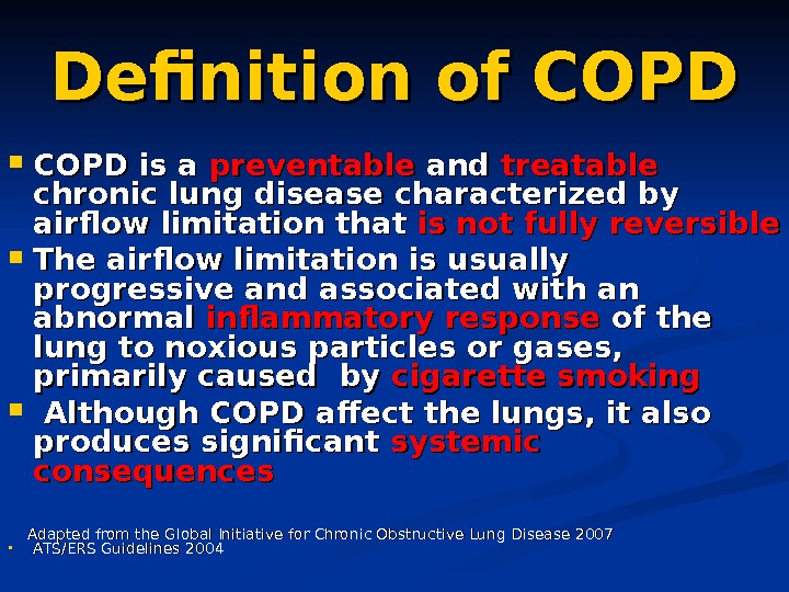
Definition of COPD is a preventable and treatable chronic lung disease characterized by airflow limitation that is not fully reversible The airflow limitation is usually progressive and associated with an abnormal inflammatory response of the lung toto noxious particles or gases, primarily caused by by cigarette smoking Although COPD affect the lungs, it also produces significant systemic consequences Adapted from the Global Initiative for Chronic Obstructive Lung Disease 2007 • ATS/ERS Guidelines
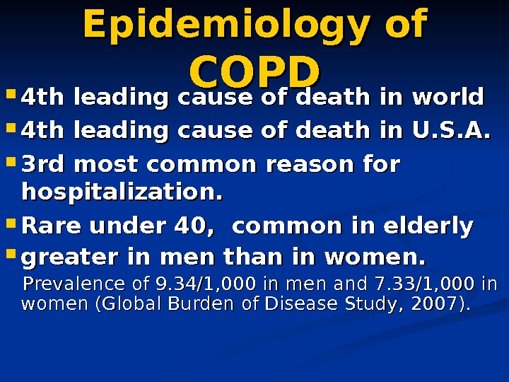
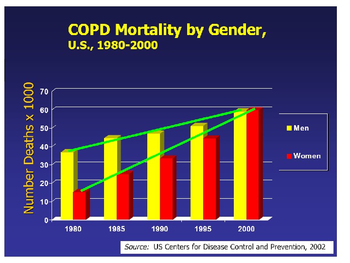
Epidemiology of COPD 44 th leading cause of death in world 4 th leading cause of death in U. S. A. 3 rd most common reason for hospitalization. Rare under 40, сс ommon in elderly greater in men than in women. Prevalence of 9. 34/1, 000 in men and 7. 33/1, 000 in women (Global Burden of Disease Study, 22 00 0707 ). ).
COPD includes: chronic bronchitis chronic bronchiolitis (small air way disease) Emphysema
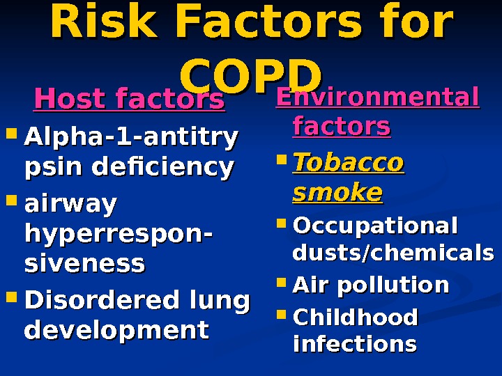
Risk Factors for COPD Host factors Alpha-1 -antitry psin deficiency airway hyperrespon- siveness Disordered lung development Environmental factors Tobacco smoke Occupational dusts/chemicals Air pollution Childhood infections
Risk factors cigarette smoking remains the most important. Susceptibility to cigarette smoke varies but both the dose and duration of smoking appear to be important and it is unusual to develop COPD with less than 10 pack years. (1 pack year = 20 cigarettes / day /year).
Alpha-1 -antitrypsin deficiency α 1 -Antitrypsin is a proteinase inhibitor which is produced in the liver, secreted into the blood and diffuses into the lung s. Mechanism of action: an inhibit ion of proteolytic enzymes such as neutrophil elastase, which are capable of destroying alveolar wall connective tissue.
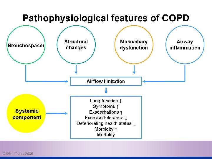
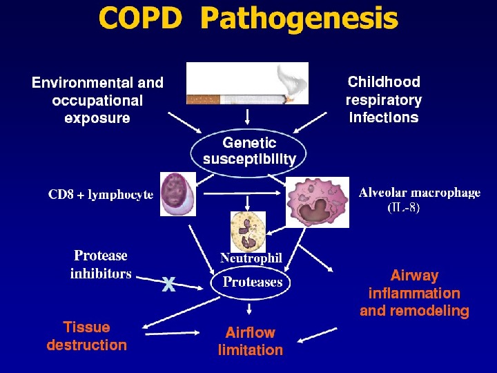
Pathophysiolog yy COPD has both Pulmonary components Systemic components
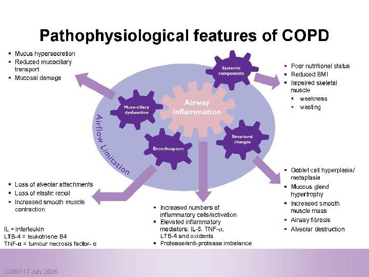
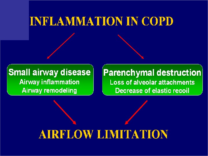
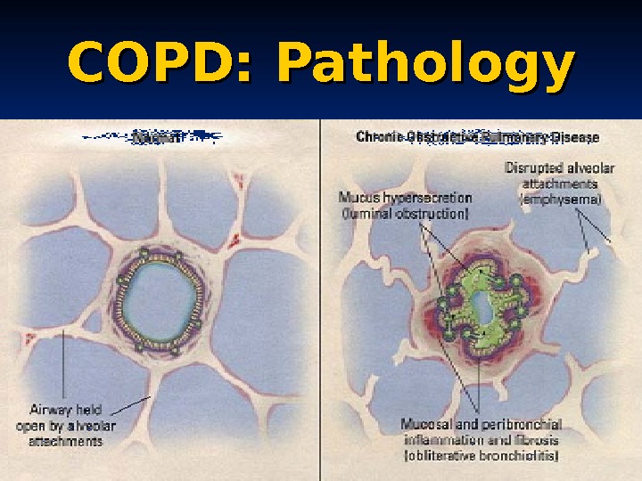
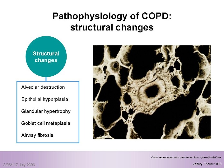
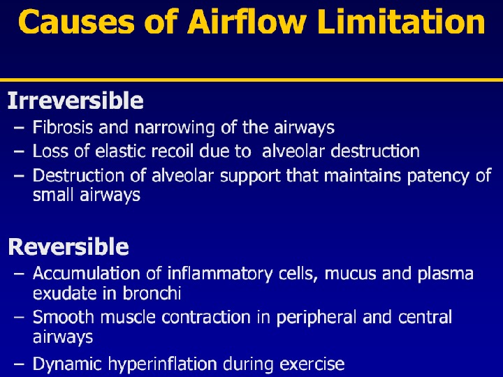
Pulmonary components: Mucus secretion An An enlargement of mucous secreting glands and an increasing number of goblet cells in the large airways → → increase mucous that causes chronic bronchitis Loss of elastic tissue surrounding the smaller airways combined by inflammation and fibrosis in the airway wall → → airflow limitation.
Pulmonary components: Premature airway closure leads to gas trapping and hyperinflation → ↓↓ pulmonary and chest wall compliance. (during exercise the time available for expiration shortens resulting in progressive hyperinflation)
Pulmonary components: Flattening of the diaphragmatic muscles and increase horizontal alignment of the intercostals muscles → mechanical disadvantage of respiratory muscles → increase work of breathing first on exercise but then at rest.
Pulmonary components: In the alveolar capillary units the unopposed action of proteases and oxidants → destruction of the alveoli → bullae formation in some individuals which → impaired gas exchange and respiratory failure.
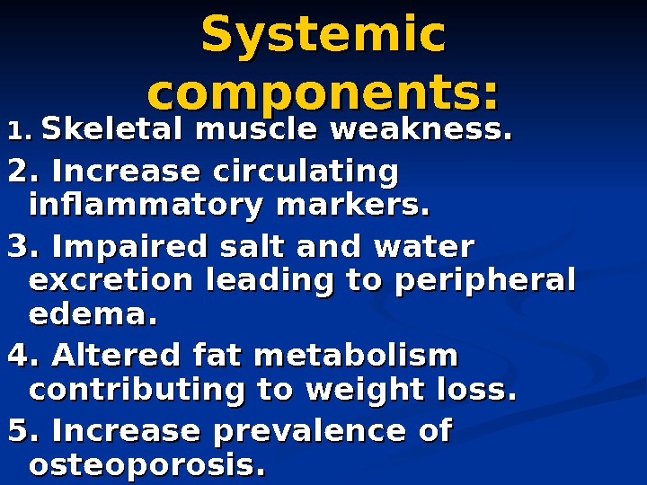
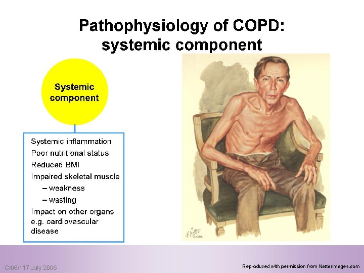
Systemic components: 1. 1. Skeletal muscle weakness. 2. Increase circulating inflammatory markers. 3. Impaired salt and water excretion leading to peripheral edema. 4. Altered fat metabolism contributing to weight loss. 5. Increase prevalence of osteoporosis.
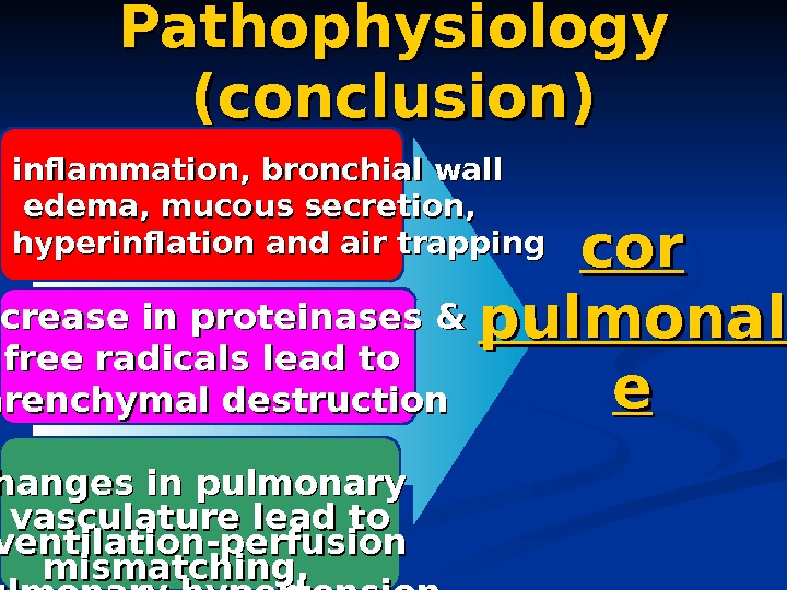
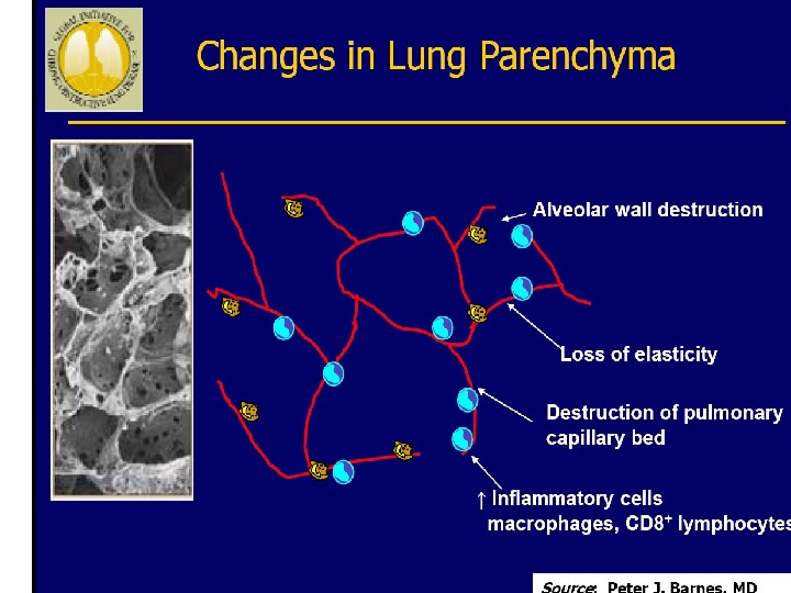
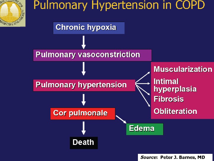
Pathophysiology (conclusion) inflammation, bronchial wall edema, mucous secretion, hyperinflation and air trapping Increase in proteinases & free radicals lead to parenchymal destruction Changes in pulmonary vasculature lead to ventilation-perfusion mismatching, pulmonary hypertension cor pulmonal ee
COPD: Pathology
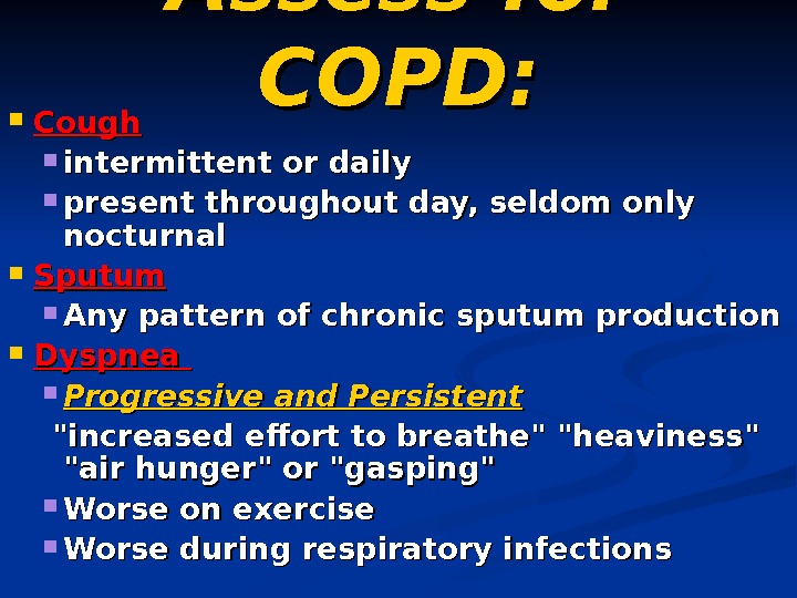
Assess for COPD: Cough intermittent or daily present throughout day, seldom only nocturnal Sputum Any pattern of chronic sputum production Dyspnea Progressive and Persistent «increased effort to breathe» «heaviness» «air hunger» or «gasping» Worse on exercise Worse during respiratory infections
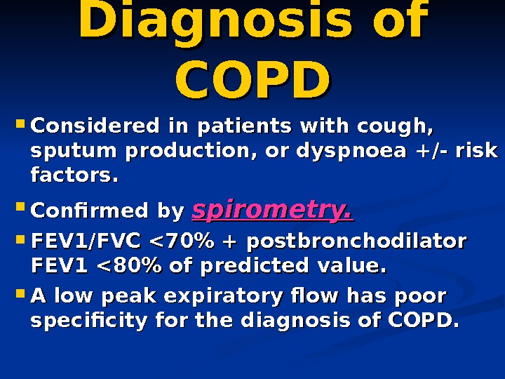
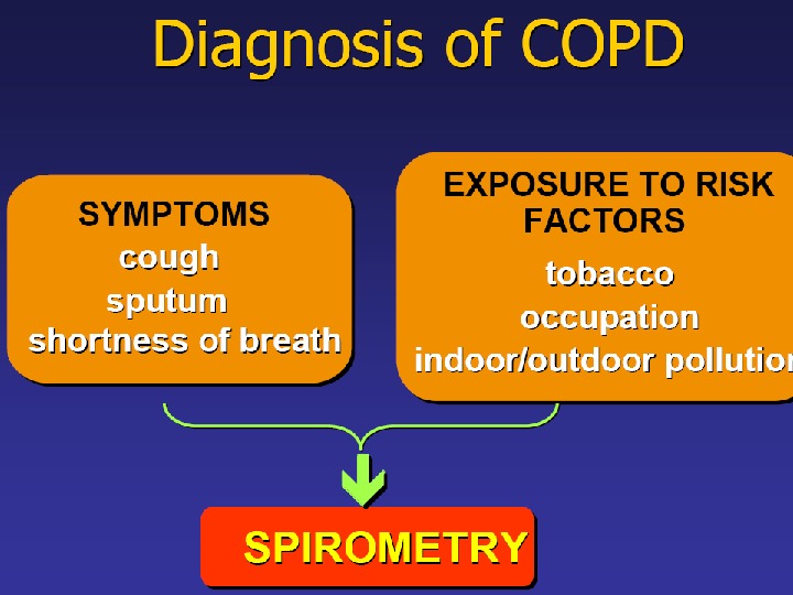
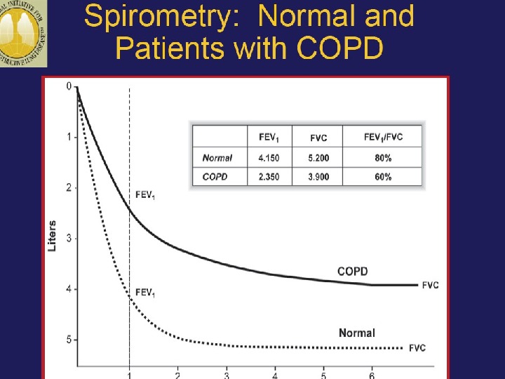
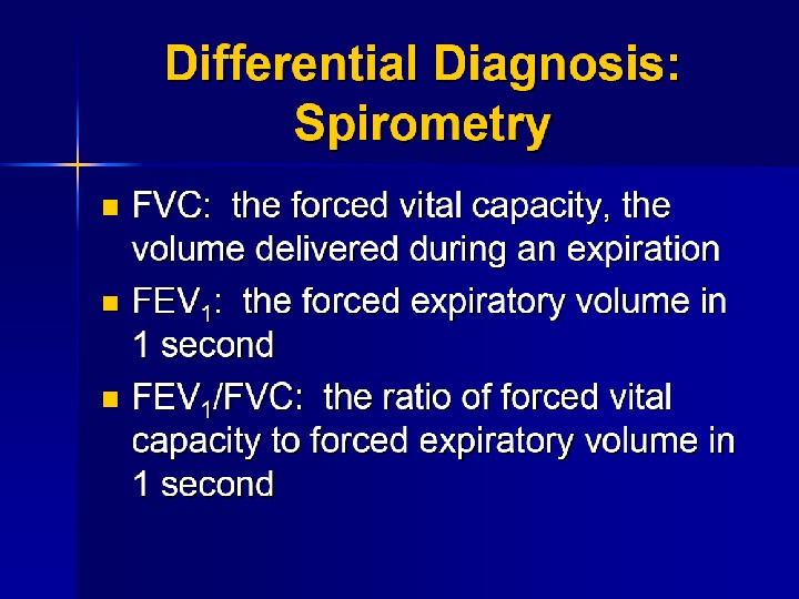
Diagnosis of COPD Considered in patients with cough, sputum production, or dyspnoea +/- risk factors. Confirmed by spirometry. FEV 1/FVC <70% + postbronchodilator FEV 1 <80% of predicted value. A low peak expiratory flow has poor specificity for the diagnosis of COPD.
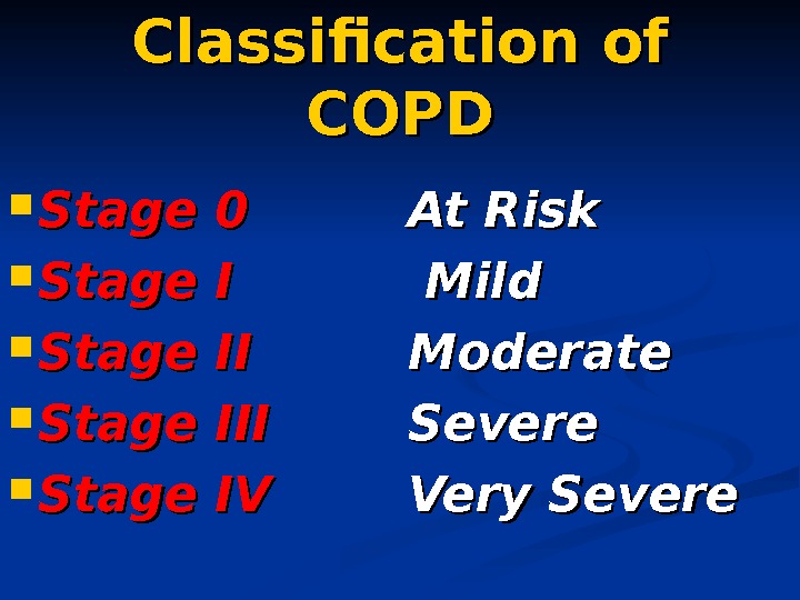
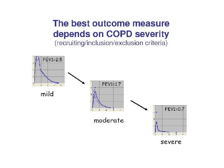
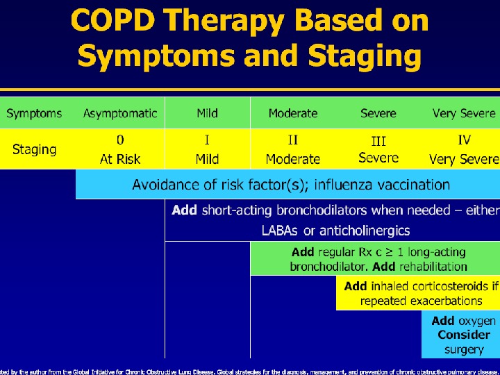
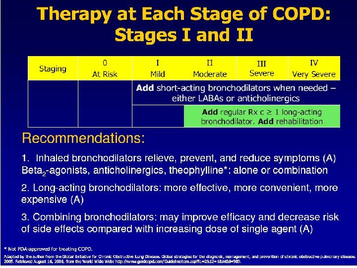
Classification of COPD Stage 0 At Risk Stage I Mild Stage II Moderate Stage III Severe Stage IV Very Severe
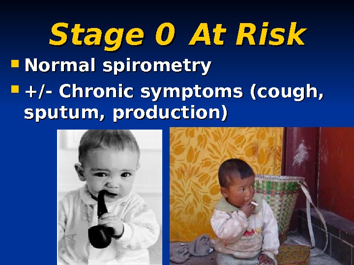
Stage 0 At Risk Normal spirometry +/- Chronic symptoms (cough, sputum, production)
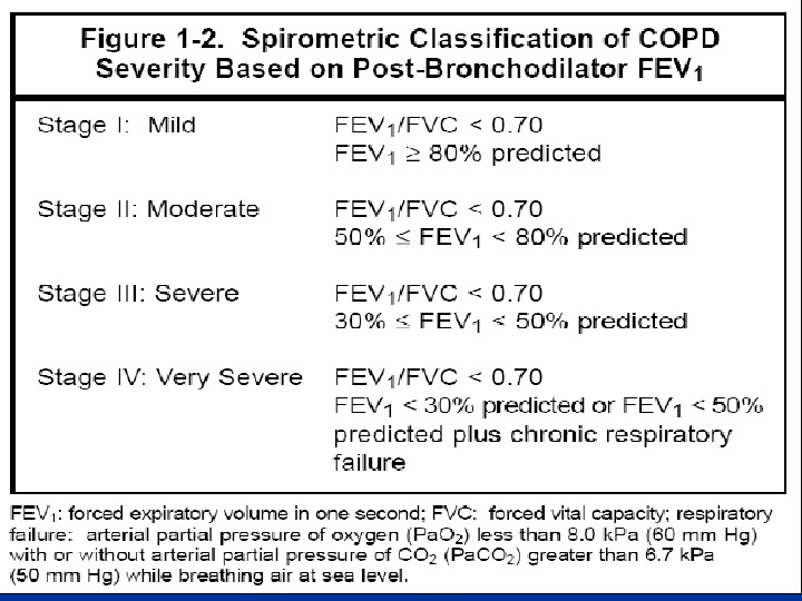
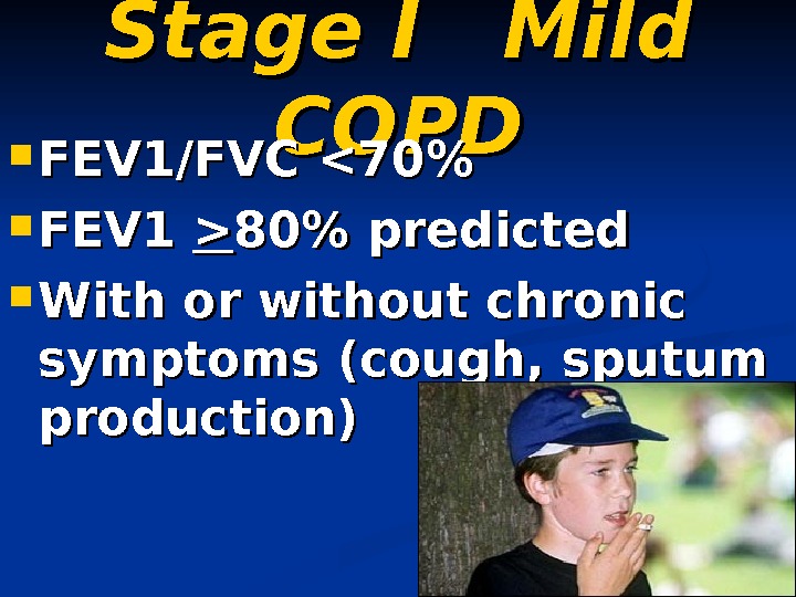
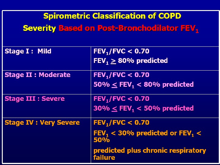
Stage I Mild COPD FEV 1/FVC > 80% predicted With or without chronic symptoms (cough, sputum production)
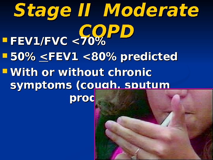
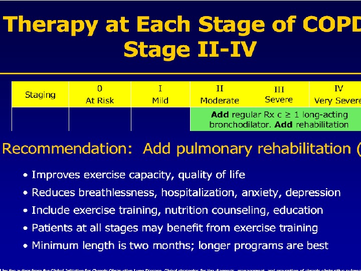
Stage II Moderate COPD FEV 1/FVC <70% 50% << FEV 1 <80% predicted With or without chronic symptoms (cough, sputum production)
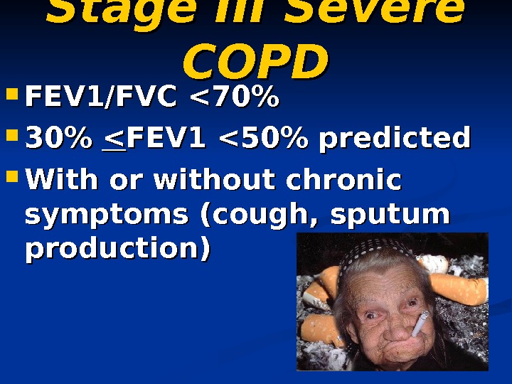
Stage III Severe COPD FEV 1/FVC <70% 30% << FEV 1 <50% predicted With or without chronic symptoms (cough, sputum production)
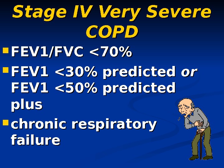
Stage IV Very Severe COPD FEV 1/FVC <70% FEV 1 <30% predicted oror FEV 1 <50% predicted plus chronic respiratory failure
Diagnosis of COPD History Symptoms Lung function Plain chest CT CT Arterial blood gases
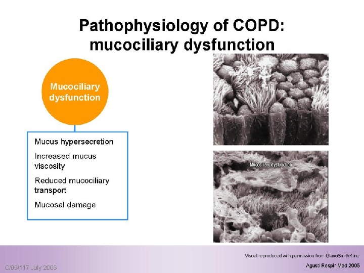
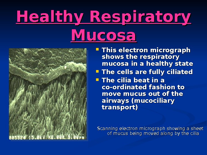
Healthy Respiratory Mucosa This electron micrograph shows the respiratory mucosa in a healthy state The cells are fully ciliated The cilia beat in a co-ordinated fashion to move mucus out of the airways (mucociliary transport) Scanning electron micrograph showing a sheet of mucus being moved along by the cilia
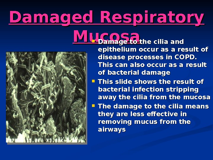
Damaged Respiratory Mucosa Damage to the cilia and epithelium occur as a result of disease processes in COPD. This can also occur as a result of bacterial damage This slide shows the result of bacterial infection stripping away the cilia from the mucosa The damage to the cilia means they are less effective in removing mucus from the airways
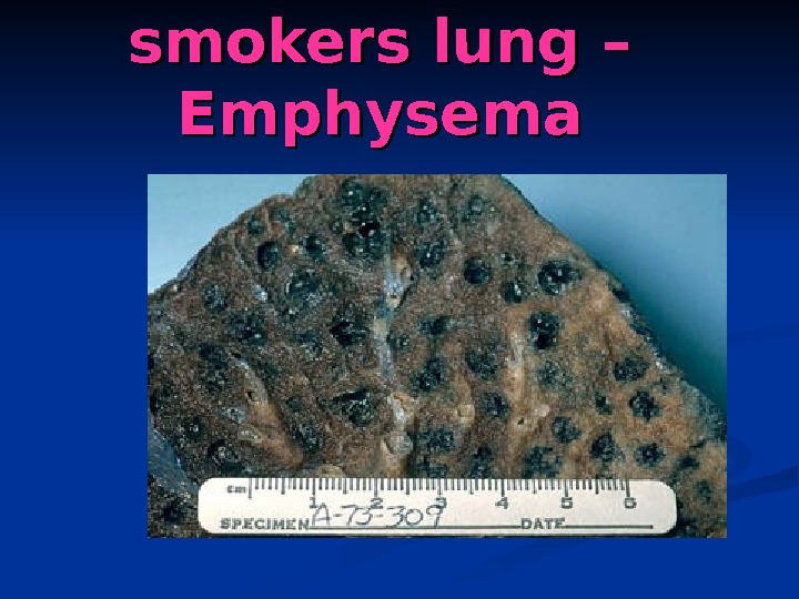
smokers lung – Emphysema
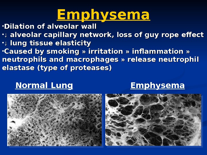
Emphysema • Dilation of alveolar wall • ↓↓ alveolar capillary network, loss of guy rope effect • ↓↓ lung tissue elasticity • Caused by smoking » » irritation » » inflammation » » neutrophils and macrophages » » release neutrophil elastase (type of proteases) Emphysema. Normal Lung
Emphysema is defined pathologically as dilatation and destruction of the lung tissue distal to the terminal bronchiole.
classifi cation Centri-acinar emphysema. Pan-acinar emphysema. Irregular emphysema.
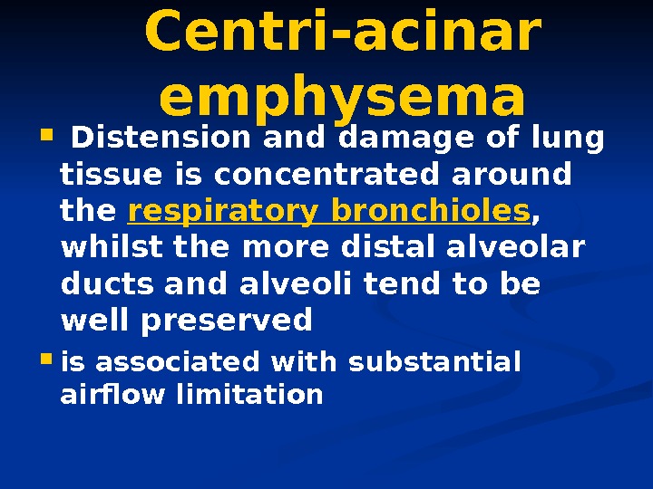
Centri-acinar emphysema Distension and damage of lung tissue is concentrated around the respiratory bronchioles , whilst the more distal alveolar ducts and alveoli tend to be well preserved is associated with substantial airflow limitation
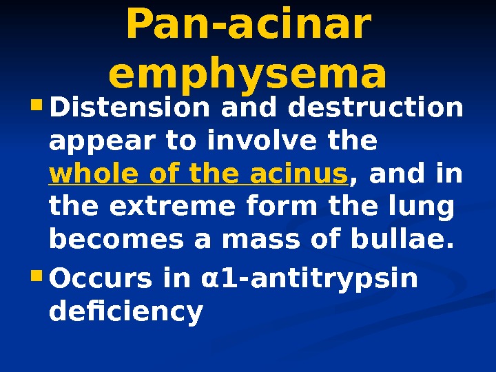
Pan-acinar emphysema Distension and destruction appear to involve the whole of the acinus , and in the extreme form the lung becomes a mass of bullae. Occurs in α 1 -antitrypsin deficiency
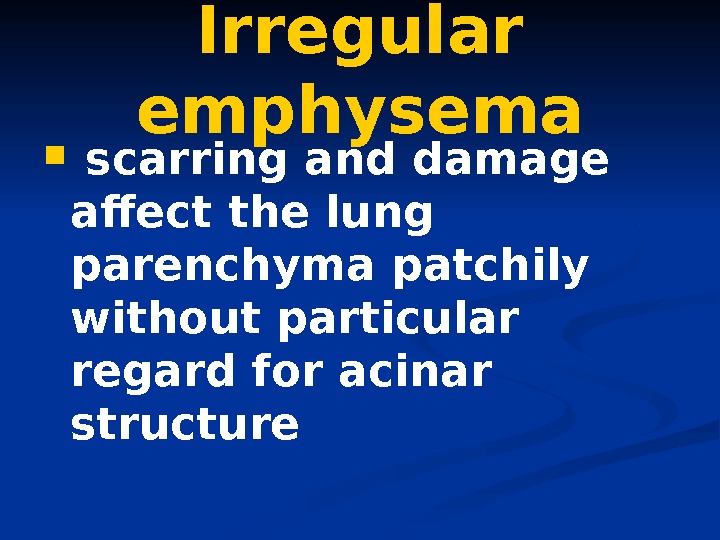
Irregular emphysema scarring and damage affect the lung parenchyma patchily without particular regard for acinar structure
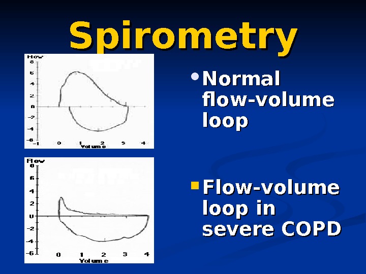
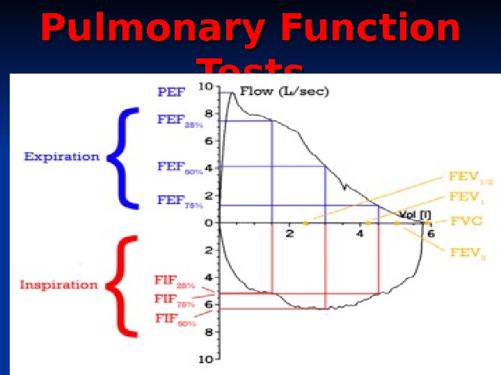
Spirometry Normal flow-volume loop Flow-volume loop in severe COP
Pulmonary Function Tests
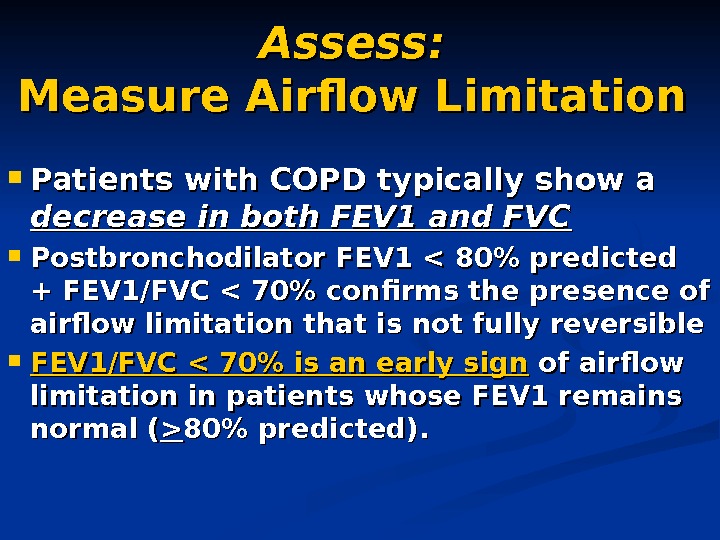
Assess: Measure Airflow Limitation Patients with COPD typically show a decrease in both FEV 1 and FVC Postbronchodilator FEV 1 < 80% predicted + FEV 1/FVC < 70% confirms the presence of airflow limitation that is not fully reversible FEV 1/FVC > 80% predicted).
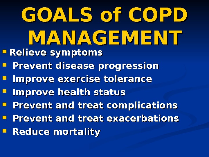
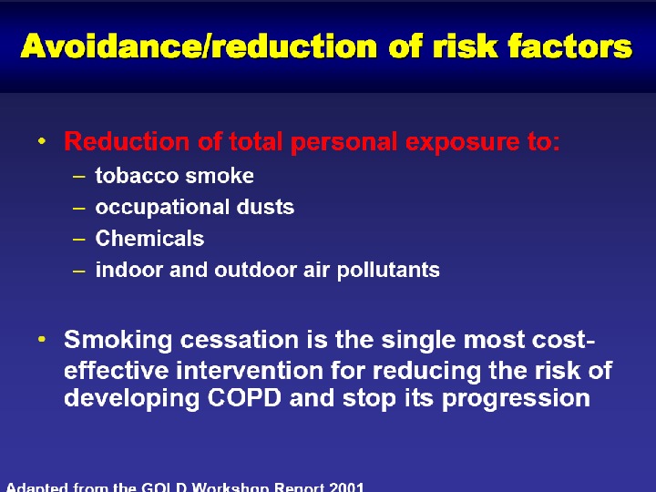
GOALS of COPD MANAGEMENT Relieve symptoms Prevent disease progression Improve exercise tolerance Improve health status Prevent and treat complications Prevent and treat exacerbations Reduce mortality
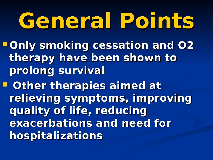
General Points Only smoking cessation and O 2 therapy have been shown to prolong survival Otherapies aimed at relieving symptoms, improving quality of life, reducing exacerbations and need for hospitalizations
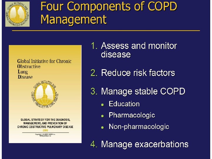
Exacerbation management Chronic stable management Adjuvant therapy
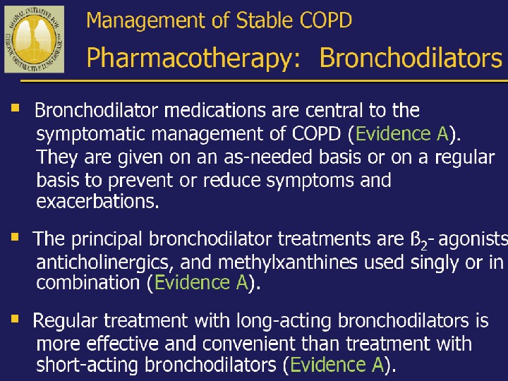
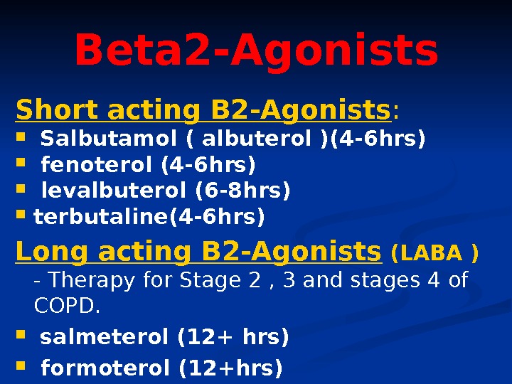
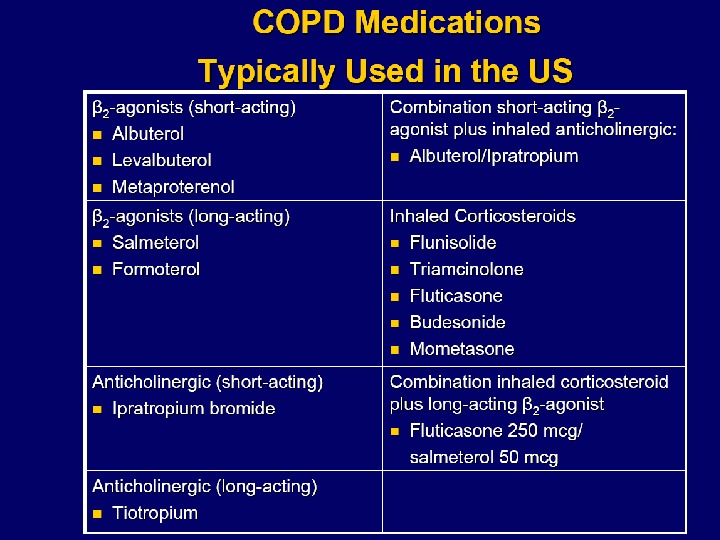
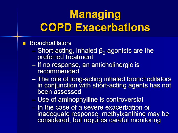
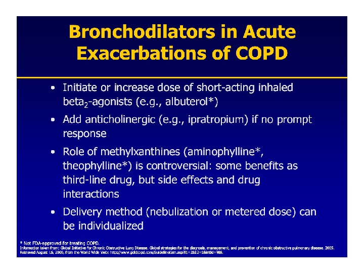
Beta 2 -Agonists Short acting B 2 -Agonists : Salbutamol ( albuterol )(4 -6 hrs) fenoterol (4 -6 hrs) levalbuterol (6 -8 hrs) terbutaline(4 -6 hrs) Long acting B 2 -Agonists (LABA ) — Therapy for Stage 2 , 3 and stages 4 of COPD. salmeterol (12+ hrs) formoterol (12+hrs)
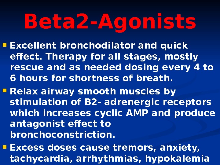
Beta 2 -Agonists Excellent bronchodilator and quick effect. Therapy for all stages, mostly rescue and as needed dosing every 4 to 6 hours for shortness of breath. Relax airway smooth muscles by stimulation of B 2 — adrenergic receptors which increases cyclic AMP and produce antagonist effect to bronchoconstriction. Excess doses cause tremors, anxiety, tachycardia, arrhythmias, hypokalemia
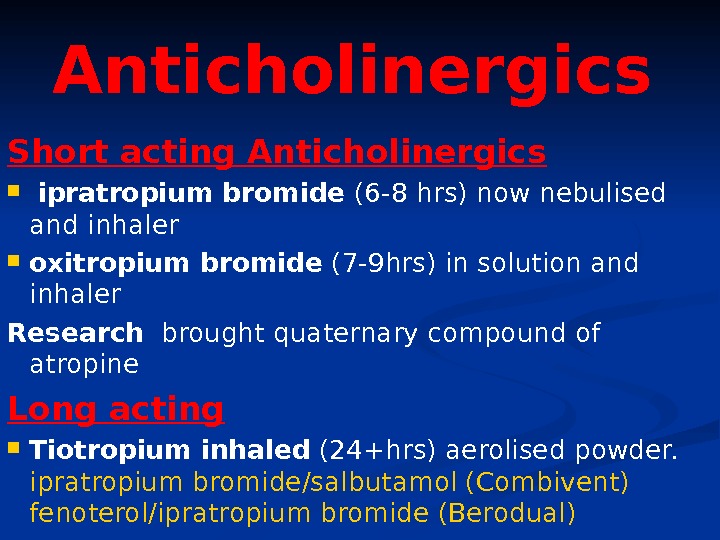
Anticholinergics Short acting Anticholinergics ipratropium bromide (6 -8 hrs) now nebulised and inhaler oxitropium bromide (7 -9 hrs) in solution and inhaler Research brought quaternary compound of atropine Long acting Tiotropium inhaled (24+hrs) aerolised powder. ipratropium bromide/salbutamol (Combivent) fenoterol/ipratropium bromide (Berodual)
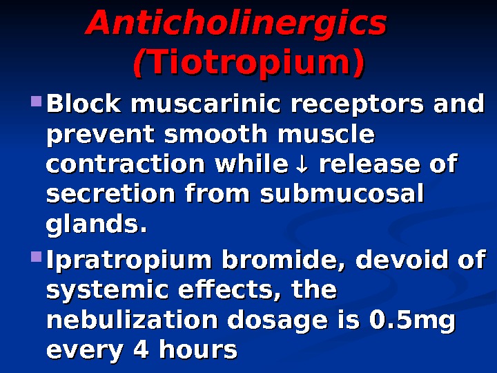
Anticholinergics (( Tiotropium) Block muscarinic receptors and prevent smooth muscle contraction while release of secretion from submucosal glands. Ipratropium bromide, devoid of systemic effects, the nebulization dosage is 0. 5 mg every 4 hours
Drug therapy for COPD begins with long acting anticholinergics and beta-2 agonist bronchodilators. These provide symptom relief but do not stop progression of the disease
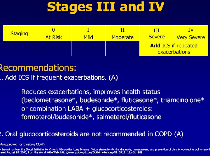
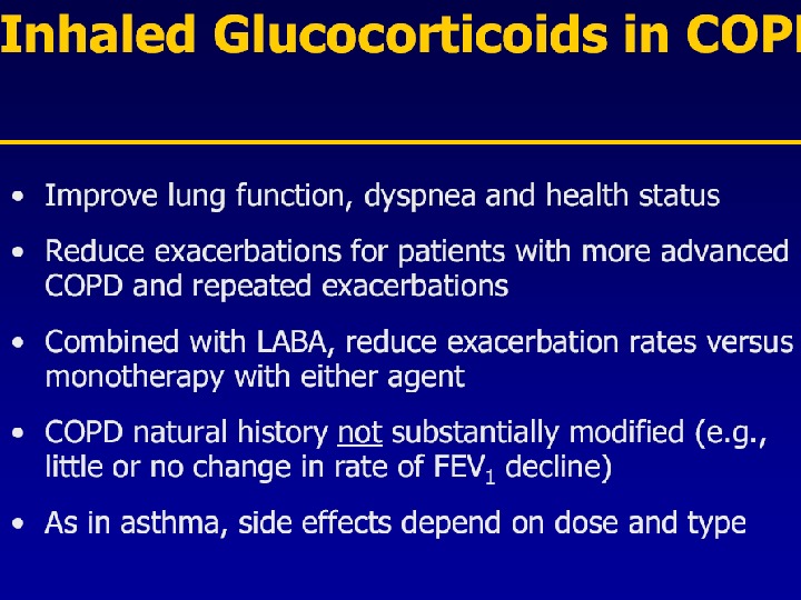
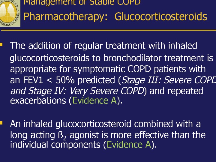
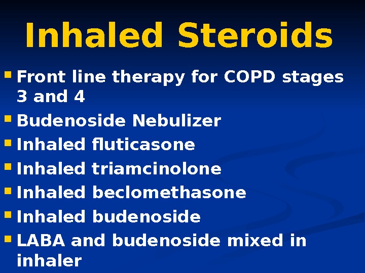
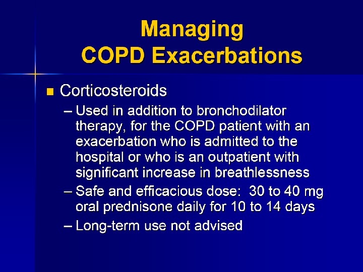
Inhaled Steroids Front line therapy for COPD stages 3 and 4 Budenoside Nebulizer Inhaled fluticasone Inhaled triamcinolone Inhaled beclomethasone Inhaled budenoside LABA and budenoside mixed in inhaler
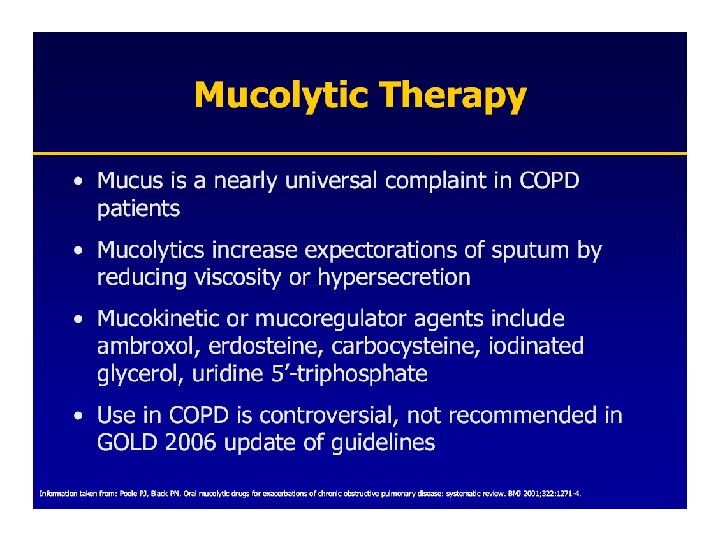
Treatment of Stable COPD Other Medications Chronic oral Prednisone Use in chronic COPD is controversial. No effect on survival. May improve symptoms and reduce hospitalizations in some patients already at maximum treatment Mucolytics & Expectorants Relives symptoms from copious, viscous secretions Oral Theophylline ( ( If inhalers not sufficient ) Side effects are common
Methylxanthines Multiple modes of action : bronchodilatation, dia-phragmatic contractility, stimulation of respiratory drive, inotropism, mucociliary clearance, and synergy with ß 2 -Agonists and Anticholinergics 5 mg/kg IV over 10 to 15 min then 0. 5 mg/kg/hr if normal liver function 1 mg/kg IV elevate a 2 g/ml in blood level (await blood level results before IV dose when patient on oral aminophylline) Lower dosing : Alcoholism, old age, chronic liver disease, CHF, fever, erythromycin. ciprofloxacin or H 2 -blocker
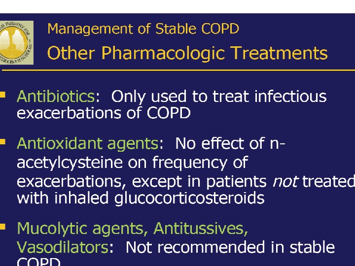
Mucokinetic Medications Nebulized water and saline and oral expectorants guaifenesin and saturated iodide are of no benefit Acetylcysteine cause reflex bronchoconstriction Clinical improvement with oral iodinated glycerol but can cause thyroid dysfunction Simple oral hydration is the easiest and safest agent
Treatment of Stable COPD: Home Oxygen Therapy >> 15 hours/day reduces mortality Criteria for O 2 therapy Pa O 2 << 55 mm Hg (O 2 saturation << 88%) at rest or during exercise or sleep oror Pa O 2 52%
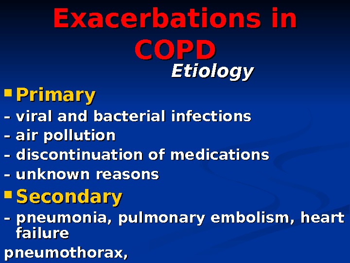
Exacerbations in COPD Etiology Primary – – viral and bacterial infections – – air pollution – – discontinuation of medications – – unknown reasons Secondary – – pneumonia, pulmonary embolism, heart failure pneumothorax,
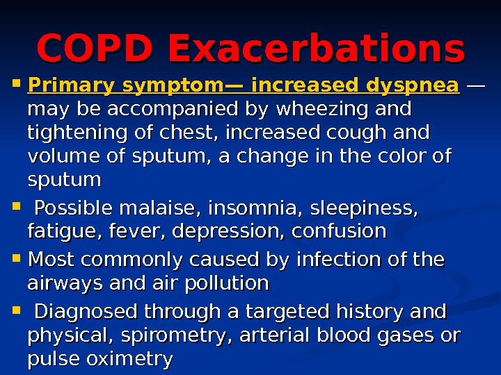
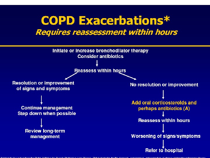
COPD Exacerbations Primary symptom— increased dyspnea —— may be accompanied by wheezing and tightening of chest, increased cough and volume of sputum, a change in the color of sputum Possible malaise, insomnia, sleepiness, fatigue, fever, depression, confusion Most commonly caused by infection of the airways and air pollution Diagnosed through a targeted history and physical, spirometry, arterial blood gases or pulse oximetry
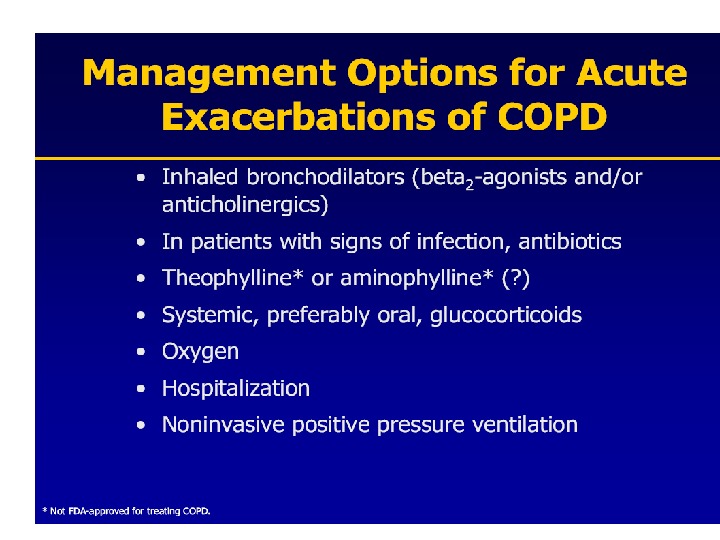
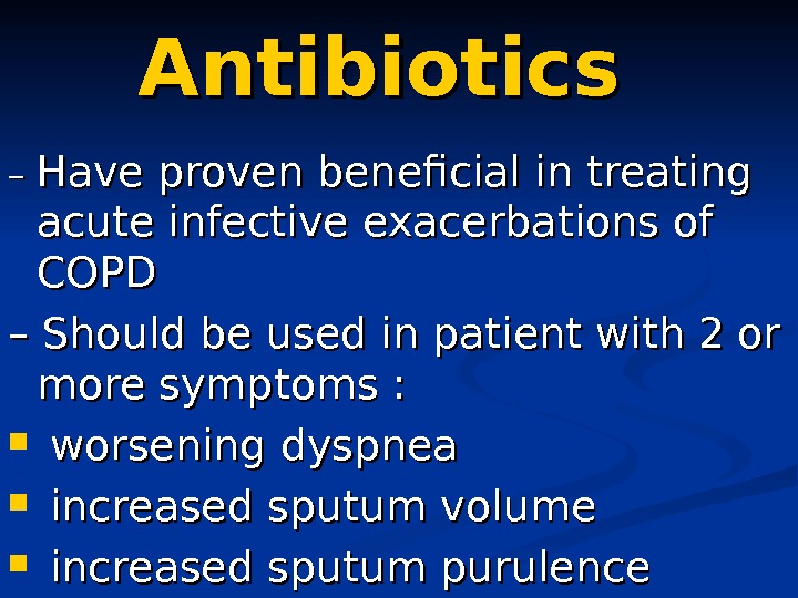
Antibiotics – – Have proven beneficial in treating acute infective exacerbations of COPD – – Should be used in patient with 2 or or more symptoms : : worsening dyspnea ii ncreased sputum volume increased sputum purulence
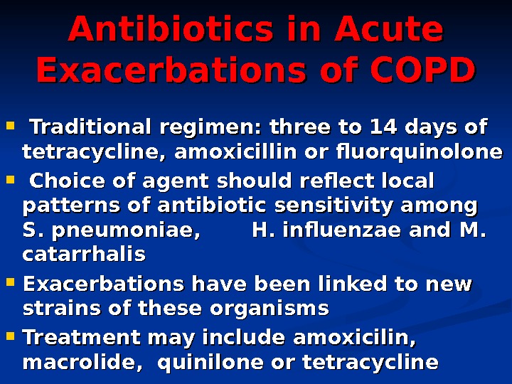
Antibiotics in Acute Exacerbations of COPD Traditional regimen: three to 14 days of tetracycline, amoxicillin or fluorquinolone Choice of agent should reflect local patterns of antibiotic sensitivity among SS. . pneumoniae, HH. . influenzae and M. . catarrhalis Exacerbations have been linked to new strains of these organisms Treatment may include amoxicilin, macrolide, quinilone or tetracycline
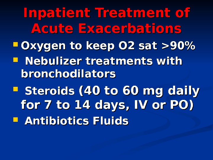
Inpatient Treatment of Acute Exacerbations Oxygen to keep O 2 sat >90% Nebulizer treatments with bronchodilators Steroids (40 to 60 mg daily for 7 to 14 days, IV or PO) Antibiotics Fluids
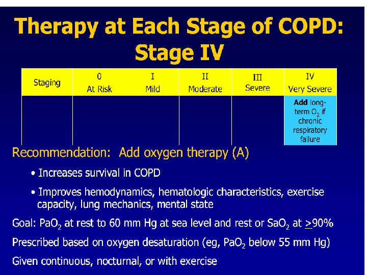
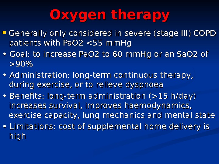
Oxygen therapy Generally only considered in severe (stage III) COPD patients with Pa. O 2 > 90%90% • • Administration: long-term continuous therapy, during exercise, or to relieve dyspnoea • • Benefits: long-term administration (>15 h/day) increases survival, improves haemodynamics, exercise capacity, lung mechanics and mental state • • Limitations: cost of supplemental home delivery is high
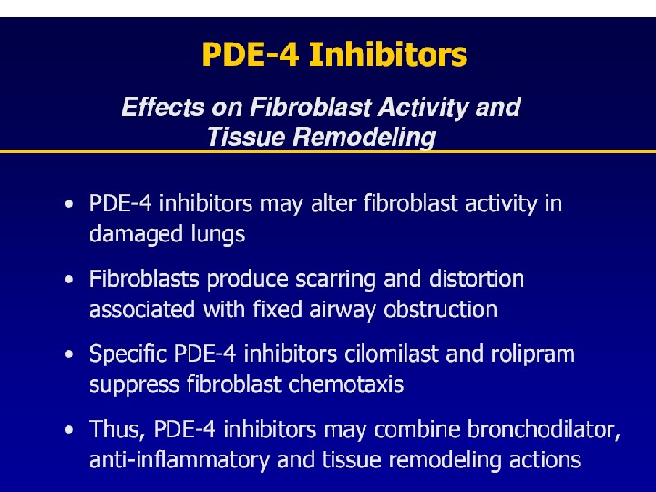
Phosphodiesterase-4 Inhibition (( Roflumilast )) Inhibition raises intracellular levels of c. AMP resulting in downregulation of signaling pathways in inflammatory cells Major isoenzyme in inflammatory cells implicated in inflammatory airway disease
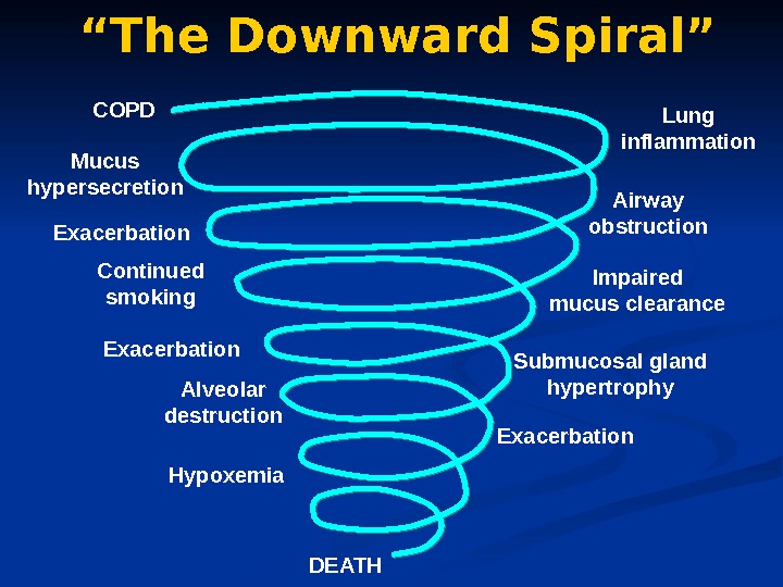
COPD Airway obstruction Exacerbation. Mucus hypersecretion Continued smoking Lung inflammation Alveolar destruction Impaired mucus clearance Submucosal gland hypertrophy Exacerbation Hypoxemia DEATH“ The Downward Spiral”
SMOKERS ““ Hope and expect for the best. Prepare for the worst. ” Back AL, Arnold RM, Quill TE. Hope for the best, and prepare for the worst. Ann Intern Med 2003; 138: 439 -43.
NEXT STAGE…
PREVENT COP
PREVENT COP