

7e8ea21ff42803e7750cad2f7def596e.ppt
- Количество слайдов: 187
 Chapter 5 Introduction to Studying Proteins
 Learning Objectives • Describe the structure of proteins • Explain transcription and translation • Discuss naturally occurring and recombinant proteins in biotechnology • Learn the function of proteins in structure, recognition and as catalysts • Explore enzymes and conditions of activity • Understand PAGE
 Structure and Function of Proteins • Proteins are essential parts of organisms and participate in virtually every process within cells • Proteins make up half the dry weight of an Escherichia coli cell, whereas other macromolecules such as DNA and RNA make up only 3% and 20%, respectively
 Structure and Function of Proteins • Biotech products are often proteins or related to proteins • Human growth hormone • BEANO (agalactosidase) • Monosodium glutamate • Amylase • ELISA • Vaccines • Toxins (black widow, cholera)
 Structure and Function of Proteins carry out the duties specified by the information encoded in genes The set of proteins expressed in a particular cell or cell type is known as its proteome
 Structure and Function of Proteins Characteristic Measured by • Molecular mass • Mass spectrometer • Normally reported in units of daltons (synonymous with atomic mass units), or the derivative unit kilodalton (k. Da). • Insulin 4 k. Da in mass • Muscle protein (myosin)250 k. D in mass – ionizing chemical compounds to generate charged molecules – sorts the ions by mass by applying electromagnetic field
 Structure and Function of Proteins Characteristic Three-Dimensional Structure Methods X-ray Crystallography • Pure crystals of proteins are generated • X-ray are directed on the crystals • Position of atoms diffract the x-ray • Diffraction pattern analyzed by computer programs to generate a 3 -D picture
 Structure and Function of Protein Chemical behavior Activity • • • Catalysis Structure Movement Defense Regulation Transport
 Structure and Function Solubility Dependant on Shape • Insoluble in water—keratins • Fibrous proteins with long, found in skin, hair and nails, rod-shaped molecules • Soluble in water—nearly all enzymes and immunoglobulin's— dynamic functions • Globular proteins are compact spherical molecules that usually bind other molecules
 Structure and Function of Proteins Charge Electrophoresis/Chromatography • Charge is determined by R groups on the amino acids in the protein • Separates on charge and size • PAGE = Polyacrylamide gel electrophoresis • Use SDS to impart a negative charge on all proteins
 To Summarize: The structure and Function of Proteins • Mass • Three dimensional structure • Activity • Solubility • Charge
 Protein Molecule Structure • Proteins are polymers of amino acids • Common structure • • • Central Carbon atom Amino Group Carboxylic acid R-group H H H 2 N – C--COOH R
 Amino Acids • Twenty different amino acids • Categorized by the nature of the R group • Zwitterions (Carry both a positive and negative charge in water solutions) H O H 2 N – C--COH R
 Amino Acids • Twenty different amino acids • Categorized by the nature of the R group H O • Zwitterions (Carry both +H N – C—CO 3 a positive and negative R charge in water solutions)
 Amino Acids and Polarity • The interaction of amino acids with a water solution (the cytosol) and with other amino acids in a protein will depend upon the polarity of the R group.
 Polar water molecule – Unequal sharing of electrons in a covalent bond leads to a polar molecule. • IN WATER: – The region around oxygen bears a partial negative charge – The region around the hydrogens bear partial positive charge – Water forms hydrogen bonds
 What does it mean to be polar? Oil and Water do not mix • Very different bonding characteristics • Must have bonding similar to water to interact with water • These interactions drive the secondary and tertiary structure of proteins
 Amino Acids • R groups may be: • Acidic…………………. . Loss of a proton (H+) Negatively charged • Basic…………………. Gain of a proton (H+) Positively charged • Polar…………………. . Characterized by OH Interact with water and other polar groups • Hydrophobic……………. Characterized by C-H » Repelled by water
 Amino acids Acidic Amino Acid Basic
 Amino Acids Nonpolar Polar
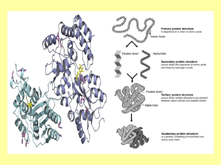
 Primary Structure The sequence of amino acids in the protein
 Secondary Structure : Hydrogen bonding between atoms in the peptide bond can produce regular repeating patterns Beta- Pleated Sheet Alpha Helix
 Secondary Structure
 GFP-Structural Barrel with Beta -Pleated Sheet
 Tertiary Structure: Overall folded pattern of a Globular Protein
 Sickle-Cell anemia due to change in molecular shape An A to T mutation of the βglobin gene results in the amino acid glutamate to be substituted by valine at position 6 of the polypeptide The genetic disorder is due to the mutation of a single nucleotide, from a GAG to GTG codon mutation.
 When deoxygenated in the capillaries, the S-hemoglobin has a hydrophobic patch on the protein The hydrophobic residues of the valine at position 6 of the beta chain in hemoglobin are able to associate with the hydrophobic patch, causing hemoglobin S molecules to aggregate and form fibrous precipitates
 Changes to primary structure lead to changes in tertiary structure and function • Hb. S molecules tend to clump together, making red blood cells sticky, stiff, and more fragile, and causing them to form into a curved, sickle shape.
 Quaternary Structure: 2 or more subunits associate to form the functional protein Hemoglobin DNA Polymerase
 http: //wpcontent. answers. com/wikipedia/commons/b/ba/Hemoglobin_tr_state_ani. gif
 Proteins grouped by function • • • Structural Enzyme Transport Contractile Hormone Pigment Recognition Toxins Antibody • • • Collagen, keratin Amylase, rennin Hemoglobin, HDL Actin, myosin, tubulin Insulin, Adrenalin Melainin, rhodopsin Glycoprotein, MHC Botox, tetanus Gamma globulin, Ig. E
 Antibodies: Structure and Function RECOGNIZE, BIND & NEUTRALIZE STRUCTURE CRITICAL TO FUNCTION • Recognize foreign proteins called antigens • Tag and aggregate antigens • Thousands of different foreign invaders need to be recognized—NEED VARIETY • Remove or neutralize antigenic proteins in the body • Molecular weapons of defense • All antigens will be recognized, bound, marked and clumped together for removal – NEED A CONSTANT REGION
 Each Antibody has Same Basic Shape • Y-shaped glycoprotein • Made of 4 polypeptide chains – Two heavy chains – Two light chains
 Antibody structure • Variable region— identifies all the many possible antigens • Constant region that is identical for all antibodies –the amino acid sequence is the same for each class of antibodies • Held together by disulfide bonds
 Types of Antibodies • There are several different types of antibody heavy chains, and several different kinds of antibodies, • Grouped into different isotypes based on which heavy chain they possess. • Five different antibody isotypes are known in mammals • Perform different roles • Direct the appropriate immune response for each different type of foreign object they encounter.
 Five Antibody Isotypes • Five antibody isotypes known as Ig. A, Ig. D, Ig. E, Ig. G and Ig. M. • They are each named with an "Ig" prefix that stands for immunoglobulin (another name for antibody) • Differ in their biological properties, functional locations and ability to deal with different antigens
 Name Description Ig. A Found in mucosal areas, such as the gut, respiratory tract and urogenital tract, and prevents colonization by pathogens. Also found in saliva, tears, and breast milk Ig. D Functions mainly as an antigen receptor on B cells that have not been exposed to antigens. It has been shown to activate basophils and mast cells to produce antimicrobial factors. Ig. E Binds to allergens and triggers histamine release from mast cells and basophils, and is involved in allergy. Also protects against parasitic worms. Ig. G In its four forms, provides the majority of antibody-based immunity against invading pathogens. The only antibody capable of crossing the placenta to give passive immunity to fetus. Ig. M Expressed on the surface of B cells and in a secreted form with very high avidity. Eliminates pathogens in the early stages of B cell mediated (humoral) immunity before there is sufficient Ig. G
 • The basic structural units—each with two large heavy chains and two small light chains. • May form monomers with one unit, dimers with two units or pentamers with five units
 Variable region allows for recogniton of various antigens • The small region at the tip of the protein is extremely variable • Known as the hypervariable region. • Each will bind to a different antigen.
 Diversity of antibodies allows the immune system to recognize a diversity of antigens • The unique part is called an epitope. • A highly specific interaction with antigen by induced fit • Recognition of an antigen by an antibody tags it for attack by other parts of the immune system. • Neutralize targets directly by binding to a part of a pathogen that it needs to cause an infection.
 Genetic Shuffle Creates Variety • The diversity of antibodies is due to random combinations of a set of gene segments • Gene encodes different antigen binding sites • Random mutations in this area of the antibody gene to create further diversity. • Antibody genes also re-organize in a process to change the base of the heavy chain to another
 Mark Antigens (AG)for Elimination • Binding of AB to AG • Enhances") Antibodies (AB) Mark Antigens (AG)for Elimination • Binding of AB to AG • Enhances phagocytosis – Neutralize the AG – Agglutinate microbe – Precipitate dissolved antigen – Activate complement • Leads to cell lysis
Antibodies (AB) Mark Antigens (AG)for Elimination • Binding of AB to AG • Enhances phagocytosis – Neutralize the AG – Agglutinate microbe – Precipitate dissolved antigen – Activate complement • Leads to cell lysis
 Antibody Variability • Creates a different isotype of the antibody but retains the antigen specific variable region • Allows a single antibody to be used by several different parts of the immune system
 Problems with Immune System • Autoimmune disorders • Antibodies against self – (Lupus, R. Arthritis, MS, Diabetes) • Immunodeficiency disease – (SCID, Hodfkin’s, AIDS) • Allergies – Abnormal Sensitivity to antigens
 Allergens • Antigens that induce the formation of Ig. E antibodies • Excess Ig. E stimulates an inflammatory response – – Allergen binds to antibodies bound to MAST cells Mast cells release histamine Blood vessels dilate and leak fliud Nasal irritation, itchy skin and tears • Aanaphylactic shock—so rapid– blood pressure falls
 Ouchterlony Test for antigen-antibody • Hundreds of AB-AG complexes form to produce a precipitation
 Poison Ivy Exaggerated Immune Response • The poison ivy plant and its relatives are common throughout the United States. • Poison ivy leaves are coated with a mixture of chemicals called urushiol
 Allergic Contact Dermatitis Allergy is an altered or unwanted immune response Dermatitis is an inflammation of the skin • The immune system • Response to something neutralizes and eliminates which came into contact foreign substances from our with the skin bodies. • Poison ivy, other things • It sometimes attacks which contact the skin such harmless substances as clothing, shampoo, vigorously, causing an jewelry, make-up, and inflammation which can be deodorants can also cause far more harmful than the allergic contact dermatitis. foreign substance alone. • Also can be caused from something we ate.
 Toxic Effects of Urushiol are indirect, mediated by an induced autoimmune response Urushiol acts as a hapten, • chemically reacting with • binding to and • changing the shape of integral membrane proteins on exposed skin cells. Affected proteins no longer are recognized as normal parts of the body, causing an immune response
 Poison Ivy • This immune response is directed towards the complex of urushiol bound with the skin proteins, attacking the cells as if they were foreign bodies.
 Proteins grouped by function • • • Structural Enzyme Transport Contractile Hormone Pigment Recognition Toxins Antibody • • • Collagen, keratin Amylase, rennin Hemoglobin, HDL Actin, myosin, tubulin Insulin, Adrenalin Melainin, rhodopsin Glycoprotein, MHC Botox, tetanus Gamma globulin, Ig. E
 Proteins for recognition • Proteins are used to identify an organism’s normal healthy cells from viral infected or tumor cells that need to eliminated • Macrophages have identified a cancer cell (the large, spiky mass). Upon fusing with the cancer cell, the macrophages (smaller white cells) will inject toxins that kill the tumor cell.
 The immune system and autoimmunity…. . Uses MHC genes • The major histocompatibility complex (MHC)genes and proteins (HLA) play an important role • A large genomic region or gene family found in most vertebrates. • Genetically. . . most genedense region of the mammalian genome
 The MHC gene is the most polymorphic in the genome • Extreme allelic diversity • Nine classical genes • Roughly 250, 500, and 300 known alleles on chromosome 6 • Proteins can be referred to as HLA proteins for the name--Human leukocyte antigens
 MHC proteins act as "signposts” • MHC proteins act as "signposts" that display fragmented pieces of an antigen on the host cell's surface
 MHC proteins act as "signposts • Expressed on the surface of cells • Display both self antigens (peptide fragments from the cell itself) and nonself antigens to a white blood cell (a T cell) that has the capacity to kill What gets displayed • fragments of invading microorganisms of pathogens • Viral protein from infected • Protein from malfunctioning cells like tumor cells
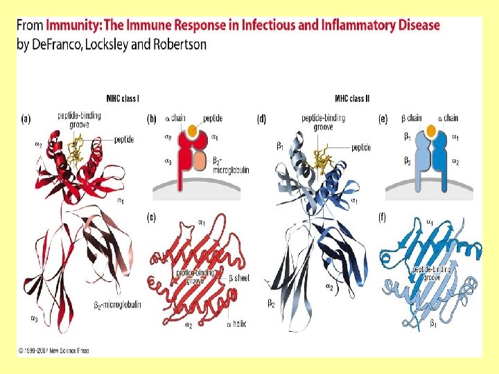
 MHC molecules retrieve polypeptides from the interior of the cell they are part of and display them on the cell's surface for recognition by T cells of the immune system
 is a transmembrane glycoprotein that serves as a") CD 8 (cluster of differentiation 8) is a transmembrane glycoprotein that serves as a co-receptor for the T cell recepter (TCR). CD 8 binds to a major histocompatibility complex (MHC) molecule.
CD 8 (cluster of differentiation 8) is a transmembrane glycoprotein that serves as a co-receptor for the T cell recepter (TCR). CD 8 binds to a major histocompatibility complex (MHC) molecule.
 MHC Molecules As Glycoproteins
 Glycoproteins • Proteins that contain sugar chains bonded to their polypeptide sidechains. • The carbohydrate is attached to the protein in a cotranslational or posttranslational modification. • Not in the genetic code!
 Two types of Glycoproteins • N-glycosylation the addition of sugar chains on the nitrogen on the side chain of the asparagine. • In O-glycosylation, the addition of sugar chains can happen on the hydroxyl oxygen on the side chain of serine, or threonine
 Sugars used include mannose, fucose and others generally not used in respiration
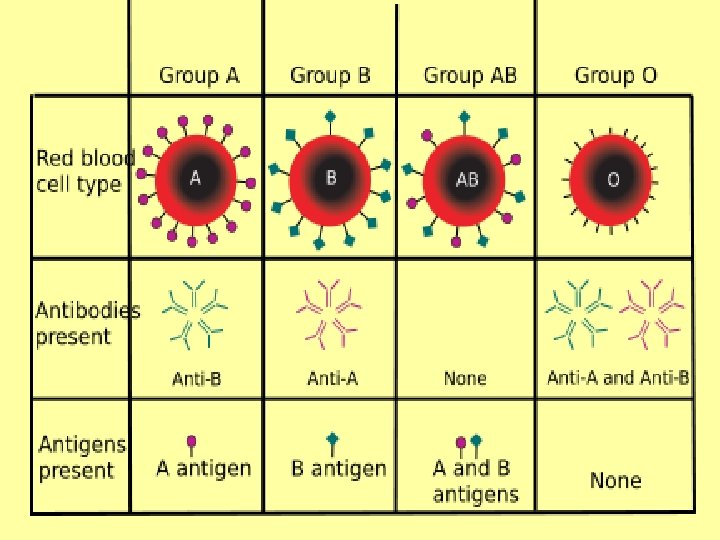
 The ABO blood group system • Based on sugars linked to the proteins found the red blood cells
 The H antigen • Essential precursor to • It encodes a the ABO blood group fucosyltransferase that antigens. produces the H antigen on RBCs. • The H gene is located on chromosome 19. It contains 3 exons that • The H antigen is a span more than 5 kb of carbohydrate sequence genomic DNA with carbohydrates linked mainly to protein
 The ABO blood group system • Based on sugars linked to the proteins found the red blood cells
 The ABO locus has three main alleleic forms: A, B, and O. • The A allele encodes a • The B allele encodes a glycosyltransferase that bonds α-Njoins α-D-galactose acetylgalactosamine to bonded to D-galactose end of H antigen, producing the creating the B antigen A antigen.
 The H antigen remaining unchanged in case of O groups • In case of O allele the gene contains a deletion that results in a loss of enzymatic activity by glycosyltransferase. • The O allele differs slightly from the A allele by deletion of a single nucleotide - Guanine at position 261. • The deletion causes a frameshift and results in translation of an almost entirely different protein that lacks enzymatic activity.
 Recognition Proteins • MHC protein for self recognition and infection • Glycoproteins as blood type proteins • Viral recognition proteins
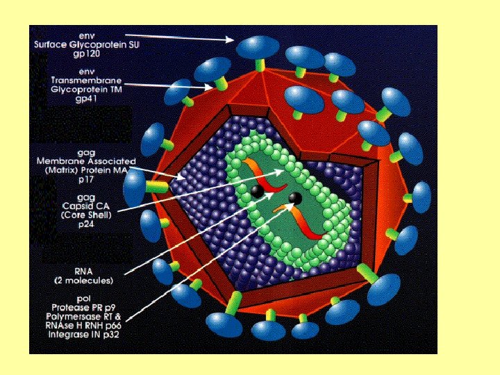
 Glycoprotein 120 and HIV • Exists on the surface of HIV • Several roles for the virus – Causes virus to attach to T-helper cells – Required for HIV to infect a T-helper cell • Role depends upon its shape – Single polypeptide – Five looped domains
 GP 120 • Gene is around 1500 nucleotides long • GP-120 is made of around 500 amino acid residues • Give HIV its spikey appearance • Also include gp 41
 GP 120 and CD 4 receptors and infection of the cell • GP 120 Binds to CD 4 receptor on a T cell bearing such receptors before infection of the cell can occur • CD 4 binding site of gp 120 • the specific region of the molecule which attaches to CD 4 via intermolecular attractions
 HIV Characteristics • Protein coat—GP 120 • Genetic information— RNA • Enzymes of replication —Reverse Transcriptase
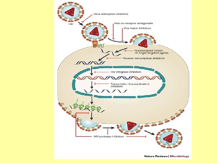
 Events of Infection • HIV “bumps” into CD 4 cell with recpetors. • Reactions take place to capture virus • Reverse transcriptase converts RNA to DNA (called c. DNA—not like genomic DNA) • c. DNA integrates into genomic DNA • Production of viral proteins and viral particles
 HIV Infection • HIV primarily infects vital cells in the human immune system such as helper T cells (specifically CD 4+ T cells), macrophages, and dendritic cells.
 HIV infection leads to low levels of CD 4+ T cells Three main mechanisms: 1. firstly, direct viral killing of infected cells; 2. secondly, increased rates of apoptosis in infected cells; and 3. thirdly, killing of infected CD 4+ T cells by CD 8 cytotoxic lymphocytes that recognize infected cells.
 Progression to AIDS • When CD 4+ T cell numbers decline below a critical level, cell-mediated immunity is lost, and the body becomes progressively more susceptible to opportunistic infections
 CD 4 receptor binding is the most obvious step in HIV infection • The protein gp 120 is necessary during the initial binding of HIV to its target cell. • GP 120 was among the first targets of HIV vaccine research • Consequently, anything which binds to gp 120's target can block gp 120 from binding to a cell by being physically in the way. • Many of these are toxic to the immune system
 Errors of Opportunity • Reverse Transcriptase makes several errors • High mutation rate is beneficial for the virus • Surface proteins are constantly changing
 ELISA • Enzyme-linked immunosorbent assay is a biochemical technique used to detect the presence of an antibody or an antigen in a sample
 In ELISA……………. . 1. An unknown amount of antigen is affixed to a surface 2. Then a specific antibody is washed over the surface The antibody binds to the antigen. 3. A secondary antibody, linked to an enzyme, is added 4. Chemically developed
 • In the final step a substance is added that the enzyme can convert to some detectable signal.
 Fluorescence ELISA • When light of the appropriate wavelength is shone upon the sample, any antigen/antibody complexes will fluorescence. • The amount of antigen in the sample can be inferred through the magnitude of the fluorescence.
 ELISA FOR DIAGNOSIS • ELISA can be performed • HIV test or West Nile to evaluate either the Virus presence of antigen or • It has also found the presence of applications in the food antibody in a sample industry in detecting potential food allergens • It is a useful tool for • In toxicology as a rapid determining serum screen for certain antibody concentrations classes of drugs.
 ELISA: Testing for HIV • In an ELISA, a person's serum is diluted 400 fold and applied to a plate to which HIV antigens are attached. • If antibodies to HIV are present in the serum, they may bind to these HIV antigens.
 ELISA: Testing for HIV • The plate is then • Followed by another washed to remove all wash. other components of • This secondary antibody the serum. is chemically linked in • A specially prepared advance to an enzyme "secondary antibody" — an antibody that binds to other antibodies — is then applied to the plate.
 • The plate will contain enzyme in proportion to the amount of secondary antibody bound to the plate. • A substrate for the enzyme is applied, and catalysis by the enzyme leads to a change in color or fluorescence
 Monoclonal antibodies (m. Ab or mo. Ab)") Source of Antibodies Polyclonal antibodies (or antisera) Monoclonal antibodies (m. Ab or mo. Ab) • are antibodies that are • are monospecific antibodies derived from different B cell that are identical because lines. They are a mixture of they are produced by one immunoglobulin molecules type of immune cell that are secreted against a specific all clones of a single parent antigen, each recognizing a cell different epitope.
Source of Antibodies Polyclonal antibodies (or antisera) Monoclonal antibodies (m. Ab or mo. Ab) • are antibodies that are • are monospecific antibodies derived from different B cell that are identical because lines. They are a mixture of they are produced by one immunoglobulin molecules type of immune cell that are secreted against a specific all clones of a single parent antigen, each recognizing a cell different epitope.
 Polyclonal antibodies These antibodies are typically produced by immunization of a suitable mammal, such as a mouse, rabbit or goat. Larger mammals are often preferred as the amount of serum that can be collected is greater.
 Polyclonal antibodies • An antigen is injected into the mammal. • This induces the Blymphocytes to produce Ig. G immunoglobulins specific for the antigen. • This polyclonal Ig. G is polyclonal purified from the mammal’s serum
 Monoclonal AB production • Antigen is injected in animal for production • Spleen cells making AB are targetd • Fuse with cancer cells • Hybridoma makes specific AB—lots of it!
 Monoclonal antibodies are derived from a single cell line Given almost any substance, it is possible to create monoclonal antibodies that specifically bind to that substance; they can then serve to detect or purify that substance
 Molecular Delivery • Such m. Ab could also be modified for delivery of a toxin, radioisotope, cytokine or other active conjugate
 Monoclonal antibodies for cancer treatment Cancer treatment using monoclonal antibodies that bind only to cancer cell-specific antigens and induce an immunological response against the target cancer cell. • Molecularly targeted drugs
 Proteins grouped by function • • • Structural Enzyme Transport Contractile Hormone Pigment Recognition Toxins Antibody • • • Collagen, keratin Amylase, rennin Hemoglobin, HDL Actin, myosin, tubulin Insulin, Adrenalin Melainin, rhodopsin Glycoprotein, MHC Botox, tetanus Gamma globulin, Ig. E
 Proteins grouped by function • • • Structural Enzyme Transport Contractile Hormone Pigment Recognition Toxins Antibody • • • Collagen, keratin Amylase, rennin Hemoglobin, HDL Actin, myosin, tubulin Insulin, Adrenalin Melainin, rhodopsin Glycoprotein, MHC Botox, tetanus Gamma globulin, Ig. E
 ENZYMES • Enzymes are proteins that catalyze chemical reactions • Increase the rate of a reaction • Allow a reaction to occur at a lower temperature
 Enzyme Catalysis In enzymatic reactions, the molecules at the beginning of a reaction are converted into different molecules. Amylase Starch Maltose ( also called amylose) Substrate Product
 In enzymatic reactions • The molecules at the beginning of the reaction are called substrates Very specific as to which reactions they catalyze • The enzyme provides an environment to convert a substrate to new molecules called products Very specific as to which substrates are involved in these reactions
 Enzymes speed up a reaction • All catalysts work by lowering the activation energy for a reaction, • This dramatically increases the rate of the reaction. • Enzyme reaction rates are millions of times faster than those of comparable uncatalyzed reactions
 Enzymes speed up a reaction • Unlike other kind of catalysts – The enzyme molecule contains a unique surface that binds the substrate………. . called the – Active site
 Enzyme Active Site • Many amino acids that line the active site participate in the catalytic process • Lock and Key model • Vs • Induced fit
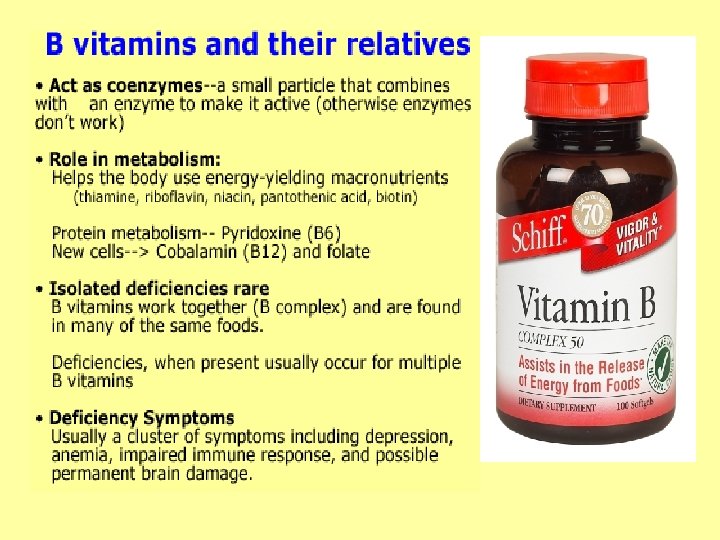
 Enzyme Activity May Require other Groups • Active site amino acids interact with substrate • Enzyme cofactors (coenzymes) required by some enzymes • Ions Mg 2+, Ca 2+ etc. . • Vitamins
 Enzyme names provide clues to function • First enzymes identified were named using common names • Rennin • Trypsin • Chymotrypsin • Pepsin
 Enzyme names provide clues to function • A system of naming enzymes was introduced • Name (telling function) • End with suffix -ASE • Cellulase – Breaks down cellulose • Protease – Breaks down protein • Pectinase – Breaks down pectin
 Plant Molecules Acted Upon by Enzymes Cellulose Pectin • Polysaccharides of glucose • Consists of a polysaccharides present primary cell walls • The structural component of the primary cell wall of green plants • In the non-woody parts of terrestrial plants • Also in the middle lamella between plant cells where it helps to bind cells together
 Enzyme Treatment Enzyme, u. L Water, u. L Juice, vol Ave 0 800 Cellulase 200 600 Cellulase 400 Cellulase 800 0 Pectinase 200 600 Pectinase 400 Pectinase 800 0 Protease 200 600 Protease 400 Protease 800 0 Rennin 200 600 Rennin 400 Rennin 800 0
 Major Enzyme Categories
 Factors that Affect Enzyme Structure
 Factors that Affect Enzyme Structure • Changes in conditions can affect the enzyme by disrupting the interactions that are responsible for 2 o, 3 o and 4 o structure. • When destroyed—the protein denatures
 Factors Affecting Enzymatic Structure • p. H • Temperature • Ionic strength • Solubility
 Structure vs Function • Loss of structure results in loss of function • Temperature • Factors affecting function are the same • Concentration of Enzyme • Other factors affect function • Concentration of Substrate (to a limit)
 Proteases are enzymes that degrade polypeptides • Cysteine proteases have a common catalytic mechanism that involves a cysteine • Cysteine proteases are used as an ingredient in meat tenderizers
 Cysteine proteases Papain • Enzyme present in the milky juice of the papaya • Catalyzes the breakdown of proteins by hydrolysis (addition of a water molecule) Bromelain • One of two protease enzymes extracted from the plant family Bromeliaceae, • Pineapple, kiwi
 Papain • 212 amino acids • 3 disulfide bridges • 3 D structure consists of 2 distinct structural domains with a cleft between them.
 Catalysis uses Cysteine • the active site which contains a catalytic triad • Made up of 3 amino acids cysteine-25 histidine 159 asparagine-158
 Papain is used in biochemical research • in tenderizing meat, Breaks down the protein toxins in the venom • in enzyme-action cleansing agents for soft contact lenses. It is also the main • Home remedy ingredient in Stop Itch treatment for jellyfish, and Stop Itch Plus, a bee (wasps) stings, Derma. Tech mosquito bites, and Laboratories first aid possibly stingray cream popular in wounds Australia
 Bromelain • It is an antiinflammatory agent, • used for sports injury, trauma, arthritis, and other kinds of swelling. • treatment of athletic injuries, digestive problems, phlebitis, sinusitis, and aiding healing after surgery. • Doses of 200 mg have proven to be effective alternative to NSAIDS
 Bromelain • As a marinade or sprinkled on uncooked meat. • The enzyme will penetrate the meat • If the enzyme is allowed to work for too long, the meat may become too "mushy" • Cooked or canned pineapple does not have a tenderizing effect, as the enzymes are heat labile
 Studying Proteins Separate Proteins based on Most common techniques used • Solubility • Chromatography • Size • Electrophoresis • Charge • Binding Affinity
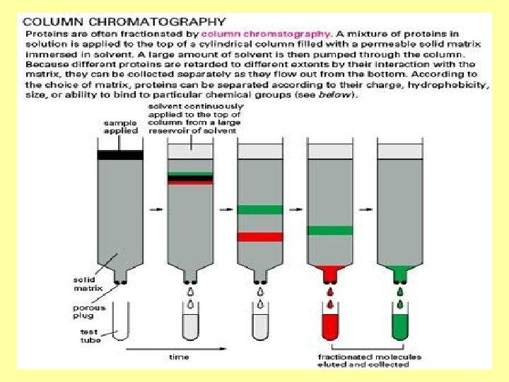
 Columns may be filled with beads to separate based on • Size—Gel filtration • Charge –Ion Exchange • Binding Affinity. Antibodies or another ligand Ex. Concavalin A = Glucose
 Poly. Acrylamide Gel Electrophoresis Gel electrophoresis is • Gel-the matrix used to contain, then separate technique used for the target molecules separation of • deoxyribonucleic acid (DNA), • ribonucleic acid (RNA), • protein molecules using an electric field applied to a gel matrix
 GELS………………. . crosslinked polymer • Vary in composition andporosity • Selection based on the weight and composition of the target to be analyzed • The gel forms a solid, yet porous matrix
 Gels………………. . crosslinked polymer Agarose Polyacrylamide • When separating larger nucleic acids the preferred matrix is purified agarose. • When separating proteins or small nucleic acids or oligonucleotides the gel is acrylamide and a crosslinker …creating polyacrylamide
 PAGE vs. Agarose • Acrylamide, in contrast • Agarose is composed of to polyacrylamide, is a long unbranched chains neurotoxin and must be of uncharged handled using carbohydrate without appropriate safety cross links resulting in a precautions to avoid gel with large pores poisoning. Pore size can allowing for the be manipulated by % separation of composition macromolecules and macromolecular complexes
 • Determined largely by •") Electrophoresis • The electromotive force Rate of movement (EMF) • Determined largely by • Moves molecules mass when the charge through a gel matrix. to mass ratio (Z) of all species is uniform • Molecules are placed wells in the gel • An electric field is • Move toward the anode applied, the molecules if negatively charged or will move through the toward the cathode if matrix at different rates positively charged
Electrophoresis • The electromotive force Rate of movement (EMF) • Determined largely by • Moves molecules mass when the charge through a gel matrix. to mass ratio (Z) of all species is uniform • Molecules are placed wells in the gel • An electric field is • Move toward the anode applied, the molecules if negatively charged or will move through the toward the cathode if matrix at different rates positively charged
 Proteins can have varying charges and complex shapes • May not migrate into the polyacrylamide gel at similar rates, or at all, when placing a negative to positive EMF on the sample.
 Proteins are subjected to SDS • Proteins therefore, are usually denatured in the presence of a detergent such as sodium dodecyl sulfate/sodium dodecyl phosphate (SDS/SDP) that coats the proteins with a negative charge
 Proteins are subjected to SDS • The amount of SDS bound is relative to the size of the • Usually 1. 4 g SDS per gram of protein • the resulting denatured proteins have an overall negative charge • all the proteins have a similar charge to mass ratio.
 Separation based only on size • Since denatured proteins act like long rods instead of having a complex tertiary shape, the rate at which the resulting SDS coated proteins migrate in the gel is relative only to its size and not its charge or shape.
 Other forms of Electrophoresis • Native gel electrophoresis • quantitative preparative native continuous polyacrylamide gel electrophoresis (QPNCPAGE • 2 -D electrophoresis
 Visualization • Stained most commonly with Coomassie Brilliant Blue R-250 • Silver stain allowing visualization of the separated proteins • Processed further (e. g. Western blot. • After staining, different proteins will appear as distinct bands within the gel.
 • Molecular markers of known molecular weight in a separate lane in the gel • Calibrate the gel and determine the weight of unknown proteins by comparing the distance traveled relative to the marker.
 Poly. Acrylamide Gel Electrophoresis • This figure has from 3, 000 ng (far left lane) to 8 ng (far right lane) of total protein loaded in the lanes. The proteins have been stained with coomassie blue.
 • The gel is actually formed because the acrylamide solution contains a small amount, generally about 1 part in 35 of bisacrylamide, which can form cross-links between two polyacrylamide molecules. • The ratio of acrylamide to bisacrylamide can be varied for special purposes. • The acrylamide concentration of the gel can also be varied, generally in the range from 5% to 25%.
 • Lower percentage gels are better for resolving very high molecular weight proteins • Determining how much of the various solutions to mix together to make gels of particular acrylamide concentration can be • While much higher done on line. percentages are needed to resolve smaller proteins.
 • http: //www. invitrogen. com/site/us/en/home/ Products-and. Services/Applications/P rotein-Expression-and. Analysis/Protein-Gel. Electrophoresis/1 DElectrophoresis/novextris-glycine-precastgels. html • http: //www. shsu. edu/~ chm_tgc/sounds/flashfil es/GE. swf
 Preparing protein samples for electrophoresis Basic 2 X Laemmli Buffer contains: • Buffer is especially formulated for protein • 4% SDS sample preparation for • 20% glycerol • 10% 2 -mercaptoethanol use in SDS-PAGE • 0. 004% bromphenol blue • 2 X sample buffer is added to each protein • 0. 125 M Tris HCl • The solution has a p. H of sample at a 1: 1 ratio, and is boiled (or heated) approximately 6. 8. on a heating block for 15 min
 Laemmli Buffer • The SDS detergent binds all positive charges giving protein overall negative charge , also denatures the proteins – subunits separate based on size, not on shape • Bromophenol blue serves as an indicator dye – functions to make it easier to see the sample during loading. • Glycerol in the Laemmli buffer increases the density – Sample will fall to the bottom of the well. • DTT or 2 ME reduces disulphide S-S bonds – disrupts secondary/tertiary structure and/or dimer formation.
 Eliminating Disulfide Bonds Beta-Mercaptoethanol Dithiothritol
 Proteins grouped by function • • • Structural Enzyme Transport Contractile Hormone Pigment Recognition Toxins Antibody • • • Collagen, keratin Amylase, rennin Hemoglobin, HDL Actin, myosin, tubulin Insulin, Adrenalin Melainin, rhodopsin Glycoprotein, MHC Botox, tetanus Gamma globulin, Ig. E
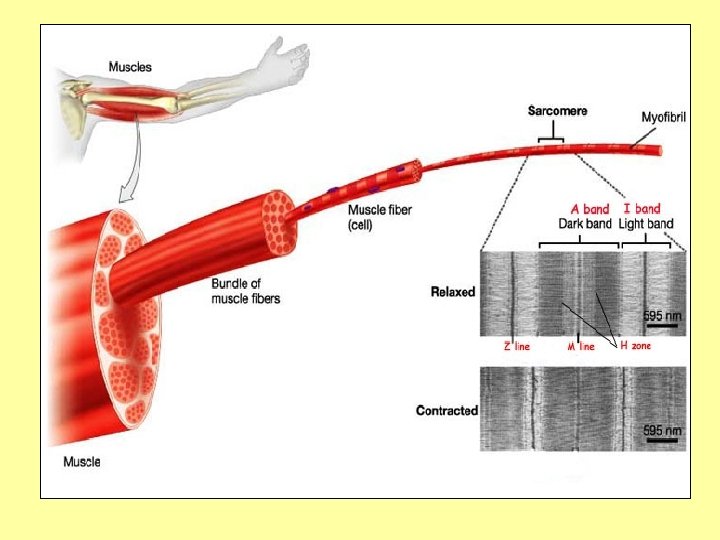
 Muscle Proteins • Movement is driven by interactions between specialized proteins in our muscle fibers • Contractile elements of muscle cells are called myofibrils • Bundled into muscle fibers
 Myofibril Sarcomeres • Each myofibril consists of a liner series of contractile units called sarcomeres • Thick myosin filaments and thin actin proteins form myofibrils that are bundled together to form a muscle fiber
 Sarcomeres are assemblies of actin and myosin Actin Myosin
 In the sarcomeres • Thin filaments of actin are aligned with thick filaments of myosin • Parallel and partly overlapping • Myosin hydrolyzes ATP • Slides along the actin filament • Ends of the sarcomeres are pulled together
 Combine contractions • Many sarcomeres contract along a fiber • Causes a contraction of the entire muscle
 Muscle Contraction • http: //video. google. com/videosearch? q=musc le+contraction+actin+myosin&hl=en&emb=0& aq=1&oq=Muscle+contraction#
 NO ATP? • Rigor mortis is one of the recognizable signs of death—no ATP • Caused by a chemical change in the muscles after death • The limbs of the corpse to become stiff (Latin rigor) and difficult to move or manipulate.
 NO ATP? • Respiration ceases • Depletes the corpse of oxygen • ATP is no longer provided to operate the sarcoplasmic movements which pump calcium
 No ATP? • Calcium ions to diffuse from the area of higher concentration • In the sarcomere, Ca binds with troponin and allowing for crossbridging to occur between myosin and actin proteins. [2] • The body is unable to complete the cycle • Creates a perpetual state of muscular contraction • Breaks-down by digestive enzymes during decomposition
 Rigor mortis is very important in meat technology. The onset of rigor mortis and its resolution partially determines the tenderness of meat. If the post-slaughter meat is immediately chilled to 15°C (59°F), a phenomenon known as cold shortening occurs, where the muscle shrinks to a third of its original size. This will lead to the loss of water from the meat along with many of the vitamins, minerals, and water soluble proteins. The loss of water makes the meat hard and interferes with the manufacturing of several meat products like cutlet and sausage. Cold shortening is caused by the release of stored calcium ions from the sarcoplasmic reticulum of muscle fibers in response to the cold stimulus. The calcium ions trigger powerful muscle contraction aided by ATP molecules. To prevent cold shortening, a process known as electrical stimulation is carried out, especially in beef carcass, immediately after slaughter and skinning. In this process, the carcass is stimulated with alternating current, causing it to contract and relax, which depletes the ATP reserve from the carcass and prevents cold shortening.
 • Numerous other proteins are required for contraction •") Other Proteins MW(k. D) • Numerous other proteins are required for contraction • Myosin and actin are highly conserved • Other muscles show greater variability • Myosin – Heavy chain=210 k. D – Light chain =19 k. D, 16 k. D • • • Titin 3, 000 Dystronphin 400 Fiamin 270 Spectrin 265 C protein 140 Actin 42 Tropomyosin 35 Troponin 19 Thymosin 5
Other Proteins MW(k. D) • Numerous other proteins are required for contraction • Myosin and actin are highly conserved • Other muscles show greater variability • Myosin – Heavy chain=210 k. D – Light chain =19 k. D, 16 k. D • • • Titin 3, 000 Dystronphin 400 Fiamin 270 Spectrin 265 C protein 140 Actin 42 Tropomyosin 35 Troponin 19 Thymosin 5
 Other Proteins • Variation in muscle protein reflect refinements of muscle function and performance • Specialization • Adaptation to particular niches, environments and stresses
 – Morphology – DNA –") Evolutionary Trees • Phylogenetic tress based on variability (similarity) – Morphology – DNA – Proteins • Complement each other, but also characteristics may evolve independently
Evolutionary Trees • Phylogenetic tress based on variability (similarity) – Morphology – DNA – Proteins • Complement each other, but also characteristics may evolve independently
 Establishing Evolutionary Trees Questions to Ask In Looking at Several Kinds of Fish • How does muscle vary from species to species • Are differences related to evolutionary history • Are similar muscle in two different species related to environment • Can SDS-PAGE provide a measure • Presence of other proteins (beyond actin and myosin) infer evolutionary relationships • Do Bioinformatic data bases help construct phylogenic trees and infer evolutionary relationships?
 DATA BASES • http: //www. fishbase. net/ • http: //animaldiversity. umm z. umich. edu/site/index. html • Biology of most of the world’s fishes. • International Center of Living Aquatic Resources Management. Question ? ? ? • What factors will cause similarities in fish muscle, or differences ?
 In what types of environments do fish live? • • Freshwater Saltwater Brackish water Still water Light currents Deep/Shallow water DO THESE FACTORS INFLUENCE MUSCLE PROTEINS?
 Common Names • • • Swordfish Salmon Grouper Snapper Founder Tuna • Country? • Yellow fin? Blue fin? • I need to find more information
 Taxonomic Classification Systems KPCOFGS • • Kingdom Phylum Class Order Family Genus Species
 Taxonomic Classification • Complete classification of the gray wolf Canis lupus. • Classification system invented in the eighteenth century by the Swedish biologist Carolus Linnaeus.
 Taxonomic Classification • Formal classification of organisms based on degrees of relatedness amongst those being considered. • Biologists do not think of species simply as a long alphabetical list. • Since Linnaeus, the father of modern taxonomy, species have been arranged in a taxonomic hierarchy:
 Gray Wolf • Species are grouped in genera. – The gray wolf species Canis lupus and the golden jackal Canis aureus , for example, are grouped in the genus Canis.
 Gray Wolf • Genera are grouped into families • The genus containing dogs and wolves combines with several other genera, such as the fox genus Vulpes , to make up the family Canidae.
 Gray Wolf • Several families combine to make up an Order (Carnivora, in this example -- any meat-eating animal). • Orders make a Class (Mammalia). • Classes make a Phylum (Chordata). • Phyla make up one of the five Kingdoms (Animalia).
 LINNAEAN CLASSIFICATION OF HUMANS Kingdom: Animalia Phylum: Chordata Subphylum: Vertebrata Class: Mammalia Subclass: Theria Infraclass: Eutheria Order: Primates Suborder: Anthropoidea Superfamily: Hominoidea Family: Hominidae Genus: Homo Species: sapiens
 Each species, therefore, is a member of a genus, a family, an order, and so on…………………. • The problem is how to group species into higher categories. This is an important theoretical issue with conflicting taxonomic schools of classification. • A major problem with classification is that it has, and is, changing. • Advanced techniques of molecular sequencing, gives more details than could ever be seen from a complete organisms
 BACK to FISH: DATA BASES http: //www. f ishbase. net/ FISH DATA SHEET Common Name: Scientific Name: Taxonomic Classification: – – – Family Order Class Size: Environment: Biology: Swim Type: Additional Factors: Definition of unfamiliar terms:
 Cladograms and Evolution • A tree-like diagram showing evolutionary relationships. • Any two branch tips sharing the same immediate node are most closely related.
 Cladograms and Evolution • Much like a family tree showing the phylogenic tree of different species • Demonstrates where species evolved from common ancestors. Once based on physical, easilyobserved characteristics • Today, use more reliable information like genetic and biochemical analysis to determine the relationships between different species.
 Cladograms and Evolution • Each branch referred to as a "clade" • Can have two or more arms • Cladograms should not be seen as evolutionary fact, but only as a possible path for speciation
 A cladogram can be made from the following set of organisms • • kangaroo, earthworm, amoeba, lizard, cat, sponge, and salmon. • Each of these creatures has a common with another. • Evolutionary relationship • They all share a common origin, and their current forms are all derived from branching events somewhere in the phylogenetic past.
 Derived Characteristics segmented jaws hair placenta multicellular limbs • • kangaroo earthworm Amoeba lizard cat sponge salmon + + - + + - + + - - - - + - - + + - + + + - - + + - -
 • In the next step each of the organisms are compared to see if they share a trait or derived character. • For example all but the amoeba share the common derived trait of 'multicellularity', but only the cat and the kangaroo share the derived trait of 'hair'.
 • Using these patterns of shared derived characters, a cladogram can be constructed as a series of Y's or branches. • At every branch, one of the organisms that does not share a common character with the rest of the group is "branched off" into its own clade. • The order, or sequence, of these branches depends on how many characters are left within the larger group.
 • http: //www. brooklyn. cuny. edu/bc/ahp/CLAS/ CLAS. Clad. html
 Using Data Bases to Find Protein Sequences • NCBI http: //www. ncbi. nlm. nih. gov/ • Under resources……. select Proteins…. • again resources……. . select Proteins • Enter Search for Protein-Organism Protein • myosin and Oncorhynchus mykiss GO
![• Select desired return: q. Myosin light polypeptide 6 [Oncorhynchus mykiss] 1. 151](https://present5.com/presentation/7e8ea21ff42803e7750cad2f7def596e/image-185.jpg "• Select desired return: q. Myosin light polypeptide 6 [Oncorhynchus mykiss] 1. 151") • Select desired return: q. Myosin light polypeptide 6 [Oncorhynchus mykiss] 1. 151 aa protein ACO 08105. 1 GI: 225704518 Scroll down and copy sequence
• Select desired return: q. Myosin light polypeptide 6 [Oncorhynchus mykiss] 1. 151 aa protein ACO 08105. 1 GI: 225704518 Scroll down and copy sequence
 1 rhqgvmvgmg qkdsyvgdea qskrgiltlk ypiehgiitn wddmekiwhh tfynelrvap 61 eehptlltaa plnpkanrek mtqimfetfn vpamyvaiqa vlslyasgrt tgivldsgdg 121 vthnvpiyeg yalphaimrl dlagrdltdy lmkiltergy sfvttaerei vrdikeklcy 181 valdfenema taasssslek syelpdgqvm tignerfrcp etlfqpsfig mesagiheta 241 ynsimkcdid irkdlyannv lsggttmypg iadrmqkeit alapstmkik iiapperkys 301 vwiggsilas lstfqamwit kqeydeagps ivhrkcf Copy sequence Select Protein Select Blast Select Protein blast Paste sequence in blast box—remove numbers BLAST!!!!!!
 • Compare sequence to match • Scroll down to compare matches