67161677204bee375239cda336baf53a.ppt
- Количество слайдов: 35


Chapter 10 Blood
 o White Blood Cells (WBC’s) o Platelets")
Formed Elements o Red Blood Cells (RBC’s) o White Blood Cells (WBC’s) o Platelets
 o Anucleated with very few organelle o Full of protein Hemoglobin (contains")
RBC’s (Erythrocytes) o Anucleated with very few organelle o Full of protein Hemoglobin (contains Iron) o Shaped like biconcave disks o Single cell contains 250 million Hg. B, each can carry 4 Oxygen molecules o Called Oxyhemoglobin when attached (bright red) o Confined to the blood stream
 Leukocytes o Less numerous than RBC’s (account for less than 1% of total")
(WBC’s) Leukocytes o Less numerous than RBC’s (account for less than 1% of total blood volume) o Contain nuclei and organelle o Can leave the blood and enter tissues (diapedesis) o Are alerted to tissue damage and infection by chemicals released by tissues
 o Body will increase production of WBC’s in response to infection, inflammation")
WBC’s (cont’d) o Body will increase production of WBC’s in response to infection, inflammation o Leukocytosis (count over 11, 000 cells/mm 3) o Corticosteriods, anticancer agents and some diseases cause Leukopenia (abnormally low count)

Types of WBC’s o Two groups: Granulocytes and Agranulocytes (based on whether or not there are visible granules in the cytoplasm)
 o Include neutrophils,")
Granulocytes o Have granules o Have lobed nuclei (several nuclear areas) o Include neutrophils, eosinophils and basophils
 o Very fine granules o Cytoplasm looks pink")
Neutrophils o Multilobed nucleus (stains purple) o Very fine granules o Cytoplasm looks pink when stained o Phagocytosis o Number increases rapidly during acute illness or infection

Eosinophils o Figure 8 shaped nucleus that stains blue-red o Granules are coarse and stain red in cytoplasm o Kill parasitic worms and are present in allergy attacks

Basophils o Granules stain dark blue o Contain Histamine o Active at site of inflammation o rarest

Granulocytes Phil G. NEB Phil = Suffix G= granulocytes NEB=neutro, eosino, baso

Agranulocytes o More ‘normal’ shaped nucleus o Lack visible cytoplasmic granules o Lymphocytes and Monocytes

Lymphocytes o Large dark purple staining nucleus that almost completely fills the cell o Formed in bone marrow & found in lymphatic tissue o Produce antibodies, fight tumors and viruses

Monocytes o Indented nucleus o Change into macrophages when migrate into tissues o Provide ‘long term’ clean up in chronic diseases o phagocytic
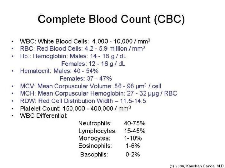

Platelets o Really not cells o Anucleated pieces of ruptured megakaryocytes o Irregular shaped o Needed for clotting process
 90% water Salts (Na,")
Plasma o o o o Liquid component of blood (matrix) 90% water Salts (Na, K, Ca, Mg, Cl, Bicarbonate) Nutrients Metabolic Wastes (urea, uric acid) Gasses Hormones

Plasma Protiens o o o Most abundant solutes Antibodies Hormones Albumin Globulins Fibrinogen

Albumin o Made by the liver o Maintains osmotic pressure of blood o Keeps water in the blood and out of the tissues o Levels are low when liver disease is present, malnutrition, Chron’s and Celiac diseases o Helps move small molecules like calcium, progesterone and medications through the blood

Globulins o Gamma globulins are antibodies o Produced by lymphocytes o Other types carry hormones and lipids

Hematopoiesis o Occurs in red bone marrow o Flat bones of skull, pelvis, ribs, sternum & proximal ends of humerus & femur o Produced in response to changing body needs o Arise from hemocytoblast (stem cell)

Hemocytoblasts o Produce two types of cells lymphoid stem cells and myeloid stem cells o Lymphoid stem cells make lymphocytes o Myeloid stem cells make all other formed elements

Red Blood Cell Formation o Have a life span of about 120 days because they don’t have a nucleus. o Dead remains are removed by phagocytosis in liver, spleen and other organs o Takes 3 -5 days from hemocytoblast to mature RBC (during this time cell synthesized Hemoglobin and ejects it’s organelle) o Rate is controlled by hormone Erythropoietin, produced by kidneys in response to decreased blood Oxygen levels

Hemostasis o Stoppage of blood flow o Three stages: vascular spasms, platelet plug formation, coagulation

Steps 1. Blood vessel is broken, exposing the underlying collage fibers 2. Platelets become sticky and cling to the damaged area 3. Platelets send chemical signals that attract more platelets, forming a platelet plug (white thrombus)

Steps 4. Platelets release serotoin, causing blood vessel to go into spasms, which narrows the vessel 5. Injured tissue releases thromboplastin 6. Thromboplastin interacts with PF 3 on the surface of the platelets, plasma clotting factors and Ca+2 to form an activator
 8. Thrombin and fibrinogen")
Steps 7. The prothrombin activator converts prothrombin to thrombin (enzyme) 8. Thrombin and fibrinogen become fibrin (long insoluble protein strands) 9. Fibrin forms a meshwork that traps the RBC’s forming the inner part of the clot 10. The clot will retract, squeezing serum out & pulling edges together

Abnormal Clots o Thrombus – clot that develops in an unbroken blood vessel o Embolus – a thrombus “on the move”

Bleeding Disorders o Thrombocytopenia – too few platelets, bruises easily, petechiae o Congenital, acquired o Treatments include medications and transfusions

Hemophilia o o o Slow clotting times Hereditary Signs show up early Different degrees Lack of clotting factors

Today: o Blood groups o Compatibility / incompatability o Lab

Blood Typing o o How many types? What are they? Why are there different types? Which ones mix / don’t mix?

Blood Groups o Experiments with blood transfusions, the transfer of blood or blood components into a person's blood stream, have been carried out for hundreds of years. Many patients have died and it was not until 1901, when the Austrian Karl Landsteiner discovered human blood groups, that blood transfusions became safer.

Frequency in the U. S. ABO Type Rh Type Percentage O + 37. 4% O - 6. 6% A + 35. 7% A - 6. 3% B + 8. 5% B - 1. 5% AB + 3. 4% AB - . 6%

Blood Cell Compatibility
67161677204bee375239cda336baf53a.ppt